of this malocclusion point to a better prognosis, it is practically impossible for the orthodontist to foresee cases that require new intervention. Many patients need retreatment, whether compensatory or orthodontic-surgical. The present study re-ports the case of a Class III patient treated at the end of the mixed dentition with the use of a face mask followed by conven-tional fixed appliances. The case remains stable 10 years after treatment completion. It was presented to the Brazilian Board of Orthodontics and Dentofacial Orthopedics (BBO) as a requirement for the title of certified by the BBO.
Keywords:Angle Class III malocclusion. Face mask. Stability.
How to cite this article: Ramos AL. Class III treatment using facial mask: Sta-bility after 10 years. Dental Press J Orthod. 2014 Sept-Oct;19(5):123-35: DOI: http://dx.doi.org/10.1590/2176-9451.19.5.123-135.bbo
Submitted: August 01, 2014 - Revised and accepted: August 17, 2014
» Patients displayed in this article previously approved the use of their facial and intraoral photographs.
Contact address: Adilson Luiz Ramos E-mail: [email protected]
» The author reports no commercial, proprietary or financial interest in the prod-ucts or companies described in this article.
1 PhD in Orthodontics, State University of São Paulo (UNESP) / Araraquara.
Adjunct professor, Department of Dentistry, State University of Maringá (UEM). Professor, Postgraduate program, Brazilian Dental Association (ABO), UNICESUMAR and UEM.
*Case report, DI 18, approved by the Brazilian Board of Orthodontics and Facial
Orthopedics (BBO).
INTRODUCTION
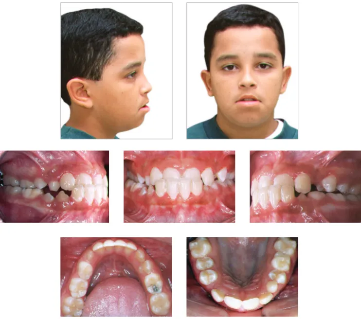
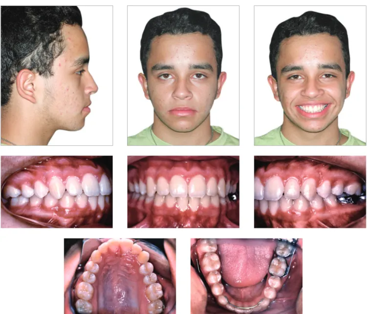
This study reports the case of a 12-year and 4-month-old patient referred to treatment with chief complaint of “crossed front teeth and protruded lower lip”.The patient sought improvements in smile and fa-cial esthetics. He was in good general health without relevant register in his medical history, and presented in regular oral hygiene with a few white spot lesions and mild gingivitis. His parents reported that anterior crossbite was not present in deciduous dentition, it only appeared ater permanent incisors eruption.
DIAGNOSIS
Facial assessment (Fig 1) revealed absence of pas-sive labial seal, a concave proile and protruded lower lip; thereby suggesting anteroposterior skeletal relation-ship (Class III).The patient was at the end of the second transitional period of the mixed dentition with Class III molar relationship (Figs 1 and 2).He presented unbal-anced maxillomandibular transverse relationship which resulted in functional unilateral posterior crossbite from tooth #12 to #16 and lower midline deviation to the right when in maximum intercuspation.
O tratamento precoce da má oclusão de Classe III pode não apresentar estabilidade em longo prazo, em decorrência do crescimento mandibular. Embora algumas características sinalizem um melhor prognóstico para o tratamento des-sa má oclusão, é praticamente impossível para o ortodontista prever qual caso irá requerer nova intervenção. Muitos pacientes precisam de retratamento, que pode ser compensatório ou mesmo ortodôntico-cirúrgico combinado. O presente artigo relata o tratamento realizado em um paciente com má oclusão de Classe III, no final da dentição mista, mediante o auxílio da máscara facial seguida de aparelhagem fixa convencional, e que continua estável após 10 anos da conclusão do tratamento. Esse caso foi apresentado à Diretoria do Board Brasileiro de Ortodontia e Ortopedia Facial (BBO) como parte dos requisitos para obtenção do título de Diplomado pelo BBO.
Figure 1 - Initial facial and intraoral photographs.
and 2 mm) as well as protruded and buccally tipped mandibular incisors (1-NB = 29o and 7 mm). These
dental features indicated dentoalveolar and skeletal components of Class III malocclusion.
In functional terms, there was little anterior man-dibular shit to the right from centric relation to maxi-mum intercuspation.
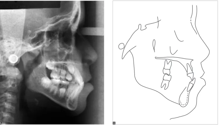
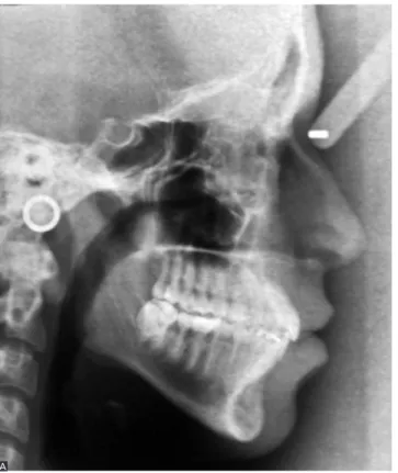
Panoramic radiograph (Fig 3), taken at the end of the second transitional period of the mixed denti-tion, did not reveal any abnormalities. Cephalometric analysis (Fig 4 and Tab 1) highlighted retrusion of the maxilla (SNA = 80o) and poor maxillomandibular
re-lationship for the patient’s age (ANB = 0.5o).He had
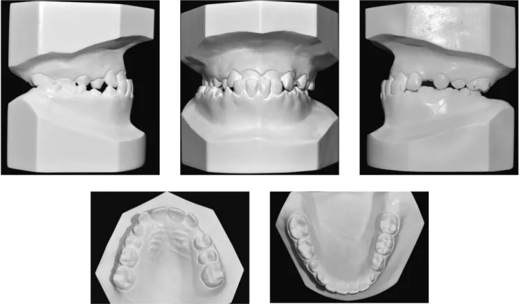
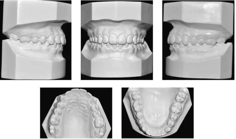
Figure 2 - Initial casts.
Figure 4 - Initial lateral cephalogram (A) and cephalometric tracing (B).
TREATMENT PLAN
Treatment planning initially aimed at correcting maxillomandibular discrepancy by means of a face mask followed by rapid expansion of the maxilla which also contributed to correct the transverse deiciency. To this end, a modiied Haas appliance associated with hooks in the region of canines used as support for protraction of the maxilla was used. Patient and parents were aware of the need for strong compliance to achieve treatment success. They were also informed that unpredictable mandibular growth could create the need for a new intervention and potential orthognathic surgery during adulthood.
Thus, they were presented with an alternative ap-proach: wait for growth completion during adolescence and have orthodontic treatment with ixed appliances and potential orthognathic surgery carried out in the fu-ture. Patient’s parents agreed on the early approach and follow-up to monitor the possibility of new interven-tions. A pre-adjusted, metallic, ixed orthodontic Roth
prescription appliance would be installed in both maxil-lary and mandibular arches. Ater treatment inishing, the retention phase would begin with the use of a re-movable wraparound retainer in the maxillary arch and an intercanine bar in the mandibular arch.
TREATMENT PROGRESS
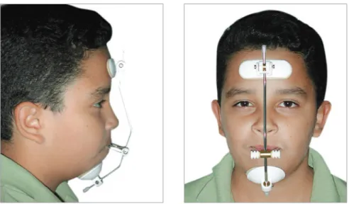
The modiied Haas appliance was manufactured based on a Hyrax expander. Maxillary expansion fol-lowed an activation protocol of ¼ turn every 12 hours for 14 days. Figure 5 illustrates the inal outcomes. Ater 14 days, the expansion appliance remained stabilized and therapy with Petit face mask began (Fig 6). A 600-g force was applied 14 hours a day for 6 to 8 months so as to over-correct overjet. However, the patient reported having ap-plied force 12 hours a day and, for this reason, 6 months were rendered necessary to correct anterior crossbite. He was then advised to wear the appliance while sleeping for 4 months so as to achieve protraction stability.
Ater removing the expander and discontinuing protraction, a removable 0.8-mm stainless steel wire palatal bar was installed to achieve transverse stability and adjust irst molars rotation. During the same ap-pointment, pre-adjusted, metallic, ixed orthodontic Roth prescription brackets were bonded on maxillary teeth, except for unerupted canines. Alignment and leveling procedures began with the use of 0.016-in
NiTi archwire followed by 0.016, 0.018 and 0.020-in stainless steel archwire associated with omega loops on molars mesial surface until canines erupted so as to preserve dental arch circumference. At treatment inishing, 0.019 x 0.025-in stainless steel rectangular archwire was used. Fluoride-varnish at 5 ppm (Du-raphat®, Colgate, Germany) was applied every three
months to prevent white spot lesions1 (Fig 7).
Figure 5 - Treatment outcomes after 14 days using the expander activated twice a day.
Figure 6 - Petit face mask with 600-g force applied.
On the mandibular arch, bracket bonding was car-ried out with individualized angulation for teeth #43 and 33 so as bracket slots were perpendicular to the root axis, thereby favoring format compensation. The sequence of archwires used for alignment and leveling was similar to that used in the maxillary arch: 0.019 x 0.025-in stainless steel rectangular archwire associated with intermaxillary Class III elastics 16 hours a day.
Importantly, maxillary canines and second molars eruption was delayed, which increased total treatment time (41 months). Let mandibular second molar was mesially and buccolingually tipped while the patient and his parents were anxious for treatment conclusion. Thus, the appliance was completely removed with only the TMA rectangular archwire remaining on teeth #36 and 37 to correct #37 positioning. This wire segment was re-moved ater 4 months when the patient brought his inal exams to the last appointment. Additionally, the patient was referred to extraction of tooth #48 which was the only mesially tipped third molar he had.
A removable wraparound retainer was continuously used in the maxillary arch for 6 months during the day and at night for 2 years during the retention phase. In the mandibular arch, however, a 0.8-mm stainless steel wire intercanine bar was installed and used for life.
RESULTS
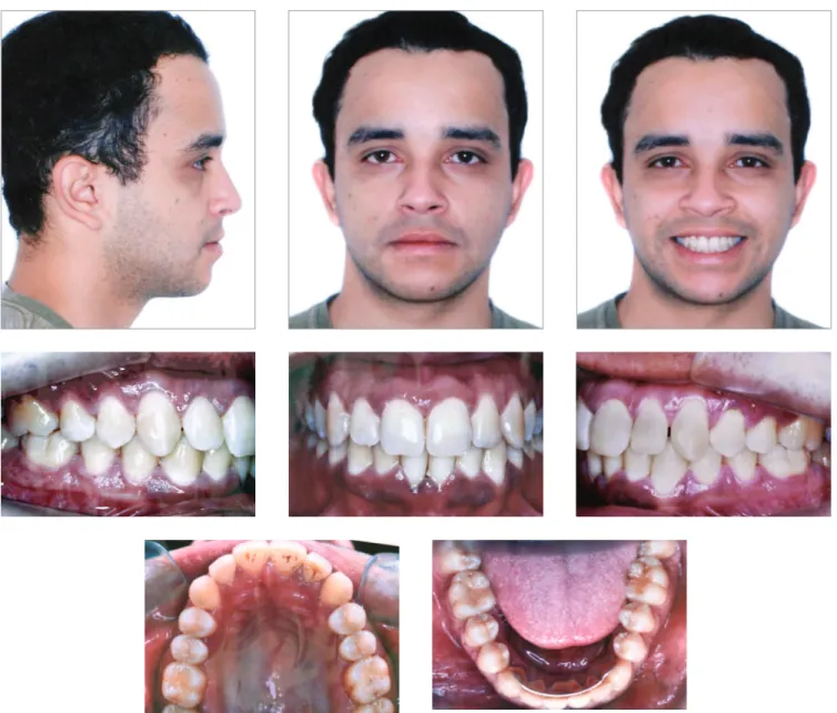
Patient’s inal exams (Figs 8 to 12)revealed har-monious lip repositioning as well as improvements in facial proile which became slightly convex. Maxillo-mandibular relationship was restored to normal-ity and ANB angle increased from 0.5o to 2o. Such
improvements were due to maxillary advancement (SNA increased in 2o).Class III relationship was
cor-rected by maxillary incisors buccal tipping (1-NA in-creased in 11o and 3 mm).Mandibular incisors
practi-cally remained in their original position. Treatment was completed without further shits between centric relation and maximum intercuspation. Occlusion guidance was restored (protrusion and laterality).
Figure 9 - Final casts.
Figure 11 - Final lateral cephalogram (A) and cephalometric tracing (B).
Figure 12 - Initial (black) and final (red) cephalometric tracings total (A) and partial (B) superimposition.
A B
In view of the above, it is reasonable to assert that treatment results were in accordance with treatment initial objectives. The patient was highly satisied with his inal facial and dental esthetics and remained aware of the need for a long-term follow-up to monitor man-dibular growth and occlusal relationship.
Total treatment time was long due to early treatment approach, delayed maxillary canines and second molars eruption as well as patient’s absence at some appoint-ments. New examinations were requested ten years
ater orthodontic treatment completion (Figs 13 to 17). They revealed excellent occlusal stability and facial bal-ance. Both panoramic radiograph (Fig 14) and lateral cephalogram (Fig 15A) revealed all aspects were with-in standards of normality. The patient was advised with-in terms of periodontal care and the need for extracting tooth #48. Control and cephalometric tracings super-imposition (Fig 16) revealed little residual mandibular growth which did not compromise facial and dental fea-tures achieved at treatment completion.
Figure 15 - Control lateral cephalogram (A) and cephalometric tracing (B) 10 years after treatment completion.
Figure 14 - Control panoramic radiograph 10 years after treatment completion.
Figure 16 - Final (red) and control cephalometric tracings superimposition 10 years after treatment (green).
Figure 17 - Initial (black), final (red) and control cephalometric tracings su-perimposition 10 years after treatment (green).
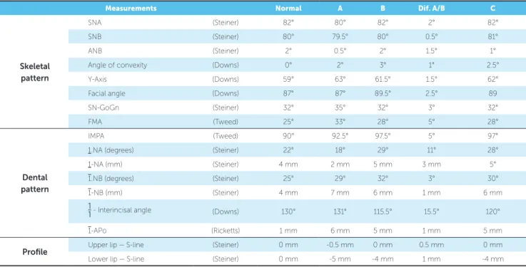
Table 1 - Initial (A), final (B) and control cephalometric values 10 years after treatment (C).
Measurements Normal A B Dif. A/B C
Skeletal pattern
SNA (Steiner) 82° 80° 82° 2° 82°
SNB (Steiner) 80° 79.5° 80° 0.5° 81°
ANB (Steiner) 2° 0.5° 2° 1.5° 1°
Angle of convexity (Downs) 0° 2° 3° 1° 2.5°
Y-Axis (Downs) 59° 63° 61.5° 1.5° 62°
Facial angle (Downs) 87° 87° 89.5° 2.5° 89
SN-GoGn (Steiner) 32° 35° 32° 3° 32°
FMA (Tweed) 25° 33° 28° 5° 28°
Dental pattern
IMPA (Tweed) 90° 92.5° 97.5° 5° 97°
1.NA (degrees) (Steiner) 22° 18° 29° 11° 28°
1-NA (mm) (Steiner) 4 mm 2 mm 5 mm 3 mm 5°
1.NB (degrees) (Steiner) 25° 29° 32° 3° 30°
1-NB (mm) (Steiner) 4 mm 7 mm 6 mm 1 mm 6 mm
1
1- Interincisal angle (Downs) 130° 131° 115.5° 15.5° 120°
1-APo (Ricketts) 1 mm 6 mm 5 mm 1 mm 5 mm
Profile Upper lip — S-line (Steiner) 0 mm -0.5 mm 0 mm 0.5 mm 0 mm
stability in 75% of cases. Treatment is considered as early when performed before permanent dentition onset, in which case better results with small chances of relapse
lent stability, patients similar to the one reported in the present study must be informed about the poten-tial need for compensatory retreatment and surgery.
1. Demito CF, Vivaldi-Rodrigues G, Ramos AL, Bowman SJ. Eicacy of a
luoride varnish in preventing white-spot lesions as measured with laser luorescence. J Clin Orthod. 2011;45(1):25-9.
2. Wells AP, Sarver DM, Proit WR. Long-term eicacy of reverse pull
headgear therapy. Angle Orthod. 2006;76(6):915-22.
3. Ngan P. Early timely treatment of Class III malocclusion. Semin Orthod. 2005;11:140-5.
4. Baccetti T, McGill JS, Franchi L, McNamara JA Jr, Tollaro I. Skeletal efects of early treatment of Class III malocclusion with maxillary expansion and face-mask therapy. Am J Orthod Dentofacial Orthop. 1998;113(3):333-43.
5. Westwood PV, McNamara JA Jr, Baccetti T, Franchi L, Sarver DM.
Long-term efects of Class III treatment with rapid maxillary expansion and face mask therapy. Am J Orthod Dentofacial Orthop. 2003;123(3):306-20.
6. Hägg U, Tse A, Bendeus M, Rabie ABM. Long-term follow-up of early
treatment with reverse headgear. Eur J Orthod. 2003;25(1):95-102. 7. Delaire J. Maxillary development revisited: relevance to the orthopaedic
treatment of Class III malocclusions. Eur J Orthod. 1997;19(3):289-311. REFERENCES
8. Oltramari PVP, Garib DG, Conti ACCF, Henriques JFC, Freitas MR.
Orthopedical treatment of Class III in diferent facial patterns. Rev Dental Press Ortod Ortop Facial. 2005;10(5):72-82.
9. Silva Filho OG, Magro AC, Capelozza Filho L. Early treatment of the Class III malocclusion with rapid maxillary expansion and maxillary protraction. Am J Orthod Dentofacial Orthop. 1998;113(2):196-203. 10. Turley P. Orthopedic correction of Class III malocclusion with
palatal expansion and custom protraction headgear. J Clin Orthod. 1988;22(5):314-25.
11. Almeida MR, Almeida RR, Oltramari-Navarro PV, Conti AC, Navarro R de L, Camacho JG. Early treatment of Class III malocclusion: 10-year clinical follow-up. J Appl Oral Sci. 2011;19(4):431-9.
12. Vaughn GA, Mason B, Moon HB, Turley PK. The efects of maxillary protraction therapy with or without rapid palatal expansion: