Does the CO
2laser reduce bond strength in different
types of ceramic brackets?
Fábio Lourenço Romano1, Giovanna Pessoti2, Rodrigo Galo3, Jaciara Miranda Gomes-Silva4,
Marília Pacífico Lucisano4, Maria Cristina Borsatto5, Paulo Nelson-Filho5
Objective: The aim of this study was to assess in vitro the influence of the CO2 laser and of the type of ceramic bracket on the shear bond strength (SBS) to enamel. Methods: A total of 60 enamel test surfaces were obtained from bovine incisors and
randomly assigned to two groups, according to the ceramic bracket used: Allure (A); Transcend (T). Each group was divided into 2 subgroups (n = 15): L, laser (10W, 3s); C, no laser, or control. Twenty-four hours after the bonding protocol using Trans-bond XT, SBS was tested at a crosshead speed of 0.5 mm/min in a universal testing machine. After deTrans-bonding, the Adhesive Remnant Index (ARI) was evaluated at 10 x magnification and compared among the groups. Data were analyzed by one-way ANOVA, Tukey’s, Mann-Whitney’s and Kruskal-Wallis tests (α = 0.05). Results: Mean SBS in MPa were: AL = 0.88 ± 0.84;
AC = 12.22 ± 3.45; TL = 12.10 ± 5.11; TC = 17.71 ± 6.16. ARI analysis showed that 73% of the specimens presented the entire ad-hesive remaining on the tooth surfaces (score 3). TC group presented significantly higher SBS than the other groups. The lased specimens showed significantly lower bond strength than the non-lased groups for both tested brackets. Conclusion: CO2
laser irradiation decreased SBS values of the polycrystalline ceramic brackets, mainly Allure.
Keywords: Carbon dioxide. Dental debonding. Lasers. Orthodontic brackets. Shear strength.
1 Professor, Universidade de São Paulo, Ribeirão Preto Dental School,
Department of Pediatric Clinics, Ribeirão Preto/SP, Brazil.
2 Dental surgeon, Universidade de São Paulo, Ribeirão Preto Dental School,
Ribeirão Preto/SP, Brazil.
3 Professor, Universidade Federal dos Vales do Jequitinhonha e Mucuri, School
of Biological and Health Sciences, Department of Dentistry, Diamantina/MG, Brazil.
4 Dental surgeon, Universidade de São Paulo, Ribeirão Preto Dental School,
Department of Pediatric Clinics, Ribeirão Preto/SP, Brazil.
5 Full professor, Universidade de São Paulo, Ribeirão Preto Dental School,
Department of Pediatric Clinics, Ribeirão Preto/SP, Brazil.
How to cite this article: Romano FL, Pessoti G, Galo R, Gomes-Silva JM, Lucisano MP, Borsatto MC, Nelson-Filho P. Does the CO2 laser reduce bond strength in different types of ceramic brackets? Dental Press J Orthod. 2017 Mar-Apr;22(2):55-60. DOI: http://dx.doi.org/10.1590/2177-6709.22.2.055-060.oar
Submitted: February 29, 2016 - Revised and accepted: October 14, 2016
» The authors report no commercial, proprietary or financial interest in the products or companies described in this article.
Contact address: Fábio Lourenço Romano Avenida do Café s/n, Monte Alegre, CEP: 14.040-904 Ribeirão Preto/SP, Brasil – E-mail: [email protected] DOI: http://dx.doi.org/10.1590/2177-6709.22.2.055-060.oar
Objetivo: o objetivo deste estudo foi avaliar in vitro a influência do laser de CO2 sobre a resistência ao cisalhamento da colagem (RCC) no esmalte dentário, usando diferentes tipos de braquetes cerâmicos. Métodos: no total, 60 superfícies de esmalte de in-cisivos bovinos foram obtidas e aleatoriamente divididas em dois grupos, de acordo com o braquete cerâmico utilizado: Allure (A) e Transcend (T). Cada grupo foi dividido em dois subgrupos (n = 15): L, laser (10W, 3s); C, sem laser, ou controle. Vinte e quatro horas após a colagem dos braquetes com o sistema Transbond XT, foi realizado o teste de resistência ao cisalhamento, com velo-cidade de 0,5 mm/min, em máquina universal de ensaios mecânicos. Após a descolagem, o Índice de Remanescente de Adesivo (IRA) foi avaliado com aumento de 10X e comparado entre os grupos. Os dados foram analisados pelo ANOVA one-way, testes de Tukey’s, Mann-Whitney’s e Kruskal-Wallis (α = 0,05). Resultados: as médias da RCC em MPa foram: AL = 0,88 ± 0,84; AC = 12,22 ± 3,45; TL = 12,10 ± 5,11; TC = 17,71 ± 6,16. A análise do IRA mostrou que 73% dos corpos de prova apresentaram todo o compósito remanescente aderido à superfície do esmalte (escore 3). O grupo TC apresentou valor significativamente maior de RCC do que os outros grupos. Os corpos de prova dos grupos com laser obtiveram valores adesivos significativamente menores do que os corpos de prova dos grupos sem laser, com ambos os tipos de braquetes. Conclusão: a irradiação com laser de CO2 diminuiu os valores de RCC dos braquetes policristalinos testados, principalmente do Allure.
INTRODUCTION
The use of ceramic brackets has become wide-spread in orthodontic treatments due to the in-creased number of adult patients seeking care and esthetic appliances.1 However, compared to
con-ventional metallic brackets, ceramic brackets are more costly, with a questionable clinical perfor-mance, since their rigid properties may cause an-tagonist tooth contact wear. Also, conventional methods for debonding ceramic brackets (pliers and drills) can cause injuries and fractures to the enam-el.2-5 The difficulties for debonding ceramic
brack-ets can be attributed to the high bond strength and to the low fracture strength of ceramics,5,6 which
can lead to iatrogenic enamel damages, bracket fractures and longer clinical chairtime.1-6
Therefore, several techniques were suggested for debonding of ceramic brackets, such as electro-thermal devices,7 ultrasound,8 solvents9 and
recent-ly the lasers.5,10-17 The use of electrothermal devices
has been an effective method in debonding ceramic brackets, however, due to irreversible heating dam-ages to the pulp, this device lost popularity among the clinicians.7
The laser debonding technique is based on con-trolled thermal softening of the adhesive resin that leads to adhesion strength degradation. Studies have shown no pulp damage when debonding brackets with a laser device.5,10,11,18
In this way, despite the good results obtained with different types of lasers, carbon dioxide (CO2) laser has been considered the best choice for removing ceramic brackets, due to the high absorption of its wavelength in ceramic surfaces.14,15
There are several variables in the studies concern-ing ceramic bracket removal by lasers, such as: laser settings, type of brackets and bonding agents, as well as the employed methodology. Therefore, in view of the increased use of ceramic brackets in orthodon-tic patients and the improvements in laser technology and adhesive dentistry in dental practice, the aim of the current investigation was to assess in vitro the in-fluence of CO2 laser use and of the type of ceramic bracket on the shear bond strength (SBS) to enamel. The null hypothesis tested was that the CO2 laser ir-radiation does not decrease SBS values of the evaluat-ed polycrystalline ceramic brackets.
MATERIAL AND METHODS
This study had no need to be submitted to an ethics committee in animal experimentation, since the sam-ples were product of commercial slaughter of animal.
Recently extracted sound bovine permanent man-dibular incisors were immersed in 0.1% thymol solu-tion at 4 oC for 1 week. Prior to use, the teeth were hand
scaled, cleaned with pumice-water slurry using Robin-son bristle brushes in a low-speed handpiece and exam-ined with a stereomicroscope (Nikon Inc. Instrument Group, Melville, NY, USA) at 10x magniication to discard those with cracks, fractures or structural abnor-malities that could interfere in the results. Sixty bovine teeth were selected, thoroughly washed in running wa-ter to eliminate storage solution traces, and maintained in distilled water at 4oC. The samples followed the ISO
TR1140519 instructions, that recommend 15 teeth per
group for shear bond strength tests.
The crowns were embedded in chemically activat-ed acrylic resin (Jet Clássico, São Paulo/SP, Brazil) and ater resin polymerization, the buccal enamel surfaces were lattened with #400- to #1200-grit silicon car-bide (SiC) papers (Buehler Ltd., Lake Bluf, IL, USA) in a low-speed polishing machine (Politriz DP-9U2; Struers, A/S, Copenhagen, Denmark) in order to ob-tain the test sites that, for standardization, were de-marcated by attaching a piece of insulating tape with a central square (5 x 5 mm) on each surface. Next, the roots were embedded in chemically activated acrylic resin (Jet Clássico, São Paulo/SP, Brazil) using polyvi-nyl chloride rings (2.1 cm diameter and 1.1 cm high) in order to facilitate the laser irradiation and the SBS test immediately ater.
The 60 enamel test surfaces were randomly as-signed to two groups (n = 30), according to the used polycrystalline ceramic bracket: A) Allure (GAC, New York, NY, USA), and T) Transcend (3M Unitek, Monrovia, CA, USA).
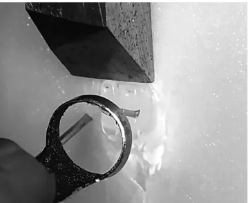
Figure 1 - Specimen with the active chisel tip acting on the upper part of the bracket base during the shear bond strength test.
CA, USA) was applied to the acid etched enamel bond-ing site in a uniform layer, slightly thinned with a mild, oil-free air stream.
The ceramic brackets for lower central incisor were bonded to the center of the specimens using Trans-bond XT composite (3M Unitek, Monrovia, CA, USA) according to the manufacturer’s recommendations. All bonding procedures were performed by the same operator, who used a pair of pliers (Ortoply, Philadel-phia, PA, USA), and the excess of material was removed with a sharp explorer (Dulex, Juiz de Fora/MG, Bra-zil). Each bracket bonding was photoactivated at a 1 mm distance between the bracket base and the light-cur-ing device for 40 s (10 s for each side of the bracket) with a visible light curing unit (XL 1500; 3M/ESPE, St. Paul, MN, USA) with a 450 mW/cm2 output
pow-er. Light intensity of each device was measured prior to each photo-activation cycle using a curing radiometer (Demetron, Danbury, CT, USA). Then the specimens were randomly divided into two subgroups (n = 15): Laser (L) and No Laser, or control (C). The specimens were stored in distilled water at 37 oC for 24 h.
Immediately before the SBS test, the samples in the laser group were irradiated. The equipment used for CO2 laser irradiation was a UM-L30 device (Shangai-Jue Hua Technology Development Shangai, PR China) emitting at 10.6 µm wavelength. The laser beam was delivered in non-contact mode. The laser tip was held perpendicularly at a 4-mm distance from the bracket surface and the light was delivered in focused mode. The parameter settings were 10 W for 3s. The laser op-erated at ultra-pulse mode with pulse duration of 100 µs (interval time: 0,01s). The time delay between laser ir-radiation and force application was up to 3s17.
The SBS was tested to failure using a knife-edge blade in a universal testing machine (Model DL 500, EMIC Ltda., São José dos Pinhais/PR, Brazil) running at a crosshead speed of 0.5 mm/min with a 50 kgf load cell (Fig 1).
Mean SBS values (in MPa) and standard deviations were calculated and data were analyzed statistically by one-way ANOVA. Tukey’s test was used for multiple comparisons at 5% signiicance level.
The debonded specimens were observed with a 10x stereomicroscope (Carl Zeiss, Goettingen, Germany) by an calibrated and experienced examiner, in order to assess the amount of resin material adhered to enamel
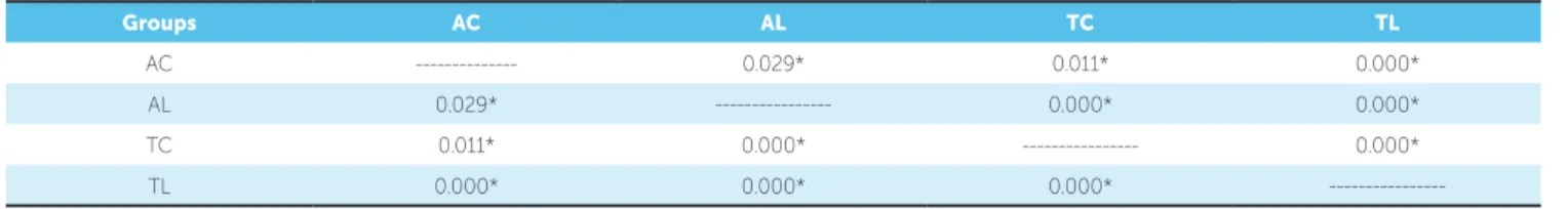
ater bracket removal, which were classiied according to the Adhesive Remnant Index (ARI) scores estab-lished by Artun and Bergland:20 0 = no adhesive
remain-ing adhered to enamel; 1 = less than half of the adhesive remaining adhered to enamel; 2 = more than half of the adhesive remaining adhered to enamel; 3 = all the ad-hesive remaining adhered to enamel. All examinations were done by a single examiner blinded to the groups to which the specimens belonged. The ARI scores data were statistically analyzed by the Kruskal-Wallis test and Mann-Whitney complementary test, at a signii-cance level of 5%.
RESULTS
SBS means (in MPa), standard deviation, and statis-tical analysis are presented in Table 1. Group TC pre-sented the highest and group AL present the lowest SBS values of all groups (p < 0.05), respectively. There was no statistically signiicant diference between groups AC and TL. Nevertheless, the SBS values were statistically lower in the CO2 laser irradiated specimens for both Al-lure and Transcend tested ceramic brackets.
DISCUSSION
Recently, Orthodontics experienced a great scien-tiic advance regarding the improvement of materials and techniques, allowing more esthetic treatments. Ce-ramic brackets ofer esthetics, durability and better col-or stability. Nevertheless, enamel cracks and fractures or bracket breakage due to the brittleness, low ductili-ty, high elastic modulus and high adhesion strength to teeth of ceramics have been observed in the convention-al debonding of these appliances.1,3
According to some authors, laser debonding of ceramic brackets eliminates damages to enamel by reducing the required force to remove it.13,14,18,21 The
lasers act softening the adhesive bonding agent by heat conductivity.11,21 Saito et al.22 concluded that
ceramic bracket debonding with CO2 laser, when adhesives containing thermal expansion microcap-sules are used, can be an effective and safe method. CO2 laser has been considered the favorite laser de-vice for debonding ceramic brackets since its wave-length is more easily absorbed by these brackets.14,15
In the present research, the CO2 laser irradiation de-creased the force required to remove the polycrys-talline ceramic brackets.
Studies have reported no pulp or enamel tissue injuries and also a decreased debonding force and operation time with CO2 laser.11,15 Lijima et al.10
have observed that the hardness and elastic modulus of enamel are not affected by CO2 laser irradiation. Nevertheless, the workable laser parameters are di-rectly related to the lasers’ ability to soften the adhe-sive resin without adversely affecting tooth tissues.
The laser settings applied (10W / 3s) in this study were based on the indings of a previous investigation done by this research group, in which these CO2 laser parameters did not increase the intrapulpal
tempera-Figure 2 - ARI scores after bracket debonding.
Groups n Mean (SD) Tukey’s test*
AC 15 12.22 (3.45) a
AL 15 0.88 (0.84) b
TC 15 17.71 (6.16) c
TL 15 12.10 (5.11) a
Table 1 - Comparison of shear bond strength means values (MPa).
* Same letters indicate no statistically significant difference.
AC = Allure Control; AL = Allure Laser; TC = Transcend Control; TL = Transcend Laser.
Table 2 - Comparison of ARI scores by p values (Mann-Whitney test).
*Changes are significant at p < 0.05.
Groups AC AL TC TL
AC --- 0.029* 0.011* 0.000*
AL 0.029* --- 0.000* 0.000*
TC 0.011* 0.000* --- 0.000*
---ture above the pulp physiologic tolerance.17 The
em-ployed ultra-pulse CO2 laser can provide short puls-es with suicient time to permit tooth tissupuls-es to cool down between pulses.15 Obata et al.11 have shown that
CO2 laser at 3W / 3s are safe settings when debonding brackets, while other authors23 reported that CO
2
la-ser irradiation at 50 w for 2 s increased 0.70oC in pulp
chamber temperatures. Yassaeiet al.5 found a 1.46°C
increase in intrapulpal temperature ater diode laser irradiation at 2.5 W for 10 s.
As regards as the type of bracket, ceramic brack-ets are constituted by alumina and are monocrystalline (sapphire) or polycrystalline structures. The monocrys-talline ceramic brackets show greater strength, but the fracture resistance is generally below that of the poly-crystalline brackets.24,25 Also, monocrystalline
brack-ets require lower laser energy for debonding than the polycrystalline brackets21 since polycrystalline units do
not allow high transmissibility, thus increasing ener-gy loss.26 Thus, in the present study, polycrystalline
brackets were used and higher values of SBS were observed for the Transcend ceramic brackets, which might be attributed to diferences in the base design, structure, and composition of the tested brackets. Per-haps if other laser parameters were used, diferent SBS values would be reported.
Under clinical conditions, adequate bracket bond strength values might be between 6 to 8 MPa in or-der to prevent risk of tooth damages at debonding.27
However, the force required to remove ceramic brack-ets can reach values of 20 MPa, which might cause tooth or bracket fractures by exceeding the cohesive strength of the enamel or the bracket.2,3,27 Confirming
these facts, in this study, Transcend ceramic bracket reached a SBS mean of 17.71 MPa, which is much higher than the ideal bracket bond strength value.
Additionally, even though the laser decreased the ce-ramic brackets bond strength, only for the Allure group this reduction (93%) reached clinically applicable values for debonding (0.88 MPa). The Transcend lased group showed a reduction in 68%, but SBS values (12.10 MPa) were still near the limit of force (11.1 MPa) that can cause tooth fractures at debonding.28 In the diferent treatments
(with laser or without laser), the adhesion of Transcend ceramic brackets were higher than Allure. However, sig-niicant reduction in the adhesion with both brackets was observed when laser was applied.
Ma et al.18 have observed that specimens
debond-ed with CO2 laser (18W / 2s) were only 25% (1.48 MPa) of the mean of the non-lased group. Other authors10 reported that CO
2 laser
irradi-ated specimens showed a 31% decrease in SBS, if they were bonded with a conventional etch and rinse adhesive system, and a 25% decrease with a self-etching adhesive system, compared with control (non-irradiated) specimens. However, these studies tested other materials and techniques.
Concerning ARI scores, the fractures ater brack-et debonding occurred predominantly at the brackbrack-et/ resin interface (73%, score 3), which reduces the risk of bracket or tooth damages.29 ARI scores 1 and 2
oc-curred as the SBS increased and score 0 was observed only in one TL group specimen, situation in which cracking of the enamel is more probable.28 These
ind-ings suggest that, in addition to sotening, the adhesive resin might have undergone thermal ablation and pho-toablation, resulting in vaporization and decomposi-tion of the material.1 The high percentage of score 3
(all composite remain ater debonding) indicated that laser irradiation didn’t cause injuries or fractures in the enamel surface, preserving the dental structure (Fig 2). In groups 1, 3 and 4, this score was predominant and in group 2, all sample obtained this same result. These re-sults are promising, and probably during laser irradia-tion procedure a slight sotening of the composite oc-curred, without heating the tooth.
In view of the above considerations and based on indings of the current investigation, the CO2 laser has been noted as a promising technology in debonding ce-ramic brackets, mainly for the Allure, since it was ob-served a greater adhesive remnant index on the tooth surface together with decreased SBS values.
CONCLUSION
The null hypothesis was rejected. The CO2 laser ir-radiation decreased the adhesion of the ceramic brackets and enhanced brackets debonding.
Acknowledgements
The authors would like to thank the National Council for Research - CNPq (#11.1.754.58.0) for the research grant and inancial support.
Authors contribution
Conception or design of the study: FLR, MCB, PNF. Data acquisition, analysis or interpretation: GP, MPL, RG. Writing the article: FLR, JMGS. Critical revision of the article: FLR, PNF. Final approval of the article: FLR. Obtained funding: GP.
1. Azzeh E, Feldon PJ. Laser debonding of ceramic brackets: a comprehensive review. Am J Orthod Dentofacial Orthop. 2003 Jan;123(1):79-83.
2. Jena AK, Duggal R, Mehrotra AK. Physical properties and clinical characteristics of ceramic brackets: a comprehensive review. Trends Biomater Artif Organs. 2007;20:201-19.
3. Jeiroudi MT. Enamel fracture caused by ceramic brackets. Am J Orthod Dentofacial Orthop. 1991 Feb;99(2):97-9.
4. Bishara SE, Fonseca JM, Boyer DB. The use of debonding pliers in the removal of ceramic brackets: force levels and enamel cracks. Am J Orthod Dentofacial Orthop. 1995 Sept;108(3):242-8.
5. Yassaei S, Soleimanian A, Nik ZE. Efects of diode laser debonding of ceramic brackets on enamel surface and pulpal temperature. J Contemp Dent Pract. 2015 Apr 1;16(4):270-4.
6. Scott GE Jr. Fracture toughness and surface cracks--the key to understanding ceramic brackets. Angle Orthod. 1988 Jan;58(1):5-8.
7. Dovgan JS, Walton RE, Bishara SE. Electrothermal debracketing: patient acceptance and efects on the dental pulp. Am J Orthod Dentofacial Orthop. 1995 Sept;108(3):249-55.
8. Boyer DB, Engelhardt G, Bishara SE. Debonding orthodontic ceramic brackets by ultrasonic instrumentation. Am J Orthod Dentofacial Orthop. 1995 Sept;108(3):262-6.
9. Larmour CJ, McCabe JF, Gordon PH. An ex vivo investigation into the efects of chemical solvents on the debond behaviour of ceramic orthodontic brackets. Br J Orthod. 1998 Feb;25(1):35-9.
10. Iijima M, Yasuda Y, Muguruma T, Mizoguchi I. Efects of CO(2) laser debonding of a ceramic bracket on the mechanical properties of enamel. Angle Orthod. 2010 Nov;80(6):1029-35.
11. Obata A, Tsumura T, Niwa K, Ashizawa Y, Deguchi T, Ito M. Super pulse CO2 laser for bracket bonding and debonding. Eur J Orthod. 1999 Apr;21(2):193-8. 12. Sarp AS, Gülsoy M. Ceramic bracket debonding with ytterbium iber laser. Lasers
Med Sci. 2011 Sept;26(5):577-84.
13. Oztoprak MO, Nalbantgil D, Erdem AS, Tozlu M, Arun T. Debonding of ceramic brackets by a new scanning laser method. Am J Orthod Dentofacial Orthop. 2010 Aug;138(2):195-200.
14. Tehranchi A, Fekrazad R, Zafar M, Eslami B, Kalhori KA, Gutknecht N. Evaluation of the efects of CO2 laser on debonding of orthodontics porcelain brackets vs. the conventional method. Lasers Med Sci. 2011 Sept;26(5):563-7.
15. Ahrari F, Heravi F, Fekrazad R, Farzanegan F, Nakhaei S. Does ultra-pulse CO(2) laser reduce the risk of enamel damage during debonding of ceramic brackets? Lasers Med Sci. 2012 May;27(3):567-74.
REFERENCES
16. Tozlu M, Ostoprak MO, Arun T. Comparison of shear bond strengths of ceramic brackets aftediferente times lags between lasing and debonding. Lasers Med Sci. 2012 Nov;27(6):1151-5.
17. Macri RT, Lima FA, Bachmann L, Galo R, Romano FL, Borsatto MC, et al. CO2 laser as auxiliary in the debonding of ceramic brackets. Lasers Med Sci. 2015 Sept;30(7):1835-41.
18. Ma T, Marangoni RD, Flint W. In vitro comparison of debonding force and intrapulpal temperature changes during ceramic orthodontic bracket removal using a carbon dioxide laser. Am J Orthod Dentofacial Orthop. 1997 Feb;111(2):203-10.
19. International Organization for Standardization. Guidance on testing of adhesion to tooth structure. ISSO/TC106/SC 1 N236, Resolution 61. CD TR 11405, Trieste, Oct 1994.
20. Artun J, Bergland S. Clinical trials with crystal growth conditioning as an alternative to acid-etch enamel pretreatment. Am J Orthod. 1984 Apr;85(4):333-40.
21. Strobl K, Bahns TL, Willham L, Bishara SE, Stwalley WC. Laser-aided debonding of orthodontic ceramic brackets. Am J Orthod Dentofacial Orthop. 1992 Feb;101(2):152-8.
22. Saito A, Namura Y, Isokawa K, Shimizu N. CO2 laser debonding of a ceramic bracket bonded with orthodontic adhesive containing thermal expansion microcapsules. Lasers Med Sci. 2015 Feb;30(2):869-74.
23. Abdul-Kader HM, Ibrahim AS. A comparative study of CO2 laser debonding of three types of ceramic brackets. J Dent Sci. 1999;2:79-84.
24. Mimura H, Deguchi T, Obata A, Yamagishi T, Ito M. Comparison of diferent bonding materials for laser debonding. Am J Orthod Dentofacial Orthop. 1995 Sept;108(3):267-73.
25. Flores DA, Caruso JM, Scott GE, Jeiroudi MT. The fracture strength of ceramic brackets: a comparative study. Angle Orthod. 1990 Winter;60(4):269-76. 26. Eliades T, Johnston WM, Eliades G. Direct light transmittance through ceramic
brackets. Am J Orthod Dentofacial Orthop. 1995 Jan;107(1):11-9.
27. Reynolds I. A review of direct orthodontic bonding. Br J Orthod. 1975;2(3):71-8. 28. Bishara SE, Fehr DE, Jakobsen JR. A comparative study of the debonding
strengths of diferent ceramic brackets, enamel conditioners, and adhesives. Am J Orthod Dentofacial Orthop. 1993 Aug;104(2):170-9.