REVIEW
I Department of Gastroenterology, Fatih University Medical School -
An-kara, Turkey
II Department of Internal Medicine, Fatih University Medical School -
An-kara, Turkey
Email: [email protected] Tel.: 90-0312-4829166 or 2126262 Received for publication on August 28, 2009 Accepted for publication on November 05, 2009
NONINVASIVE METHODS IN EVALUATION OF
INFLAMMATORY BOWEL DISEASE: WHERE DO WE
STAND NOW? AN UPDATE
Cansel Turkay,I Benan KasapogluII
doi: 10.1590/S1807-59322010000200015
Turkay C, Kasapoglu B.Noninvasive methods in evaluation of inlammatory bowel disease: where do we stand now? An updat. Clinics. 2010;65(2):221-31.
The inlammatory bowel diseases, consisting of Crohn’s disease, ulcerative colitis and indeterminate colitis, are distinguished by idiopathic and chronic inlammation of the digestive tract. The distinction between inlammatory bowel diseases and functional bowel disorders, such as irritable bowel syndrome, can be complex because they often present with similar symptoms. Rapid and inexpensive noninvasive tests that are sensitive, speciic and simple are needed to prevent patient discomfort, delay in diagnosis, and unnecessary costs. None of the current commercially available serological biomarker tests can be used as a stand-alone diagnostic in clinics. Instead, these are used as an adjunct to endoscopy in diagnosis and prognosis of the disease.Along these lines,, fecal lactoferrin and calprotectin tests seem to be one step further from other tests with larger number of studies, higher sensitivity and speciicity and wider availability.
KEYWORDS: Inlammatory bowel disease; Diagnosis; Serology; Fecal markers.
INTRODUCTION
The inlammatory bowel diseases (IBD), consistsCrohn’s disease (CD), ulcerative colitis (UC) and indeterminate colitis (IC) which are distinguished by idiopathic and chronic inlammation of the digestive tract. These diseases have been shown to result from an aberrant innate and acquired immune response to commensal microorganisms in genetically susceptible individuals.1 Currently, the incidence
of IBD is increasing worldwide, especially in Northern Europe and North America. Ethnic origin, lifestyle, presence of susceptibility regions on at least 12 chromosomes and geographical factors play a central role in the epidemiology of these diseases.2,3
The distinction between IBD and functional bowel disorders, such as irritable bowel syndrome (IBS), can be complex since they often present with similar symptoms, including abdominal distention, pain and diarrhea, and therefore, invasive and expensive tests may be necessary. The diagnoses of IBDs depend on the clinical indings after radiological, endoscopic and histological examinations. Although the division between UC and CD is generally clear, indeterminate colitis is present in 10-20% of patients with isolated colitis.4 Noninvasive tests for both the
diagnosis and follow-up of IBD have gained increasing attention. Rapid and inexpensive noninvasive tests that are sensitive, speciic and simple are necessary to prevent patient discomfort, delay in diagnosis and unnecessary costs. The biomarkers of IBD, including serological tests, fecal markers and genetically predisposed gene polymorphisms, are tools for disease diagnosis, estimation of activity, follow-up and disease prognosis.5-7 Moreover, in
and single photon emission computed tomography (SPECT) are also deined.8
Therefore, the purpose of the present study was to critically review the current literature on the diagnosis and follow-up of inflammatory bowel diseases. We systematically searched Medline and the Cochrane Database, with no language restrictions, for studies of humans on the topic of IBD diagnosis that were published between January 1960 and August 2009. The key words inflammatory bowel diseases, Ulcerative colitis, Crohn’s disease, fecal
calprotectin, lactoferrin, serology and their equivalent
Medical Subject Heading terms were used.
SEROLOGICAL MARKERS
Serological testing has been used for many years in the diagnosis of IBDs. Serological biomarkers are primarily produced upon intestinal exposure to normal commensal bacteria9,10 and might reflect a disregulated
immune inflammatory response.11,12 Most of the major
serological biomarkers utilized in IBD clinics are antibodies to microbial antigens, including yeast oligomanna
(anti-Saccharomyces cerevisiae, ASCA), bacterial outer
membrane porin C (OmpC), Pseudomonas fluorescens
bacterial sequence I2 (anti-I2) and, most recently, bacterial lagellin (CBir 1).13
All of these antibodies are predominantly found in CD but are not found in UC, except ASCA, which is identiied in 5% of UC patients. On the other hand, the human antibody,
perinuclear antineutrophil cytoplasm antibody (pANCA) is considered to be an autoantibody, although the speciic antigenic stimulation for its production remains imprecise. PANCA has currently been found in up to 70% of patients with UC and in up to 20% of patients with CD.14
Five new anti-glycan antibodies anti-chitobioside IgA (ACCA), laminaribioside IgG (ALCA), anti-manobioside IgG (AMCA) and antibodies against chemically synthesized (Σ) two major oligomannose epitopes, Man α-1,3 Man α-1,2 Man (ΣMan3) and Man α-1,3 Man α-1,2 Man α-1,2 Man (ΣMan4) are recognized recently.13,15 Since these new biomarkers have been shown
to be present only in IBD, they might signify an intestinal inlammation that is speciic to UC or CD. Moreover, these antibodies have been primarily studied in CD and have a high speciicity but poor sensitivity.
Joossens et al. investigated 86 families from Belgium and Northern France to test whether a combination of CD-associated genes and/or antibody responses to microbial antigens might be valuable in identifying healthy relatives at risk. Genetic (NOD2, NOD1, TLR4, CARD8) and new serologic markers (ASCA, ACMA, ALCA, ACCA,
ASigmaMA, OmpC, CBir1, I2) were analyzed in all of the subjects. After a follow-up of 54 months, the authors found that there was an additive risk for CD in subjects from multi-case families per additional affected relative and per additional positive antibody, and this was independent of NOD2 genetic marker.16 These new antibodies might
be important in complicated disease phenotype and might predict the need for surgery.
Recently, Mokrowiecka studied 125 IBD patients (71 UC, 31CD and 23 IC) and 45 patients with functional intestinal disorders to determine the accuracy of pANCA and ASCA in patients with IBD subgroups. In UC patients, the prevalence of pANCA was 68%, which was signiicantly higher than in CD (29%). ASCA were found signiicantly more often in CD (80.6%) than in UC patients (26.8%). The sensitivity, speciicity, positive predictive value (PPV) and negative predictive value (NPV) of pANCA for UC diagnosis were 68%, 84%, 75% and 78%, respectively, and of ASCA for CD diagnosis were 81%, 78%, 45.5% and 95%, respectively. Moreover, the combined use of these two markers provided changes in diagnostic accuracy, such that for pANCA+/ASCA- in UC the sensitivity, speciicity, PPV and NPV of results were 42%, 100%, 100% and 43%, respectively, and for pANCA-/ASCA+ in CD the results were 52%, 98.6% 94% and 82%, respectively. The authors concluded that the speciicity of these combined serological markers tended to be higher than their sensitivity, and thus, these markers are more useful in the differentiation of IBD subtypes than in screening the population.17
Anand et al. evaluated 98 adults with IBD and found that ASCA and pANCA had a 32% sensitivity and 100% specificity for Crohn’s disease, while there was a 50% sensitivity and 90% speciicity for UC.18
Interestingly, in another study, the presence of ASCA was found to be associated not only with the existence of Crohn’s disease but also with markers of disease severity and oral involvement.19
Two novel immunoglobulin A (IgA) cell wall polysaccharide antibodies, anti-laminarin (anti-L) and anti-chitin (anti-C), were analyzed during the diagnosis and phenotype differentiation of Crohn’s disease and UC. A cohort of 818 individuals with IBD (517 CD and 301 UC) were analyzed for seven anti-glycan antibodies (gASCA (anti-Saccharomyces cerevisiae) IgG, gASCA IgA, chitobioside (GlcNAc(beta1,4)GlcNAc(beta)), anti-laminaribioside (Glc(beta1,3)Glb(beta)), anti-mannobioside (Man(alpha1,3)Man(alpha)), anti-L and anti-C) and for pANCA. 20 The authors found that all of the glycan markers
anti-L and anti-C improved the ability to differentiate between CD and UC and that these antibodies were independently associated with a more aggressive CD phenotype.Chen et al. described the use of a whole E. coli proteome microarray as a novel high-throughput approach to screen and identify new serological biomarkers for IBD. With the use of protein arrays containing 4,256 E. coli K12 proteins, Chen et al. have identiied novel sets of serological biomarkers for the diagnosis of IBD that have a >80% overall accuracy and sensitivity in differentiating CD from UC.21
It is important to keep in mind that the diagnostic value of serological biomarkers can show a discrepancy among different ethnic or geographic groups. For instance, both ASCA and pANCA were found to be less sensitive in Chinese and Japanese patients, while the positivity of pANCA was shown to be higher in Mexican-American UC patients.22,23
It is also essential to emphasize that none of the current commercially available serological biomarker tests can be used alone as a diagnostic in clinics. Instead, they are used in addition to endoscopy in diagnosis and prognosis of the disease. Whether or not serologic markers have a role in screening for IBD remains controversial. However, due to the generally low sensitivity and speciicity of these markers for distinguishing IBD from non-IBD, they are generally not recommended for use as a screening test. As a consequence, speciic and sensitive IBD serologic biomarkers are desired, as well as future studies to evaluate the eficacy of current and newly identiied biomarkers.
BLOOD INFLAMMATORY MARKERS
Erythrocyte sedimentation rate (ESR), white blood cell count (WBC) and C-reactive protein (CRP) are known to be good predictors of disease activity in IBD. CRP, with its short half-life, becomes rapidly elevated soon after the onset of the inlammatory process and decreases after its resolution. Moreover evaluating CRP is simple, easily available and inexpensive. ESR is also inexpensive and easily available, but since it has a longer half-life it differs from CRP and causes a prolonged latency period after changes in IBD activity. In clinical practice, because ESR, WBC and CRP are non-speciic, they sometimes are not helpful for the differential diagnosis and follow-up of IBD.24,25
In addition, ESR has been found to be more reliable to be correlated with the disease activity.26 The pro-inlammatory
cytokines (TNF-alpha, IL-1beta IL-6, and IL-8) are also found to be elevated in IBD patients.27 However, these
are not widely available and are not speciic for intestinal inlammation.
FECAL MARKERS
Fecal markers comprise a heterogeneous group of substances that either pour out from, or are generated by, the inlamed intestinal mucosa.28 The fecal excretion of Indium
111-labeled leukocytes is considered to be the gold standard fecal marker of inlammation, with a sensitivity of 97% for the diagnosis of IBD.29 Even though the use of
radio-labeling techniques remains very important for research studies, they are not recommended for routine use due to high cost, exposure to radiation and the need for 4 days of fecal collection.
Fecal levels of Alpha1 1-antitrypsin, which is a protease inhibitor produced by the liver, macrophages and intestinal epithelium, are a useful indicator of IBD. Random levels of fecal Alpha1-antitrypsin levels are revealed to be useful in measuring CD activity, while testing a 72-h fecal clearance of Alpha1-antitrypsin is a useful method for quantiication of intestinal protein loss.30,31 Although
fecal α1-antitrypsin has been generally accepted as a useful marker of IBD, it is not routinely available and cost-effective.
Fecal excretion of another serum anti-proteinase, alpha2-macroglobulin, is also increased in IBD patients. The levels of alpha2-macroglobulin in the feces have a positive relationship with the activity index in CD but not in subjects with UC.32
T h e n e u t r o p h i l - d e r iv e d p r o t e i n s , l y s o z y m e , myeloperoxidase, calprotectin, lactoferrin, and PMN-elastase, are generally elevated in the feces of IBD patients.33-39 However, fecal lactoferrin and calprotectin are
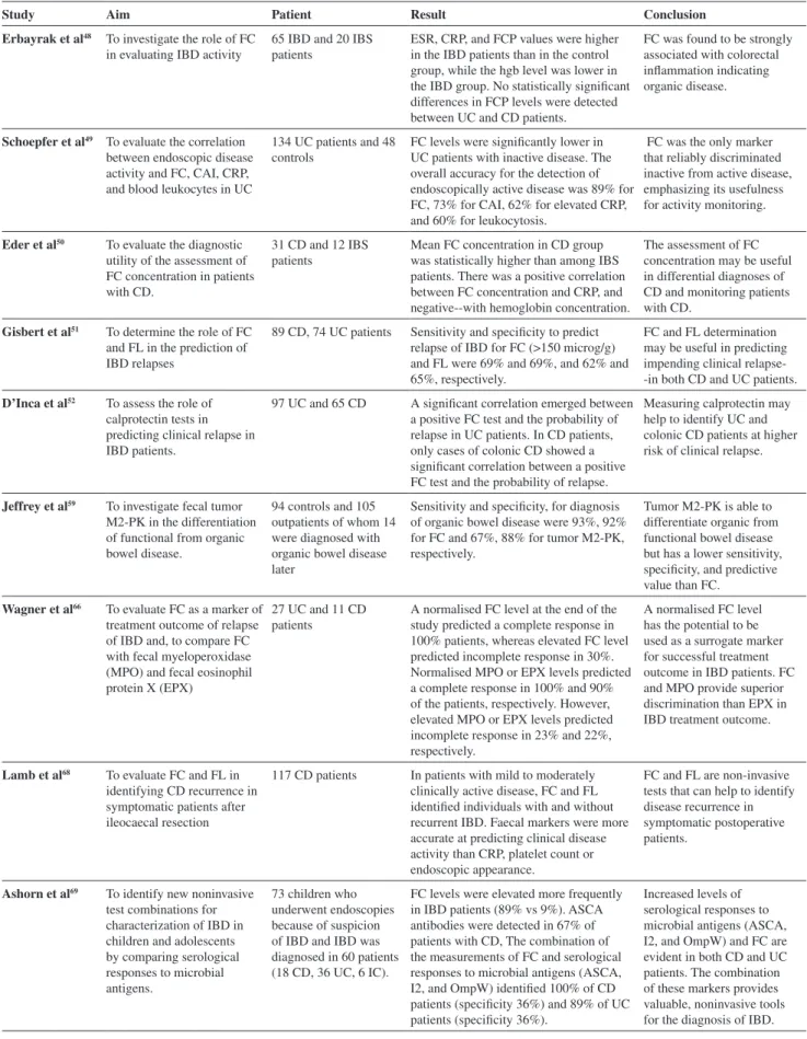
more appropriate for the differentiation of chronic IBD from IBS, and their increased levels show a positive relationship with the severity of inflammation. Some recent studies that deal with the relationship of fecal markers in IBD are summarized in Table 1.48-52, 59, 66, 68-74
Fecal Lactoferrin
Table 1 - Some recent studies about the fecal markers in the evaluation of IBD
Study Aim Patient Result Conclusion
Erbayrak et al48 To investigate the role of FC
in evaluating IBD activity
65 IBD and 20 IBS patients
ESR, CRP, and FCP values were higher in the IBD patients than in the control group, while the hgb level was lower in the IBD group. No statistically signiicant differences in FCP levels were detected between UC and CD patients.
FC was found to be strongly associated with colorectal inlammation indicating organic disease.
Schoepfer et al49 To evaluate the correlation
between endoscopic disease activity and FC, CAI, CRP, and blood leukocytes in UC
134 UC patients and 48 controls
FC levels were signiicantly lower in UC patients with inactive disease. The overall accuracy for the detection of endoscopically active disease was 89% for FC, 73% for CAI, 62% for elevated CRP, and 60% for leukocytosis.
FC was the only marker that reliably discriminated inactive from active disease, emphasizing its usefulness for activity monitoring.
Eder et al50 To evaluate the diagnostic
utility of the assessment of FC concentration in patients with CD.
31 CD and 12 IBS patients
Mean FC concentration in CD group was statistically higher than among IBS patients. There was a positive correlation between FC concentration and CRP, and negative--with hemoglobin concentration.
The assessment of FC concentration may be useful in differential diagnoses of CD and monitoring patients with CD.
Gisbert et al51 To determine the role of FC
and FL in the prediction of IBD relapses
89 CD, 74 UC patients Sensitivity and speciicity to predict relapse of IBD for FC (>150 microg/g) and FL were 69% and 69%, and 62% and 65%, respectively.
FC and FL determination may be useful in predicting impending clinical relapse- -in both CD and UC patients.
D’Inca et al52 To assess the role of
calprotectin tests in predicting clinical relapse in IBD patients.
97 UC and 65 CD A signiicant correlation emerged between a positive FC test and the probability of relapse in UC patients. In CD patients, only cases of colonic CD showed a signiicant correlation between a positive FC test and the probability of relapse.
Measuring calprotectin may help to identify UC and colonic CD patients at higher risk of clinical relapse.
Jeffrey et al59 To investigate fecal tumor
M2-PK in the differentiation of functional from organic bowel disease.
94 controls and 105 outpatients of whom 14 were diagnosed with organic bowel disease later
Sensitivity and speciicity, for diagnosis of organic bowel disease were 93%, 92% for FC and 67%, 88% for tumor M2-PK, respectively.
Tumor M2-PK is able to differentiate organic from functional bowel disease but has a lower sensitivity, speciicity, and predictive value than FC.
Wagner et al66 To evaluate FC as a marker of
treatment outcome of relapse of IBD and, to compare FC with fecal myeloperoxidase (MPO) and fecal eosinophil protein X (EPX)
27 UC and 11 CD patients
A normalised FC level at the end of the study predicted a complete response in 100% patients, whereas elevated FC level predicted incomplete response in 30%. Normalised MPO or EPX levels predicted a complete response in 100% and 90% of the patients, respectively. However, elevated MPO or EPX levels predicted incomplete response in 23% and 22%, respectively.
A normalised FC level has the potential to be used as a surrogate marker for successful treatment outcome in IBD patients. FC and MPO provide superior discrimination than EPX in IBD treatment outcome.
Lamb et al68 To evaluate FC and FL in
identifying CD recurrence in symptomatic patients after ileocaecal resection
117 CD patients In patients with mild to moderately clinically active disease, FC and FL identiied individuals with and without recurrent IBD. Faecal markers were more accurate at predicting clinical disease activity than CRP, platelet count or endoscopic appearance.
FC and FL are non-invasive tests that can help to identify disease recurrence in symptomatic postoperative patients.
Ashorn et al69 To identify new noninvasive
test combinations for characterization of IBD in children and adolescents by comparing serological responses to microbial antigens.
73 children who underwent endoscopies because of suspicion of IBD and IBD was diagnosed in 60 patients (18 CD, 36 UC, 6 IC).
FC levels were elevated more frequently in IBD patients (89% vs 9%). ASCA antibodies were detected in 67% of patients with CD, The combination of the measurements of FC and serological responses to microbial antigens (ASCA, I2, and OmpW) identiied 100% of CD patients (speciicity 36%) and 89% of UC patients (speciicity 36%).
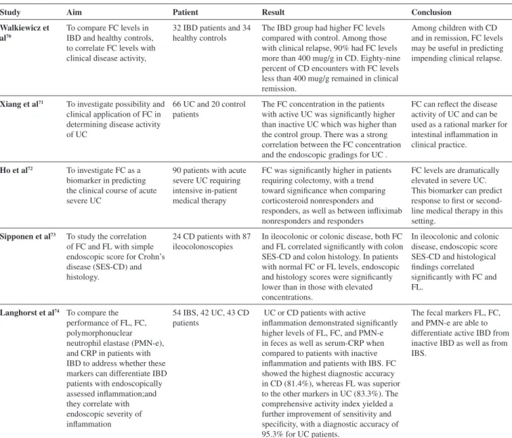
Study Aim Patient Result Conclusion
Walkiewicz et al70
To compare FC levels in IBD and healthy controls, to correlate FC levels with clinical disease activity,
32 IBD patients and 34 healthy controls
The IBD group had higher FC levels compared with control. Among those with clinical relapse, 90% had FC levels more than 400 mug/g in CD. Eighty-nine percent of CD encounters with FC levels less than 400 mug/g remained in clinical remission.
Among children with CD and in remission, FC levels may be useful in predicting impending clinical relapse.
Xiang et al71 To investigate possibility and
clinical application of FC in determining disease activity of UC
66 UC and 20 control patients
The FC concentration in the patients with active UC was signiicantly higher than inactive UC which was higher than the control group. There was a strong correlation between the FC concentration and the endoscopic gradings for UC .
FC can relect the disease activity of UC and can be used as a rational marker for intestinal inlammation in clinical practice.
Ho et al72 To investigate FC as a
biomarker in predicting the clinical course of acute severe UC
90 patients with acute severe UC requiring intensive in-patient medical therapy
FC was signiicantly higher in patients requiring colectomy, with a trend toward signiicance when comparing corticosteroid nonresponders and responders, as well as between inliximab nonresponders and responders
FC levels are dramatically elevated in severe UC. This biomarker can predict response to irst or second-line medical therapy in this setting.
Sipponen et al73 To study the correlation
of FC and FL with simple endoscopic score for Crohn’s disease (SES-CD) and histology.
24 CD patients with 87 ileocolonoscopies
In ileocolonic or colonic disease, both FC and FL correlated signiicantly with colon SES-CD and colon histology. In patients with normal FC or FL levels, endoscopic and histology scores were signiicantly lower than in those with elevated concentrations.
In ileocolonic and colonic disease, endoscopic score SES-CD and histological indings correlated signiicantly with FC and FL.
Langhorst et al74 To compare the
performance of FL, FC, polymorphonuclear neutrophil elastase (PMN-e), and CRP in patients with IBD to address whether these markers can differentiate IBD patients with endoscopically assessed inlammation;and they correlate with endoscopic severity of inlammation
54 IBS, 42 UC, 43 CD patients
UC or CD patients with active inlammation demonstrated signiicantly higher levels of FL, FC, and PMN-e in feces as well as serum-CRP when compared to patients with inactive inlammation and patients with IBS. FC showed the highest diagnostic accuracy in CD (81.4%), whereas FL was superior to the other markers in UC (83.3%). The comprehensive activity index yielded a further improvement of sensitivity and speciicity, with a diagnostic accuracy of 95.3% for UC patients.
The fecal markers FL, FC, and PMN-e are able to differentiate active IBD from inactive IBD as well as from IBS.
FC: Fecal calprotectin, IBD:Inlammatory bowel disease, IBS: Irritable bowel syndrome, ESR: Erythrocyte sedimentation rate, CRP: C-reactive pro-tein, UC: Ulcerative colitis, CD: Crohn disease, CAI: Clinical activity index, FL: Fecal lactoferrin, IC: Indetermined colitis.
Table 1 - Some recent studies about the fecal markers in the evaluation of IBD (cont.)
Dai et al. studied a total of 177 fresh stool samples collected from 42 active UC, 17 inactive UC, 13 active CD, 5 inactive CD, 41 infectious bowel diseases, 25 IBS and 34 healthy volunteers to evaluate the relationship between fecal lactoferrin and intestinal inlammation by quantitative analysis. Fecal lactoferrin was found to be signiicantly higher in active IBD than in inactive IBD, IBS and infectious bowel disease. The sensitivity and speciicity of fecal lactoferrin for UC were 92% and 88%, respectively, and for CD were 92% and 80%, respectively. As a result of this study, fecal lactoferrin was found to be a sensitive and speciic marker in measuring the activity of IBD and a valid method for discriminating between inlammatory and non-inlammatory bowel diseases.42
Kane et al. compared 104 CD, 80 UC and 31 IBS patients with 56 healthy controls to determine the sensitivity and speciicity of fecal lactoferrin concentrations for IBD or IBS. The study found that fecal lactoferrin was 90% speciic for identifying inlammation in patients with active IBD, and elevated levels of lactoferrin were 100% speciic in ruling out IBS.43
Hexagon-OBTI (immunochromatographic test for detection of human hemoglobin), and LEUKO-TEST (lactoferrin latex-agglutination test). Also, the blinded serum samples were measured for the ASCA (ELISA) and pANCA (immunofluorescence) antibodies. The authors found that fecal calprotectin and lactoferrin could accurately discriminate between IBD and IBS. Moreover, there was only a marginal improvement in diagnostic accuracy when ASCA and pANCA were also involved.44
Another study of 20 patients with IBS, 36 with IBD (24 CD, 12 UC) and 18 with other forms of colitis (8 infectious colitis, 5 ischemic colitis, 5 medication-induced colitis) was conducted to evaluate the accuracy of four different fecal markers in discriminating between IBS, IBD and other forms of colitis. In this study, blinded fecal samples were measured for calprotectin ( with PhiCal-Test, ELISA), lactoferrin (with IBD-SCAN, ELISA), with Hexagon OBTI (immunochromatographic test for detection of human hemoglobin) and with LEUKO-TEST (lactoferrin latex-agglutination test). The overall accuracies for discriminating IBS from IBD or other forms of colitis were as follows: IBD-SCAN, 91%; PhiCal-Test, 89 %; LEUKO-TEST, 92%; Hexagon OBTI, 91%; C-reactive protein, 89%; and blood leukocytes, 92%. The differentiation of IBD from other forms of colitis usingfecal markers had an overall accuracy ranging from 43 to 50%. The feasibility of fecal sampling in outpatients was high, with an acceptance rate of 95%. In conclusion, the IBD-SCAN and PhiCal-Test had the best overall accuracy for the detection of colitis, followed by the LEUKO-TEST, Hexagon OBTI, C-reactive protein and blood leukocytes.45
Fecal lactoferrin might be a helpful noninvasive diagnostic tool for the detection of colitis; however, since it is unspeciic, its role in the diagnosis and monitoring of IBD is still questionable. Further studies are necessary to determine its exact place in routine clinical practice.
Fecal Calprotectin
Calprotectin is a calcium-binding protein that inhibits metalloproteinases, hasantibacterial and antifungal activities and induces apoptosis in malignant and nonmalignant cell cultures.46 Calprotectin constitutes 60% of neutrophil
cytosolic proteins and is an abundant protein found in all body luids in proportion to the degree of inlammation. Calprotectin has many clinical advantages. It is resistant to bacterial degradation in the gut and is stable in stool for up to one week at room temperature, allowing delays in transporting the sample to the laboratory. Furthermore, calprotectin can be readily quantified using ELISA. Notably, random stool samples of <5 g show calprotectin
concentrations equivalent to 24-hour homogenized specimens, demonstrating that calprotectin is uniformly scattered throughout the feces.47
Since calprotectin is primarily derived from neutrophils, its concentration is directly proportional with neutrophil migration toward the intestinal tract. Many studies have dealt with the role of calprotectin in IBD diagnosis and follow-up (Table 1). The leukocyte proteins calprotectin, lactoferrin, lysozyme, myeloperoxidase, and PMN-elastase were compared in fecal samples of three consecutive feces (e.g., three days) in 40 healthy persons, 39 patients with chronic IBD (21 with CD and 18 with UC) and 40 patients with IBS. From this comparison, levels of all of the fecal leukocyte markers in IBS were found to be in the range of healthy patients. Moreover, fecal PMN-elastase and calprotectin still differentiated between chronic IBD and IBS and still correlated with the severity of inlammation.34
In our study of 65 IBD patients (14 CD and 51 UC) and 20 outpatients diagnosed with IBS according to Roma II criteria, fecal calprotectin was found to be strongly associated with colorectal inflammation, indicating the presence of organic disease.48
Another study was conducted to evaluate the correlation between endoscopic disease activity and fecal calprotectin. The results of the Clinical Activity Index (CAI), CRP and blood leukocytes in 134 UC patients found that endoscopic disease activity correlated closest with the presence of calprotectin. The overall accuracy for the detection of endoscopically active diseases (score >/=4) was 89% for calprotectin, 73% for CAI, 62% for elevated CRP and 60% for leukocytosis. In conclusion, fecal calprotectin was the only marker that reliably discriminated an inactive disease from mild, moderate and highly active diseases, highlighting its usefulness for monitoring activity.49
In a different study of 31 patients diagnosed with CD, the mean calprotectin concentration in the CD group was statistically higher than that of the IBS patients. A concentration of 16.01 mg/l calprotectin had 67.7% sensitivity and 66.7% speciicity in distinguishing between CD and IBS. In this respect,the assessment of fecal calprotectin concentration might also be useful for differentiating CD from IBSCD and IBS.50
µg/g) calprotectin concentrations (30% versus 7.8%; P < 0.001) or positive lactoferrin (25% versus 10%; P < 0.05). The sensitivity and speciicity of fecal calprotectin (>150 µg/g) to predict relapse were 69% and 69%, respectively. The corresponding values for lactoferrin were 62% and 65%, respectively. As a result, it was concluded that the determination of fecal calprotectin and lactoferrin might be useful in predicting an impending clinical relapse, especially during the following 3 months of remission, in both CD and UC patients.51
Similarly, in another study with 97 UC and 65 CD patients in clinical remission, a significant correlation was found between a positive calprotectin test and the probability of relapse in UC patients (P= 0.000). However, in CD patients, only cases of colonic CD had a signiicant correlation between a positive calprotectin test and the probability of relapse (P= 0.02).52 Although fecal
calprotectin levels are considered to change with age, 50 µg/g of the suggested cut-off level is considered to be useful for all age groups over 4 years old.53
However, there are 4 main handicaps of fecal calprotectin to be kept in mind:
• In some studies, low-dose aspirin treatment did not in-crease fecal calprotectin levels, although the use of non-steroidal anti-inlammatory drugs (NSAIDs) might cause an increase in calprotectin levels due to NSAID-induced enteropathy in patients without IBD.54,55
• Any bleeding in the body over 100 ml, including men-strual bleedings, might increase fecal calprotectin lev-els.56
• Some authors suggest that, although fecal calprotectin is considered to be evenly distributed,factors other than disease might contribute to the signiicant intraindividual biological variations of it57.
• Since levels of fecal calprotectin increase in any
condition that causes neutrophil migration to the gut, including neoplasms and infections, the sensi-tivity of fecal calprotectin is not as high as desired.
Fecal calprotectin is an easy, inexpensive, sensitive and speciic way to evaluate IBD. Despite the fact that levels of fecal calprotectin have an important role in diagnosis, follow-up, prediction of relapses and assessment of response to treatment, it still has some disadvantages and can only be used as a complementary test.
Fecal Pyruvate Kinase
The dimeric isoform of pyruvate kinase (tumor M2-PK), suggested to be a marker of colorectal cancer, has also recently been suggested to be a marker of gastrointestinal inlammation.58
Jeffery et al. studied 105 gastroenterology outpatients with a possible diagnosis of organic bowel disease and 94 controls to investigate the role of fecal tumor M2-PK in the differentiation of functional disease from organic bowel disease. The sensitivity, speciicity and positive and negative likelihood ratios for diagnosis of organic bowel disease were found to be, respectively, 93%, 92%, 11.6 and 0.07 for calprotectin, and, respectively, 67%, 88% 5.6 and 0.18 for tumor M2-PK. Calprotectin, in combination with tumor M2-PK, had a sensitivity of 64%, a speciicity of 98% and likelihood ratios of 32 and 0.03. Tumor M2-PK was useful for the differentiation of organic disease from functional bowel disease but had a lower sensitivity, speciicity and predictive value than calprotectin.59
The clinical value of fecal pyruvate kinase in IBD patients requires further study.
Rectal Nitric Oxide
Nitric oxide (NO) is an endogenously produced gas with numerous physiological roles. In response to acute proinlammatory cytokines, leukocytes and epithelial cells express inducible nitric oxide synthase (NOS), which leads to the production and accumulation of signiicant quantities of NO.60
The level of rectal NO correlates with disease activity in IBD patients and it markedly decreases in response to anti-inlammatory treatment. This minimally invasive and rapid test is shown to be useful for discriminating between active bowel inlammation and IBS.61 Reinders et al. also studied
23 healthy volunteers and 32 patients with IBD to compare calprotectin and rectal NO levels. These authors found that patients with IBD had greatly increased NO and calprotectin levels compared to healthy volunteers (p <0.001). Moreover, there was a weak correlation between rectal NO levels, disease activity and the number of loose stools in IBD patients (Spearman’s rho 0.37 and 0.51, respectively; p <0.05); interestingly, there was no correlation between NO and calprotectin levels.62
Ljung et al. studied 22 UC and 24 CD patients to explore rectal nitric oxide (NO) as a biomarker for the treatment response in IBD. Patients with active UC and CD displayed markedly increased rectal NO levels compared to the controls. Rectal NO correlated weakly with disease activity in both UC and CD. Interestingly, the patients’ refractory to steroid treatment only slightly increased NO levels compared to those with a therapeutic response. In this respect, the rectal NO level might be a useful biomarker for the treatment response in IBD, since low NO levels are predictive of a poor clinical response to steroid treatment.63
test and more expensive than many other fecal tests. More studies are necessary to reveal the exact role of rectal NO levels in IBD patients.
Fecal Myeloperoxidase
Myeloperoxidase, an enzyme that functions in the oxygen-dependent killing of microorganisms, is released from the primary granules of neutrophils during acute inflammation. The concentration of myeloperoxidase is also proportional to the number of neutrophils within that region.64
Silberer et al. compared ive different leukocyte proteins, calprotectin, lactoferrin, lysozyme, myeloperoxidase and PMN-elastase and determined their levels by immunoassay in the feces of patients with IBD and IBS and of healthy persons. The areas under the ROC curves of PMN-elastase and calprotectin were not signiicantly different (p = 0.327), whereas PMN-elastase or calprotectin vs. the other proteins were signiicantly different (p < 0.001). The results suggest that fecal PMN-elastase and calprotectin are important for the differentiation of chronic IBD from IBS. The authors also found that PMN-elastase and calprotectin levels were correlated with the endoscopically classiied severity of inlammation but not the myeloperoxidase.34
However, Peterson et al. found a relationship between fecal myeloperoxidase levels and the histological indices of disease activity in UC.65
Similarly, Wagner et al. showed that normalized MPO levels predicted a complete response to treatment to treatmentin 100% of the patients, as did normalized fecal calprotectin levels. However, elevated MPO levels predicted an incomplete response in 23% patients.66
In this respect, myeloperoxidase might potentially be used as a surrogate marker for a successful treatment outcome in IBD patients, similar to calprotectin. Further investigations are necessary to identify the clinical role of fecal myeloperoxidase in IBD.
Fecal Eosinophil Protein X
Eosinophil protein X (EPX) is released by activated eosinophil granulocytes, which are abundant in the mucosa in active IBD.67 Fecal EPX levels are mainly studied as
an indicator of the treatment outcome in relapses of IBD. Wagner et al. showed that normalized EPX levels have predicted a complete response to treatment in 90%; however, an incomplete response was predicted in 22% of the patients. In this respect, FC and MPO provide superior discrimination compared to EPX in IBD treatment outcome.66 Moreover,
fecal EPX levels are also beneficial complements to
endoscopical and histopathological evaluations in the daily care of patients with UC.65 Still, more studies are necessary
to reveal the clinical role of fecal EPX in IBD.
CONCLUSION
Since inlammatory bowel diseases are chronic, fast, easily available and inexpensive noninvasive tests that are sensitive, speciic and simple are necessary for diagnosis and follow-up. A differential diagnosis of organic and inorganic diseases is also important since they might have similar symptoms. Along these lines, fecal lactoferrin and calprotectin tests seem to be one step further from other tests with larger number of studies, higher sensitivity and speciicity and wider availability.
Take-home points:
Ø None of the current commercially available serological biomarker tests can be used by themselvesin clinics for diagnosis and follow up. Instead, the tests are used as an adjunct to endoscopy in diagnosis and prognosis of the disease.
Ø The erythrocyte sedimentation rate (ESR), white blood cell count and C-reactive protein (CRP) are good predic-tors of disease activity in irritable bowel diseases (IBDs). However, since they are non-speciic, they are sometimes not helpful for the differential diagnosis and follow-up of IBD.
Ø Indium 111-labeled leukocytes are considered to be the gold standard fecal marker of inlammation, with a 97% sensitivity for the diagnosis of IBD. However, due to their high cost, the exposure to radiation and the need for prolonged fecal collections of 4 days, they are not recommended for routine use.
Ø Even though fecal α1-antitrypsin and alpha2-macroglob-ulin are generally accepted as useful markers of IBD, they are not routinely available or cost-effective. Ø Fecal lactoferrin might be a helpful as a noninvasive
di-agnostic tool for the detection of colitis; however, since it is unspeciic, its role in diagnosis and monitoring of IBD remains questionable. Fecal calprotectin is an easy, inexpensive, sensitive and speciic method with which to evaluate IBD. Although levels of fecal calprotectin are important in all diagnoses, follow-ups, predictions of relapses and assessment of response to the treatment, it still can only be used as a complementary test. Ø Tumor M2-PK differentiates organic disease from
func-tional bowel disease but has a lower sensitivity, speciic-ity and predictive value than does fecal calprotectin. Ø Rectal nitric oxide is a minimally invasive test and is
Ø Fecal myeloperoxidase and eosinophil protein X have potential as a surrogate marker for the determination of successful treatment outcomes in IBD patients, similar to calprotectin.
Ø Further studies are necessary to elucidate the exact role of fecal markers in IBD evaluation.
REFERENCES
1. Wirtz,S, Neurath MF. Mouse models of inlammatory bowel disease, Adv. Drug Deliv. Rev. 2007;59:1073-83.
2. Hildebrand H, Finkel Y, Grahnquist L, Lindholm J, Ekbom A, Askling J. Changing pattern of paediatric inlammatory bowel disease in northern Stockholm 1990–2001. Gut 2003;52:1432-34.
3. Baumgart DC. The Diagnosis and Treatment of Crohn’s Disease and Ulcerative Colitis. Dtsch Arztebl Int. 2009;106:123-33.
4. G u i n d i M , R i d d e l l R H . I n d e t e r m i n a t e c o l i t i s . J . C l i n . Pathol.2004;57:1233-44.
5. Nikolaus S, Schreiber S. Diagnostics of inlammatory bowel disease. Gastroenterology. 2007;133:1670-89.
6. Langhorst J, Elsenbruch S, Koelzer J, Rueffer A, Michalsen A, Dobos GJ. Noninvasive markers in the assessment of intestinal inlammation in inlammatory bowel diseases: performance of fecal lactoferrin, calprotectin, and PMNelastase, CRP, and clinical indices. Am J Gastroenterol. 2008;103:162-9.
7. Van Limbergen J, Russell RK, Nimmo ER, Ho GT, Arnott ID, Wilson DC, et al. Genetics of the innate immune response in inlammatory bowel disease. Inlamm Bowel Dis. 2007; 13:338-55.
8. Bruining DH, Loftus EV. Current and future diagnostic approaches: from serologies to imaging. Curr Gastroenterol Rep.2007;9:489-96. 9. Li X, Conklin L, Alex P. New serological biomarkers of inlammatory
bowel disease, World J. Gastroenterol.2008;14:5115-24.
10. Vermeire S, Vermeulen N, Van AG, Bossuyt X., and Rutgeerts P. (Auto) antibodies in inlammatory bowel diseases. Gastroenterol. Clin. North Am. 2008; 37:429-38.
11. Strober W, Fuss IJ, Blumberg RS. The immunology of mucosal models of inlammation, Annu. Rev. Immunol. 2002;20:495-549.
12. Papp M, Altorjay I, Norman GL, Shums Z, Palatka K, Vitalis Z, et al. Seroreactivity to microbial components in Crohn’s disease is associated with ileal involvement, noninlammatory disease behavior and NOD2/ CARD15 genotype, but not with risk for surgery in a Hungarian cohort of IBD patients. Inlamm Bowel. Dis. 2007;13,984-92.
13. Papp M, Altorjay I, Dotan N, Palatka K, Foldi I, Tumpek J, et al. New serological markers for inlammatory bowel disease are associated with earlier age at onset, complicated disease behavior, risk for surgery, and NOD2/CARD15 genotype in a Hungarian IBD cohort, Am J Gastroenterol. 2008;103,665-81.
14. Peyrin-Biroulet L, Standaert-Vitse A, Branche J, Chamaillard M. IBD serological panels: facts and perspectives. Inlamm Bowel Dis. 2007;13:1561-6.
15. Ferrante M, HenckaertsL, Joossens M, Pierik M, Joossens S, Dotan N, et al. New serological markers in inlammatory bowel disease are associated with complicated disease behaviour. Gut. 2007;56:1394-403.
16. Joossens M, Van Steen K, Branche J, Sendid B, Rutgeerts P, Vasseur F,et al. Familial aggregation and antimicrobial response dose-dependently affect the risk for Crohn’s disease. Inlamm Bowel Dis. 2009 Jun 5. 17. Mokrowiecka A, Daniel P, Słomka M, Majak P, Malecka-Panas E.
Clinical utility of serological markers in inlammatory bowel disease. Hepatogastroenterology. 2009; 56:162-6.
18. Anand V, Russell AS, Tsuyuki R, Fedorak R. Perinuclear antineutrophil cytoplasmic autoantibodies and anti-Saccharomyces cerevisiae antibodies as serological markers are not speciic in the identiication of Crohn’s disease and ulcerative colitis. Can J Gastroenterol. 2008;22:33-6.
19. Russell RK, Ip B, Aldhous MC, MacDougall M, Drummond HE, Arnott ID, et al. Anti-Saccharomyces cerevisiae antibodies status is associated with oral involvement and disease severity in Crohn disease.J Pediatr Gastroenterol Nutr. 2009;48:161-7.
20. Seow CH, Stempak JM, Xu W, Lan H, Grifiths AM, Greenberg GR, et al. Novel anti-glycan antibodies related to inlammatory bowel disease diagnosis and phenotype. Am J Gastroenterol. 2009;104:1426-34. 21. Chen CS, Sullivan S, Anderson T, Tan AC, Alex PJ, Brant SR, Cuffari
C, et al. Identiication of novel serological biomarkers for inlammatory bowel disease using Escherichia coli proteome chip. Mol Cell Proteomics. 2009;8:1765-76.
22. Hisabe T, Matsui T, Sakurai T, Murakami Y, Tanabe H, Matake H, et al. Anti- Saccharomyces cerevisiae antibodies in Japanese patients with inlammatory bowel disease: diagnostic accuracy and clinical value. J Gastroenterol. 2003;38:121-6.
23. Lawrance IC, Murray K, Hall A, Sung JJ, Leong R. A prospective comparative study of ASCA and pANCA in Chinese and Caucasian IBD patients. Am J Gastroenterol 2004; 99: 2186-94.
24. Osada T, Ohkusa T, Okayasu I, Yoshida T, Hirai S, Beppu K, et al. Correlations among total colonoscopic indings, clinical symptoms, and laboratory markers in ulcerative colitis. J Gastroenterol Hepatol. 2008;23 Suppl 2:S262-7.
25. Solem CA, Loftus EV Jr, Tremaine WJ, Harmsen WS, Zinsmeister AR, Sandborn WJ. Correlation of C-reactive protein with clinical, endoscopic, histologic, and radiographic activity in inlammatory bowel disease. Inlamm Bowel Dis. 2005;11:707-12.
26. Ha JS, Lee JS, Kim HJ, Moon TG, Chang DK, Lee JH, et al. Comparative usefulness of erythrocyte sedimentation rate and C-reactive protein in assessing the severity of ulcerative colitis.Korean J Gastroenterol. 2006;48:313-20.
28. Poullis A, Foster R, Northield TC, Mendall MA. Review article: faecal markers in the assessment of activity in inlammatory bowel disease. Aliment Pharmacol Ther 2002;16:675-81.
29. Saverymuttu SH, Peters AM, Lavender JP, Pepys MB, Hodgson HJ, Chadwick SV. Quantitative fecal indium 111-labeled leukocyte excretion in the assessment of disease in Crohn’s disease.Gastroenterology. 1983;85:1333-9.
30. Karbach U, Ewe K, Bodenstein H. Alpha 1-antitrypsin, a reliable endogenous marker for intestinal protein loss and its application in patients with Crohn’s disease. Gut 1983;24:718-23.
31. Becker K, Berger M, Niederau C, Frieling T. Individual fecal alpha 1- antitrypsin excretion relects clinical activity in Crohn’s disease but not in ulcerative colitis. Hepatogastroenterology. 1999;46:2309-14. 32. Becker K, Niederau C, Frieling T. Fecal excretion of alpha 2
macroglobulin: a novel marker for disease activity in patients with inlammatory bowel disease. Z Gastroenterol 1999;37:597-605. 33. Crama-Bohbouth GEBI, Pena AS. Signiicance of faecal lysozme
excretion and alph-1-antitrypsin clearance in the assessment of activity of inlammatory bowel disease. In: Ge B, editor. Activity Related Abnormalities in Inlammatory Bowel Disease. Woerden-Huybregts Press; 1998. p. 89-103.
34. Silberer H, Küppers B, Mickisch O, Baniewicz W, Drescher M, Traber L, et al. Fecal leukocyte proteins in inlammatory bowel disease and irritable bowel syndrome. Clin Lab 2005;51:117-26.
35. Langhorst J, Elsenbruch S, Mueller T, Rueffer A, Spahn G, Michalsen A, et al. Comparison of 4 neutrophilderive proteins in feces as indicators of disease activity in ulcerative colitis. Inlamm Bowel Dis. 2005;11:1085-91.
36. Saiki T. Myeloperoxidase concentrations in the stool as a new parameter of inlammatory bowel disease. Kurume Med J. 1998;45:69-73. 37. Dwarakanath AD, Finnie IA, Beesley CM, O’Dowd GM, Nash J, Tsai
HH, et al. Differential excretion of leucocyte granule components in inlammatory bowel disease: implications for pathogenesis. Clin Sci. 1997;92:307-13.
38. Adeyemi EO, Hodgson HJ. Faecal elastase relects disease activity in active ulcerative colitis. Scand J Gastroenterol. 1992;27:139-42. 39. Sugi KSO, Hiarata I, Katsu K. Faecal lactoferrin as a marker of disease
activity in inlammatory bowel disease: comparison with other neutrophil derived proteins. Am J Gastroenterol 1996;91:927-34.
40. Guerrant RL, Araujo V, Soares E, Kotloff K, Lima AA, Cooper WH, et al. Measurement of fecal lactoferrin as a marker of fecal leukocytes. J Clin Microbiol. 1992;30:1238-42.
41. Angriman I, Scarpa M, D’Incà R, Basso D, Ruffolo C, Polese L, et al. Enzymes in feces: useful markers of chronic inlammatory bowel disease. Clin Chim Acta 2007;381:63-8.
42. Dai J, Liu WZ, Zhao YP, Hu YB, Ge ZZ. Relationship between fecal lactoferrin and inlammatory bowel disease. Scand J Gastroenterol. 2007;42:1440-4.
43. Kane SV, Sandborn WJ, Rufo PA, Zholudev A, Boone J, Lyerly D, et al. Fecal lactoferrin is a sensitive and speciic marker in identifying intestinal inlammation.Am J Gastroenterol. 2003;98:1309-14.
44. Schoepfer AM, Trummler M, Seeholzer P, Seibold-Schmid B, Seibold F. Discriminating IBD from IBS: comparison of the test performance of fecal markers, blood leukocytes, CRP, and IBD antibodies. Inlamm Bowel Dis. 2008;14:32-9.
45. Schoepfer AM, Trummler M, Seeholzer P, Criblez DH, Seibold F. Accuracy of four fecal assays in the diagnosis of colitis. Dis Colon Rectum. 2007;50:1697-706.
46. Steinbakk M, Naess-Andresen CF, Lingaas E, Dale I, Brandtzaeg P, Fagerhol MK. Antimicrobial actions of calcium binding leucocyte L1 protein, calprotectin. Lancet. 1990;336:763-5.
47. Røseth AG, Fagerhol MK, Aadland E, Schjønsby H. Assessment of the neutrophil dominating protein calprotectin in feces. A methodologic study. Scand J Gastroenterol 1992;27:793-8.
48. Erbayrak M, Turkay C, Eraslan E, Cetinkaya H, Kasapoglu B, Bektas M. The role of fecal calprotectin in investigating inlammatory bowel diseases. Clinics. 2009;64:421-5.
49. Schoepfer AM, Beglinger C, Straumann A, Trummler M, Renzulli P, Seibold F. Ulcerative colitis: Correlation of the Rachmilewitz endoscopic activity index with fecal calprotectin, clinical activity, c-reactive protein, and blood leukocytes. Inlamm Bowel Dis. 2009 May 21.
50. Eder P, Stawczyk-Eder K, Krela-Ka mierczak I, Linke K. Clinical utility of the assessment of fecal calprotectin in Le niowski-Crohn’s disease. Pol Arch Med Wewn. 2008;118:622-6.
51. Gisbert JP, Bermejo F, Pérez-Calle JL, Taxonera C, Vera I, McNicholl AG, et al. Inlamm Bowel Dis. Fecal calprotectin and lactoferrin for the prediction of inlammatory bowel disease relapse.2009;15:1190-8. 52. D’Incà R, Dal Pont E, Di Leo V, Benazzato L, Martinato M, Lamboglia
F, et al. Can calprotectin predict relapse risk in inlammatory bowel disease?Am J Gastroenterol. 2008;103:2007-14.
53. Fagerberg UL, Lööf L, Merzoug RD, Hansson LO, Finkel Y. Fecal calprotectin levels in healthy children studied with an improved assay.J Pediatr Gastroenterol Nutr. 2003;37:468-72.
54. Montalto M, Curigliano V, Santoro L, Lombardi M, Covino M, Cammarota G, et al. Prophylactic aspirin therapy does not increase faecal calprotectin concentrations.Eur J Gastroenterol Hepatol. 2006;18:965-7. 55. Shiotani A, Kamada T, Haruma K. Low-dose aspirin-induced
gastrointestinal diseases: past, present, and future.J Gastroenterol. 2008;43:581-8.
56. Røseth AG, Kristinsson J, Fagerhol MK, Schjønsby H, Aadland E, Nygaard K, et al. Faecal calprotectin: a novel test for the diagnosis of colorectal cancer? Scand J Gastroenterol. 1993;28:1073-6.
57. Husebye E, Tøn H, Johne B. Biological variability of fecal calprotectin in patients referred for colonoscopy without colonic inlammation or neoplasm. Am J Gastroenterol. 2001;96:2683-7.
58. Czub E, Herzig KH, Szalarska-Popawska A, Kiehne K, Socha P, Wo H, et al. Fecal pyruvate kinase: a potential new marker for intestinal inlammation in children with inlammatory bowel disease.Scand J Gastroenterol. 2007;42:1147-50.
60. Lundberg JO, Hellström PM, Fagerhol MK, Weitzberg E, Røseth AG. Technology insight: calprotectin, lactoferrin and nitric oxide as novel markers of inlammatory bowel disease. Gastroenterol Hepatol. 2005;2:96-102.
61. Reinders CI, Herulf M, Ljung T, Hollenberg J, Weitzberg E, Lundberg JO,et al. Rectal mucosal nitric oxide in differentiation of inlammatory bowel disease and irritable bowel syndrome.Clin Gastroenterol Hepatol. 20053:777-83.
62. Reinders CA, Jonkers D, Janson EA, Stockbrügger RW, Stobberingh EE, Hellström PM, et al. Rectal nitric oxide and fecal calprotectin in inlammatory bowel disease. Scand J Gastroenterol. 2007;42:1151-7. 63. Ljung T, Lundberg S, Varsanyi M, Johansson C, Schmidt PT, Herulf M,
et al. Rectal nitric oxide as biomarker in the treatment of inlammatory bowel disease: responders versus nonresponders.World J Gastroenterol. 2006;12:3386-92.
64. Peterson CG, Eklund E, Taha Y, Raab Y, Carlson M. A new method for the quantiication of neutrophil and eosinophil cationic proteins in feces: establishment of normal levels and clinical application in patients with inlammatory bowel disease. Am J Gastroenterol. 2002;97:1755-62. 65. Peterson CG, Sangfelt P, Wagner M, Hansson T, Lettesjö H, Carlson
M. Fecal levels of leukocyte markers relect disease activity in patients with ulcerative colitis. Scand J Clin Lab Invest. 2007;67:810-20. 66. Wagner M, Peterson CG, Ridefelt P, Sangfelt P, Carlson M. Fecal
markers of inlammation used as surrogate markers for treatment outcome in relapsing inlammatory bowel disease.World J Gastroenterol. 2008;14:5584-9.
67. Bischoff SC, Wedemeyer J, Herrmann A, Meier PN, Trautwein C, Cetin Y, et al. Quantitative assessment of intestinal eosinophils and mast cells in inlammatory bowel disease. Histopathology. 1996;28:1-13.
68. Lamb CA, Mohiuddin MK, Gicquel J, Neely D, Bergin FG, Hanson JM, et al. Faecal calprotectin or lactoferrin can identify postoperative recurrence in Crohn’s disease.Br J Surg. 2009;96:663-74.
69. Ashorn S, Honkanen T, Kolho KL, Ashorn M, Välineva T, et al. Fecal calprotectin levels and serological responses to microbial antigens among children and adolescents with inlammatory bowel disease. Inlamm Bowel Dis. 2009;15:199-205.
70. Walkiewicz D, Werlin SL, Fish D, Scanlon M, Hanaway P, Kugathasan S. Fecal calprotectin is useful in predicting disease relapse in pediatric inlammatory bowel disease. Inlamm Bowel Dis. 2008;14:669-73. 71. Xiang JY, Ouyang Q, Li GD, Xiao NP. Clinical value of fecal
calprotectin in determining disease activity of ulcerative colitis.World J Gastroenterol. 2008;7;14:53-7.
72. Ho GT, Lee HM, Brydon G, Ting T, Hare N, Drummond H, et al. Fecal calprotectin predicts the clinical course of acute severe ulcerative colitis. Am J Gastroenterol. 2009;104:673-8.
73. Sipponen T, Kärkkäinen P, Savilahti E, Kolho KL, Nuutinen H, Turunen U, et al. Correlation of faecal calprotectin and lactoferrin with an endoscopic score for Crohn’s disease and histological indings. Aliment Pharmacol Ther. 2008;28:1221-9.