AR
TIGO ORIGINAL / ORIGINAL AR
TICLE
INTRODUCTION
The clinical presentation of Crohn’s disease (CD) is complex and characterised by manifestations that are sometimes subclinical but sometimes severe, with extensive or multiple sites of stenosis, free perforation, intra-abdominal masses, or istula formation being observed(9). Due to this complexity, the diagnosis is
dificult and requires clinical, endoscopic, and histo-pathological correlation(2).
After the diagnosis, it is crucial to monitor the disease via clinical, endoscopic, and laboratory indi-ces of inlammatory activity. Among the indiindi-ces of clinical activity, the CDAI (Crohn’s disease activity index) and the Harey-Bradshow index (HDI) are the most widely used(13, 29).
The CDAI is considered the gold standard for
ASSESSMENT OF THE RESPONSE OF
PATIENTS WITH CROHN’S DISEASE TO
BIOLOGICAL THERAPY USING
NEW NON-INVASIVE MARKERS:
lactoferrin and calprotectin
Islaine Martins NOGUEIRA, Sender Jankiel MISZPUTEN,
Orlando AMBROGINI Jr., Ricardo ARTIGIANI-NETO,
Cláudia Teresa
CARVENTE
and Maria Ivani
ZANON
ABSTRACT – Context - The use of fecal markers to monitor Crohn’s disease is crucial for assessing the response to treatment. Objective
– To assess the inlammatory activity of Crohn’s disease by comparing fecal markers (calprotectin and lactoferrin), colonoscopy combined with biopsy, and the Crohn’s disease activity index (CDAI), as well as serum markers, before treatment with inliximab, after the end of induction, and after the end of maintenance. Methods – Seventeen patients were included who had been previously diagnosed with Crohn’s disease and were using conventional treatment but required the introduction of biological therapy with inliximab. Each patient underwent a colonoscopy with biopsy, serum, and fecal (calprotectin and lactoferrin) tests to assess inlam-matory activity, and CDAI assessments before treatment with inliximab, after induction (week 8), and after maintenance (week 32). Results - The calprotectin levels exhibited signiicant reductions (P = 0.04) between the assessment before treatment with inliximab and the end of induction, which did not occur after the end of the maintenance phase. Lactoferrin remained positive throughout the three phases of the study. Regarding the histological assessment, a signiicant difference was found only between the assessment before treatment and after the end of maintenance (P = 0.036), and 60% of the patients exhibited histological improvements after the completion of the follow-up period. The CDAI exhibited a signiicant difference between the assessment before treatment with inliximab and after induction, as well as before treatment and after maintenance (P<0.01). Conclusion – Calprotectin and lactoferrin are not useful for monitoring inlammatory activity in Crohn’s disease patients who are subjected to biological therapy.
HEADINGS – Crohn disease. Biological markers. Biological therapy.
Declared conflict of interest of all authors: none
Federal University of São Paulo – UNIFESP, São Paulo, SP, Brazil.
Correspondence: Dr. Islaine Martins Nogueira – Rua Pedro, 336 – 02371-000 – São Paulo, SP, Brazil. E-mail [email protected]
assessing the activity of the disease because this validated index has been widely used for more than 25 years in clinical protocols and in studies seeking drug approval(26). However, the CDAI has certain
lim-itations, such as observer-dependent assessments of patient wellbeing and the intensity of their abdominal pain, which are subjective elements that are estimated based on information supplied by the patients regard-ing symptoms occurrregard-ing 7 days earlier. In addition, the CDAI lacks precision in cases of istula or stenosis and is useless in patients with extensive resections or stomas(29).
CDEIS is easy to calculate, addresses the factors that endos-copists rate as important during the study, and exhibits low interobserver disagreement, which makes it reproducible(19).
Unspeciic laboratory assays measuring, for instance, the haemoglobin level, the C-reactive protein (CRP), and the erythrocyte sedimentation rate (ESR), are also widely used to assess inlammatory activity in patients with CD(35). Because
the measurement of fecal calprotectin and lactoferrin levels is simple and non-invasive, these proteins are considered to be promising markers for intestinal inlammation(1, 28).
The calprotectin concentration correlates with the excretion of indium-111-labelled granulocytes, which is considered to be highly sensitive in assessing inlammatory activity(23). High levels of this calcium-binding protein are
found in inlammatory bowel disease (IBD), colon cancer, and treatment with non-steroidal anti-inlammatory drugs (NSAIDs). Therefore, calprotectin is a sensitive, albeit non-speciic, marker(10, 11).
Lactoferrin is an iron-binding protein found in the gran-ules of neutrophils(15). During intestinal inlammation, the
polymorphonuclear cells iniltrate the mucosa, resulting in increased fecal lactoferrin concentrations(1).
As fecal markers of inlammatory activity, calprotectin and lactoferrin appear to be highly useful due to their low cost and simplicity of measurement(1). Both proteins are
stable in the feces(16, 22), and in CD, these markers might serve
to monitor the disease activity in addition to supplying prog-nostic information. Therefore, these putative markers might contribute to the assessment of the response to treatment and to the prediction of relapse in patients with sub-clinical inlammation(30).
The use of anti-tumour nuclear factor (anti-TNF) drugs is often necessary based on the severity of the CD. In such cases, the use of inlammatory markers—particularly, non-invasive markers—is crucial for assessing the response to treatment. Therefore, the aim of the present study was to assess the inlammatory activities in patients with CD by comparing fecal markers (calprotectin and lactoferrin), colonoscopy with biop-sy, and the CDAI, as well as serum markers before treatment, after inliximab induction, and after maintenance therapy.
METHODS
Patients
Seventeen patients who had previous diagnoses of CD that were confirmed by clinical examinations, as well as laboratory, endoscopic, histological, and imaging tests, who were using conventional treatments, and who were indicated for a change to anti-TNF drugs were included.
The inclusion criteria were as follows: CD diagnosis; age 16 years or older; naïve to treatment with biological agents; signing of an informed consent form; seronegative for hepa-titis B and C, as well as for the human immunodeiciency virus (HIV); and tuberculosis ruled out by a previous chest radiograph and tuberculosis skin test (PPD).
Individuals were not included who had undergone a colectomy; patients who had severe heart disease; pregnant
women; patients who were seropositive for hepatitis B, hepa-titis C, or HIV; individuals who had active or latent tuber-culosis; or patients with a previous history of colon cancer. During the study, patients who exhibited any intolerance to the drug or who did not match any of the indicated requisites were excluded.
The patients invited to participate were provided all of the information necessary to dispel their doubts regarding the study. The individuals who agreed to participate signed an informed consent form and were subsequently subjected to the following tests: complete blood count (CBC); ESR; CRP; alpha 1-acid glycoprotein; serology for hepatitis B, hepatitis C, and HIV; PPD test; chest radiograph; and colonoscopy. The participants were also asked to perform fecal collections. For this purpose, the feces had to be collected no later than the night before the medical visit and stored in a refrigerator until delivery on the morning of the medical visit.
The clinical activity was assessed via the CDAI in all the patients who were included in the protocol. Scores below 150 were rated as having no activity; 150 to 219, as mild activity; 220 to 450, as moderate activity; and above 450, as severe activity. All the tests were performed before the patients took their irst dose of inliximab, according to the traditional phase of induction (5 mg/kg) on weeks 0, 2, and 6. On week 8 (i.e., at the end of induction), blood samples were collected for CBC, ESR, CRP, and alpha 1-acid glycoprotein determinations; colonoscopy was again performed; another fecal sample was requested; and the CDAI of the previous week was calculated. The patients were indicated for the conventional inliximab maintenance regimen and were subjected to a clinical as-sessment on the days the drug was administered (i.e., every 8 weeks until the end of the protocol on week 32). At the end of the treatment, the laboratory tests (CBC, ESR, CRP, and alpha 1-acid glycoprotein, as well as the inal colonoscopy) were repeated, and an additional fecal sample was requested. The CDAI of the previous 7 days was also calculated during week 32. As in the initial stage, the patients were required to collect the fecal samples no later than the night prior to the medical visit and to store the samples in a refrigerator until delivery on the morning of the medical visit.
Fecal tests
The assessment of the fecal markers included two tests: a quantitative test to measure calprotectin (Phical®
Calpro-tectin Elisa Kit, Immundiagnostik AG, Bensheim, Germany) and a qualitative test for detecting lactoferrin (IBD-CHEK®,
Techlab, Blacksburg, VA, USA).
diluted, and the ELISA test was performed in duplicate. An appropriate reader with a 450-nm ilter was used. The opti-cal density of all of the standards (included in the kit) was calculated, and a standard curve was obtained. The values corresponding to each sample were located on that curve, and the concentrations were calculated as ng/mL, which were multiplied by 2.5 to obtain the equivalents expressed as mg/kg. In this test, values up to 50 mg/kg of fecal calprotectin were considered to be normal, and values above 200 mg/kg deined inlammatory activity.
To perform the lactoferrin test, the samples were weighed because approximately 50 mg of each were required; subse-quently, the samples were successively diluted 1:20 and 1:400 and were mixed in a vortex. The samples were subsequently placed in the reaction plate, conjugate was added, and this step was subsequently followed by a rinse, the addition of the substrate, another rinse, and subsequent steps. Duplicate samples, i.e., negative and positive controls, were used in each reaction. The measurement was performed using an ELISA reader with a 450-nm ilter. The tests were considered to be negative if the optical density (OD) was lower than 0.200 and positive if the OD was above 0.200.
Colonoscopy
All the colonoscopy procedures were performed at the Endoscopy Unit of São Paulo Hospital, Federal University of São Paulo, SP, Brazil, always by the same team of en-doscopists who were blinded to the results of the fecal and serum tests. Serial biopsies were performed, independent of the presence or absence of lesions, except for the cases in which stenosis was found. To standardise the endoscopic reports, the CDEIS was used, with scores lower than 3 be-ing considered inactive disease; 3 to 9, mild disease; 9 to 12, moderate disease; and over 12, severe disease.
Histology
The following criteria were applied to the histological analysis: presence of neutrophils in the lamina propria, pres-ence of erosion and/or ulceration, and crypt involvement(33).
The slides were analysed by a single pathologist who was blinded to the results of the colonoscopy, laboratory, and fecal tests.
Application of biological therapy
All the participants were administered inliximab at a dosage of 5 mg/kg on weeks 0, 2, and 6 (induction), and later every 8 weeks thereafter (maintenance). The induction therapy was performed at the Gastroenterology Clinic Ward of São Paulo Hospital, and the maintenance therapy was ad-ministered at the outpatient clinic under medical supervision. The drug was acquired by the patients through the high-cost pharmacy service of the State Health Secretary of São Paulo.
Statistical analysis
The analysis was performed using the statistical software SPSS 18 (IBM, Armonk, NY, USA) and Minitab 15 (Minit-ab, State College, PA, USA).
The patients were assessed for their clinical, serum, fecal, endoscopic, and histological activities before the onset of biological therapy (time 0), after induction (8 weeks), and after maintenance (32 weeks).
An ANOVA was used to compare the means of the 3 times that were assessed, and the chi-square test (x2) was used to
compare the frequencies. A P value <0.05 was considered statistically signiicant.
The Bonferroni test was used for pairwise comparisons of the groups that exhibited signiicantly different averages, i.e., 0 vs 8, 0 vs 32, and 8 vs 32 weeks.
The variables calprotectin, lactoferrin, the CDAI, the CDEIS, the histological score, the CRP, the ERS, alpha 1-acid glycoprotein, haemoglobin (Hb), the hematocrit (Ht), and the platelet and white cell count were correlated pairwise at the 3 times assessed using Pearson’s correlation.
In the analysis of calprotectin, CD was considered active if the values were above 200 mg/kg.
For the analysis of correlation, the results of the histolog-ical examination (categorhistolog-ical variable) were translated into a numeric scale as follows: inactive = 0; mild = 1; moderate = 2; and severe = 3. A similar procedure was adopted in the case of lactoferrin: negative = 0 (absence of inlammation), and positive = 1 (presence of inlammation).
Ethics
All the participants signed an informed consent form, and the study was approved by the research ethics committee of the Federal University of São Paulo (UNIFESP 1726/07).
RESULTS
The characteristics of the patients are shown in Table 1. Patients were assessed for clinical infammatory activity,
TABLE 1. Characteristics of the patients
VARIABLE Frequency Percentage (%)
GENDER
Male 8 47.1
Female 9 52.9
ETHNICITY
East Asian 1 5.9
Pardo 5 29.4
White 11 64.7
SMOKING
No 16 94.1
Yes 1 5.9
EXTENT *
L1 2 11.8
L2 7 41.2
L3 8 47.1
BEHAVIOUR *
B1 9 52.9
B2 4 23.5
B3 4 23.5
EXTRA-INTESTINAL SYMPTOMS
No 11 64.7
Yes (joint pain**) 6 35.3
SURGERY
No 8 47.1
Yes 9 52.9
* Montreal classiication L1-ileum; L2-colon; L3-ileum-colon; B1-non-stenosing/non-istuli-sing; B2-stenoB1-non-stenosing/non-istuli-sing; B3-istulising
serum, fecal, endoscopy and histological before biological therapy (time 0), after induction (time 8) and after mainte-nance (time 32). Therefore, all comparative analyzes were performed taking into account the three phases of the study.
Calprotectin
Upon comparing the calprotectin levels at the three as-sessed times, we found a signiicant difference between times 0 and 8, with a reduction in the calprotectin fecal levels, thus indicating an improvement in the inlammation (P = 0.03). However, the levels of this marker increased again on week 32, thus denoting a worsening of the inlammatory process, as shown in Table 2.
Based on the comparison of the average calprotectin levels at times 0, 8, and 32, pairwise comparisons were per-formed via the Bonferroni test, as described in Table 3. This analysis revealed a signiicant difference only between times 0 and 8 (P = 0.041).
Lactoferrin
Lactoferrin was positive at all of the assessed time-points without signiicant differences among them, as described in Table 4.
Histology
Comparisons of the histological exams among the 3 assessed times revealed signiicant differences (P = 0.021). As in the case of the calprotectin levels, the histology also improved at 8 weeks when most of the patients exhibited mild inflammatory activities. On week 32, most of the patients exhibited mild-to-moderate activity, as described in Table 5.
Upon pairwise comparisons, we found a signiicant dif-ference only between times 0 and 32 (P = 0.036).
Pairwise comparisons Time 0 vs time 8: (x2):
P = 0.121 Time 0 vs time 32: (x2): P = 0.036
Time 8 vs time 32: (x2) :
P = 0.189
Table 6 describes the histological progression of the dis-ease after treatment.
CDEIS
There was no difference between the averages throughout the three phases of the study as shown in Table 7.
CDEIS exhibited a signiicant correlation with lactofer-rin only on week 0, with histology only on week 8 and with
TABLE 2. Comparison of the average calprotectin values at the three treatment times assessed in the study
TIME N Mean Standard deviation Standard error 95% CI lower 95% CI upper Minimum Maximum
0 17 682.5294 294.32043 71.38319 531.2038 833.8550 202.00 1199.00
8 16 414.5000 230.41007 57.60252 291.7231 537.2769 93.00 935.00
32 15 646.6667 364.63223 94.14764 444.7401 848.5933 83.00 1130.00
ANOVA: P = 0.030
TABLE 3. Pairwise comparison of calprotectin among the groups via the Bonferroni test
(I) time (J)time Mean difference (I-J) Standard error 95% CI lower 95% CI upper P
0 8 268.0294 104.4135 8.3759 527.6829 0.041
0 32 35.86275 106.1913 -228.211 299.9373 1.000
8 32 -232.1666 107.7356 -500.081 35.7482 0.110
TABLE 4: Comparison of the frequency of lactoferrin positivity at the three treatment times assessed in the study
Time 0 n (%)
Time 8 n (%)
Time 32 n (%)
Negative 1 (5.9%) 4 (25%) 4 (26.7%)
Positive 16 (94.1%) 12 (75%) 11 (73.3%)
Total 17 (100%) 16 (100%) 15 (100%)
x2: P=0.189
TABLE 5: Comparison of the frequency of characteristics upon the anatomopathological analysis at the three treatment times assessed in the study
Time 0 n (%)
Time 8 n (%)
Time 32 n (%)
Inactive 0 (0.0%) 0 (0.0%) 3 (18.75%)
Mild 4 (25.0%) 8 (50.0%) 5 (31.25%)
Moderate 3 (18.75%) 3 (31.25%) 6 (37.50%)
Severe 9 (56.25%) 3 (18.75%) 2 (12.50%)
Total 16 (100%) 16 (100%) 16 (100%)
x2: P=0.021
TABLE 6. Histological progression
Number Percentage
Histological progression
No change 5 33.3%
Improvement 9 60.0%
Worsening 1 6.7%
TABLE 7. Comparison of the averages analysis at the three treatment times assessed in the study
Time 0
Mean Time 8 Mean Time 32 Mean
CDAI 231.5606 123.6306 104.8750
CDEIS 8.0000 5.6250 6.1250
PCR 15.7712 8.6324 5.8337
VHS 28.7647 21.6471 23.9375
Alpha 1-acid
glycoprotein 98.5412 79.8353 79.8813
HB 12.6235 12.8882 13.4375
HT 38.6529 38.9000 40.0250
calprotectin only on week 32, as described in Table 8, 9 and 10, respectively.
The most relevant information was that high calprotectin levels on week 0 correlated with endoscopic worsening on week 32, as shown in Table 11.
Other parameters
Upon comparisons among the CDAI and CDEIS averag-es and the serum (CRP, ERS, and alpha 1-acid glycoprotein) and laboratory (Hb, Ht, platelet, and white blood cell count) measurements of inlammatory activity, only the CDAI ex-hibited a signiicant difference, as shown in Tables 7 and 12.
The CDAI exhibited signiicant differences (P<0.001) between times 0 and 8 and between times 0 and 32, indicating the clinical remission of the disease.
The Tables 8 and 10 describe the correlation between lactoferrin and the measure variables and calprotectin and the measure variables respectively.
Correlation between calprotectin, lactoferrin and histology
Based on multiple linear regression model, calprotectin and lactoferrin are not able to predict histological improve-ment/worsening, as shown Table 13.
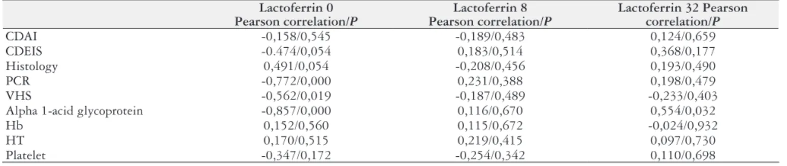
TABLE 8. Correlation analysis between lactoferrin and variables (Pearson correlation)
Lactoferrin 0 Pearson correlation/P
Lactoferrin 8 Pearson correlation/P
Lactoferrin 32 Pearson correlation/P
CDAI -0,158/0,545 -0,189/0,483 0,124/0,659
CDEIS -0.474/0,054 0,183/0,514 0,368/0,177
Histology 0,491/0,054 -0,208/0,456 0,193/0,490
PCR -0,772/0,000 0,231/0,388 0,198/0,479
VHS -0,562/0,019 -0,187/0,489 -0,233/0,403
Alpha 1-acid glycoprotein -0,857/0,000 0,116/0,670 0,554/0,032
Hb 0,152/0,560 0,115/0,672 -0,024/0,932
HT 0,170/0,515 0,219/0,415 0,097/0,730
Platelet -0,347/0,172 -0,254/0,342 0,110/0,698
TABLE 9. Correlation analysis between CDEIS, CDAI and histology (Pearson correlation) at the three treatment times CDEIS 0
Pearson/P correlation
CDEIS 8
Pearson/P correlation
CDEIS 32
Pearson/P correlation
Histology 0,167/0,535 0,507/0,045 0,445/0,084
CDAI 0,289/0,261 -0,097/0,722 0,367/0,163
TABLE 10. Correlation analysis between calprotectin and variables (Pearson correlation)
Calprotectin 0 Pearson/P correlation
Calprotectin 8 Pearson/P correlation
Calprotectin 32 Pearson/P correlation
CDAI 0,094/0,721 -0,288/0,280 0,003/0,991
Lactoferrin 0,421/0,093 0,420/0,105 0,789/0,000
CDEIS 0,255/0,323 0,032/0,910 0,597/0,019
Histology 0,471/0,066 -0,262/0,346 0,416/0,123
PCR -0,305/0,234 0,357/ 0,174 0,188/0,503
Alpha 1-acid glycoprotein -0,386/0,126 0,428/ 0,098 0,385/0,156
Hb 0,089/0,734 0,085/0,754 0,126/0,654
Ht 0,104/0,691 0,110/0,686 0,231/0,408
Platelet -0,068/0,796 -0,165/0,541 0,144/0,610
TABLE 13. Model of multiple binary logistic regression to assess the calprotectin and lactoferrin effect at time 0 in improvement histological after 32 weeks
Model Coeficient Standard error P Odds ratio
calprotectin 0 0.02 .002 .337 1.002
lactoferrin 0 20.778 40192.982 1.000 1.056E9
Constant -21.634 40192.982 1.000 .000
TABLE 11. Multiple linear regression model to assess calprotectin and lactoferrin time 0 in the condiction endoscopic after 32 weeks
Model Coeficient Coeficient standard error t
Constant 3.768 5.567 .677
calprotectin 0 .011 .005 2.154
lactoferrin 0 -5.553 6.235 -.891
TABLE 12. Pairwise comparison of the CDAI among the groups via the Bonferroni test
(I) time (J) time Mean difference (I-J) standard error 95% CI lower 95% CI upper P
0 8 107.93000* 23.70159 49.0862 166.7738 <0.001
0 32 126.68559 24.06908 66.9294 186.4418 <0.001
DISCUSSION
Although endoscopy with biopsy is considered to be the best method for assessing the localisation, extent, and severity of inlammation, it is an invasive method that is prone to risks and complications(8, 12, 33).
The participants in the present study were subjected to three endoscopic exams with biopsy at 3 different times. This protocol allowed a comparison of the histological and en-doscopic indings with the clinical, serum, and fecal results.
CDEIS
In the present study, the CDEIS exhibited a signiicant correlation with the histological score only on week 8; by contrast, there was no signiicant correlation between weeks 0 and 32, although signiicant differences have been described in two studies performed by Sipponen et al.(27, 28).
Another interesting analysis was that high calprotectin levels on week 0 were correlated with worsening endoscopy on week 32 therefore a inlammation predictor.
CDAI
From the clinical perspective, the CDAI is widely used to assess the degree of CD activity and the response to treatment(20). In the present study, no signiicant
correla-tion was found between the endoscopic, histological, and fecal variables at any time, but the CDAI exhibited a signiicant difference between weeks 0 and 8 and between weeks 0 and 32, indicating that the clinical status of the patients had improved. After induction (week 8) and after maintenance (week 32) of treatment, the CDAI exhibited a remarkable reduction, reaching levels associated with clinical remission of the disease, in spite of the persistence of the endoscopic, histological, serum, and fecal indicators of inlammation. That the intestinal mucosa might remain inlamed in patients with CD in a state of clinical remission is a well-known fact. For this reason, inlammation of the intestinal mucosa is not the only parameter that is used to determine if the disease is inactive. In addition, a portion of the items assessed in the CDAI are subjective, i.e., they depend on each patient’s individual perception of his or her own disease(26). Another criticism of the CDAI is the
fact that previous surgical procedures and the presence of istulas and ibrotic stenosis, which are not inlammatory processes, inluence the score(32).
Serum markers
Regarding the serum markers, the CRP exhibited a signiicant correlation with lactoferrin the week before induction (week 0) and with the ERS and alpha 1-acid glycoprotein during weeks 0 and 32. The average CRP value the week before induction (week 0) was 15.7 and decreased to 8.6 on the week after induction (week 0) and to 5.8 after the end of maintenance (week 32). Although we did not ind a signiicant correlation between the CRP and the histological activity, the latter improved, and the former decreased on weeks 8 and 32.
Serum measurements of the activity, such as the ERS and the platelet and white cell count, among others, are considered to be unspeciic and might be affected by a wide range of non-intestinal diseases. Consequently, these mea-surements are unable to measure the intestinal inlammatory activity directly. Patients with the active disease might ex-hibit normal levels of the serum markers, whereas patients with the quiescent disease might exhibit abnormal values(32).
Calprotectin
Roseth et al.(23) validated calprotectin as a marker of
intestinal inlammation by comparing it to the excretion of indium-111-labelled neutrophils during 4 days in patients with CD and by inding a signiicant correlation between the markers. Consequently, several other authors suggested the calprotectin assay as a non-invasive test for assessing intestinal inlammation in patients with IBD(17, 18).
Tibble et al.(32) observed that patients in clinical
remis-sion exhibit low calprotectin levels, whereas the patients with high levels exhibit an increased risk of relapsing with-in 1 year, suggestwith-ing that calprotectwith-in might be used not only as a marker of clinical remission but also to predict relapse(5). Thus, calprotectin might serve both as a marker
of mucosal healing and as a sign of future inlammatory relapse(24).
All of the patients who were invited to participate in the present study had been indicated for inliximab and thus were considered to have a more aggressive form of the disease, which was conirmed in most of these subjects by their high calprotectin levels and positive lactoferrin tests. The reduction of the calprotectin levels and the negative lactoferrin results on week 8 did not correlate with the decrease in histological inlammation on week 32. In some of the patients who exhibited a remarkable reduction of calprotectin and had negative lactoferrin tests on week 8, the calprotectin levels had risen, and lactoferrin had become positive again by week 32. Therefore, in our study, there was no signiicant correlation between calprotectin and the histological scores during the last two phases of the study, but there was a signiicant correlation on week zero. In addition, high calprotectin levels before induction (week 0) did not correlate with a histological deterioration after the maintenance (week 32). Despite having submitted calprotectin on week 8 a drop in their levels (P = 0.041) but did not achieve remission rates or close to it perhaps explained by the fact that 50% of patients had moderate to severe histological activity. Furthermore, some patients had high calprotectin levels (up to 1000 mg/kg) before induction and others showed no change in proile. This feature was not observed in the studies reviewed.
Several studies have revealed a satisfactory correlation between calprotectin and the endoscopic and histological indings, such as the extent and degree of inlammation, unlike the indices of clinical and laboratory activity. In addition, measurement of calprotectin is inexpensive, non-invasive, sensitive, and rather easy to perform(11, 27).
calpro-tectin levels on week 0 correlated with poorer endoscopic indings on week 32 and were thus predictive of inlamma-tion. Calprotectin and the histological scores exhibited a signiicant correlation only before induction (week 0). In addition, between weeks 0 and 8, the calprotectin levels ex-hibited a signiicant difference with a remarkable reduction, and the histological score improved.
The correlation found in several studies between calpro-tectin levels and the degree of inlammation suggests that calprotectin levels are due to inlammation at the tissue level. Several investigations have demonstrated that fecal calprotectin correlates in particular with the histology, rather than the colonoscopy indings, which means that in many cases, inlammation might not be macroscopically assessed(4).
In the present study, this correlation occurred sig-niicantly only on week 0. Tables 2 and 5 indicate high calprotectin levels at week 0 when more than 50% of the patients exhibited severe inlammation on the histological assessment. During week 8, the calprotectin levels exhibited a signiicant reduction, which correlated with a histological improvement in which 50% of the patients exhibited mild degrees of inlammation; however, this correlation was not statistically signiicant. During week 32, the calprotectin again increased and reached levels similar to those of week 0. Moreover, from the histological perspective, 50% of the patients still exhibited moderate-to-severe inlammation; however, this correlation was not statistically signiicant.
Some studies have been questioning that there is a better correlation of fecal markers with colonoscopy and histology in UC than CD because the presence of restricted lesions in the small intestine could inluence the dosage of fecal markers(5, 7, 31). However, in a study by Bunn et al.(4) observed
that calprotectin also correlated with disease in the small intestine through its correlation with technetiun-labelled neutrophils. In another study made by Jensen et al.(14) also
observed that calprotectin is sensitive in assessing disease in small intestine. The majority of the studies use as popula-tion patients with Crohn’s disease with involvement of the small bowel and colon which was not different to this study.
Lactoferrin
Like calprotectin, lactoferrin is also a non-invasive fecal marker of intestinal inlammation. Lactoferrin has been used to assess the clinical response to treatment with inliximab in children with severe CD, and the response to treatment has been demonstrated by clinical improvement (Pediatric CDAI – PCDAI) and reduction of the lactoferrin levels. These results suggest that lactoferrin might serve as a marker for the response to treatment with inliximab(3). A
study conducted by Kane et al.(15) revealed that the
lacto-ferrin concentration is signiicantly higher among patients with active or inactive IBD compared with healthy controls and patients with irritable bowel syndrome. In addition, the patients with active IBD exhibit higher lactoferrin con-centrations compared to the patients with inactive disease, suggesting that lactoferrin is a sensitive and speciic marker
for inlammation in IBD(15, 34). Our study used a
qualita-tive lactoferrin kit, rather than the quantitaqualita-tive kit that is also commercially available; thus, we could not assess the correlation between the lactoferrin level and the degree of inlammation. Lactoferrin remained positive in most of the patients during the three stages of the study. Perhaps, lac-toferrin did not become negative because the test used was qualitative, and most of the patients exhibited continuous inlammatory activity on histological assessment.
The main goal of treatment in IBD is to achieve remis-sion of the disease (improvement of the signs and symp-toms, as well as healing of the mucosa). Consequently, biological agents have been the focus of much attention because they induce rapid improvement of CD symptoms and mucosal healing 4 weeks after administration(25).
In our study, during the phase prior to treatment with inliximab, all of the patients exhibited a degree of histolog-ical activity (mild, moderate, or severe). After the period of maintenance treatment (week 32), histological improvement was detected in 60% of the patients. However, this result was not conirmed by the calprotectin and lactoferrin levels.
The major surprise in the present study was the high number of patients who relapsed during week 32, exhibiting an increase in the calprotectin levels and a positive lactofer-rin test. Furthermore, these indings were not conirmed at the histological level because although most of the patients continued exhibiting histological activity, improvement at the microscopic level was remarkable (60%).
The statistical analysis revealed that in the present study, calprotectin and lactoferrin did not act as markers of re-mission and relapse in CD patients who were subjected to biological therapy. Although the high calprotectin levels during week 0 were predictive of the poorer endoscopic indings on week 32, this correlation was not found regard-ing the histological score. Therefore, because we consider the combination of endoscopy and histology to be the gold standard for assessing the inlammatory activity, we do not recommend that calprotectin and lactoferrin be used as markers to monitor CD patients who are undergoing biological therapy.
CONCLUSIONS
The major limitation of the present study was the small number of participants associated with losses. Although smaller samples are associated with greater inaccuracies in the results, we can often accept results that are indicative of a conclusion, rather than the conclusion itself.
REFERENCES
1. Angriman I, Scarpa M, D’Incà R, Basso D, Ruffolo C, Polese L, Sturniolo GC, D’amico DF, Plebani M. Enzymes in feces: useful markers of chronic inlammatory bowel disease. Clin Chim Acta. 2007;381:63-8.
2. Benevento G, Avellini C, Terrosu G, Geraci M, Lodolo I, Sorrentino D. Diag-nosis and assessment of Crohn’s disease: the present and the future. Expert Rev Gastroenterol Hepatol. 2010;4:757-66.
3. Buderus S, Boone J, Lyerly D, Lentze MJ. Fecal Lactoferrin: a new parameter to monitor inliximab therapy. Dig Dis Sci. 2004;49:1036-9.
4. Bunn SK, Bisset WM, Main MJ, Gray ES, Olson S, Golden BE. Fecal calpro-tectin: validation as a noninvasive measure of bowel inlammation in childhood inlammatory bowel disease. J Pediatr Gastroenterol Nutr. 2001;33:14-22. 5. Costa F, Mumolo MG, Ceccarelli L, Bellini M, Romano MR, Sterpi C, Ricchiuti
A, Marchi S, Bottai M. Calprotectin is a stronger predictive marker of relapse in ulcerative colitis than in Crohn’s disease. Gut. 2005;54:364-8.
6. Cottone M, Criscuol V. Inliximab to treat Crohn’s disease: an update. Clin Exp Gastroenterol. 2011;4:227-38.
7. D’Incà R, Dal Pont E, Di Leo V, Ferronato A, Fries W, Vettorato MG, Martines D, Sturniolo GC. Calprotectin and lactoferrin in the assessmentof intestinal inlammation and organic disease. Int J Colorectal Dis. 2007;22:429-37. 8. Fell JM. Update of the management of inlammatory bowel disease. Arch Dis
Child. 2012;97:78-83.
9. Freeman HJ. Long-term natural history of Crohn’s disease. World J Gastroen-terol. 2009;15:1315-8.
10. Gaya DR, Mackenzie JF. Faecal calprotectin: a bright future for assessing disease activity in Crohn’s disease. QJM. 2002;95:557-8.
11. Gaya DR, Lyon TD, Duncan A, Neilly JB, Han S, Howell J, Liddell C, Stanley AJ, Morris AJ, Mackenzie J. F. Faecal calprotectin in the assessment of Crohn’s disease activity. QJM. 2005;98:435-41.
12. Gisbert JP, McNicholl AG. Questions and answers on the role of faecal cal-protectin as a biological marker in inlammatory bowel disease. Dig Liver Dis. 2009;41:56-66.
13. Harvey RF, Bradshaw JM. A simple index of Crohn’s-disease activity. Lancet. 1980;1:514.
14. Jensen MD, Kjeldsen J, Nathan T. Fecal calprotectin is equally sensitive in Crohn’s-disease affecting the small bowel and colon. Scand J. Gastroenterol. 2011;46:694–700.
15. Kane SV, Sandborn WJ, Rufo PA, Zholudev A, Boone J, Lyerly D, Camilleri M, Hanauer SB. Fecal lactoferrin is a sensitive and speciic marker in identifying intestinal inlammation. Am J Gastroenterol. 2003;98:1309-14.
16. Kayazawa M, Saitoh O, Kojima K, Nakagawa K, Tanaka S, Tabata K, Matsuse R, Uchida K, Hoshimoto M, Hirata I, Katsuk. Lactoferrin in whole gut lavage luid as a marker for disease activity in inlammatory bowel disease: comparison with other neutrophil-derived proteins. Am J Gastroenterol. 2002;97:360-9. 17. Lewis JD. The utility of biomarkers in the diagnosis and therapy of inlammatory
bowel disease. Gastroenterology. 2011;140:1817-26.
18. Limburg PJ, Ahlquist DA, Sandborn WJ, Mahoney DW, Devens ME, Harrington JJ, Zinsmeister AR. Fecal calprotectin levels predict colorectal inlammation among patients with chronic diarrhea referred for colonoscopy. Am J Gastro-enterol. 2000;95:2831-7.
19. Mary JY, Modigliani R. Development and validation of an endoscopic index of the severity for Crohn’s disease: A prospective multicentre study. Groupe d’Etudes Therapeutiques des Affections Inlammatoires du Tube Digestif (GETAID). Gut. 1989;30:983-9.
20. Naber AH, de Jong DJ. Assessment of disease activity in inlammatory bowel disease; relevance for clinical trials. Neth J Med. 2003;61:105-10.
21. Peyrin–Biroulet L, Deltenre P, Suray N, Branche J, Sandborn WJ, Colombel JF. Eficacy and safety of tumor necrosis factor antagonists in Crohn’s dis-ease: meta-analysis of placebo-controlled trials. Clin Gastroenterol Hepatol. 2008;6:644-53.
22. Røseth AG, Fagerhol MK, Aadland E, Schjønsby H. Assessment of the neu-trophil dominating protein calprotectin in feces: a methodologic study. Scand J Gastroenterol. 1992;27:793-8.
23. Røseth AG, Schimidt PN, Fagerhol MK. Correlation between faecal excretion of indium-111-labelled granulocytes and calprotectin, a granulocyte marker Protein, in patients with inlammatory bowel disease. Scand J Gastroenterol. 1999;34:50-4. 24. Røseth AG, Aadland E, Grzyb K. Normalization of faecal calprotectin: a pre-dictor of mucosal healing in patients with inlammatory bowel disease. Scand J Gastroenterol. 2004;39:1017-20.
25. Rutgeerts P, Vermeire S, Van Assche G. Mucosal healing in inlammatory bowel disease: impossible ideal or therapeutic target? Gut. 2007;56:453-5.
26. Sandborn WJ, Feagan BG, Hanauer SB, Lochs H, Löfberg R, Modigliani R, Present DH, Rutgeerts P, Schölmerik J, Stange EF, Sutherland LR. A review of activity indices and eficacy endpoints for clinical trials of medical therapy in adults with Crohn’s disease. Gastroenterol. 2002;122:512-30.
27. Sipponen T, Kärkkäinen P, Savilahti E, Kolho KL, Nuutinen H, Turunen U, Färkkilä M. Correlation of a faecal calprotectin and lactoferrin with an endo-scopic score for Crohn’s disease and histological indings. Aliment Pharmacol Ther. 2008;28:1221-9.
28. Sipponen T, Savilahti E, Kärkkäinen P, Kolho KL, Nuutinen H, Turunen U, Färkkilä M. Fecal calprotectin, lactoferrin, and endoscopic disease activity in monitoring anti-TNF-alpha therapy for Crohn’s disease. Inlamm Bowel Disease. 2008;14:1392-1398.
29. Sostegni R, Daperno M, Scaglione N, Lavagna A, Rocca R, Pera A. Review article: Crohn’s disease: monitoring disease activity. Aliment Pharmacol Ther. 2003;17:11-7.
30. Sutherland AD, Gearry RB, Frizelle F A. Review of fecal biomarkers in inlam-matory bowel disease. Dis Colon Rectum. 2008;51:1283-91.
31. Tibble JA, Teahon K, Thjodleifssonb, Roseth A, Sigthorsson G, Bridger S, Foster R, Sherwood R Fagerhol M, Bjarnason I. A simple method for assessing intestinal inlammation in Crohn’s disease. Gut. 2000;47:506-13.
32. Tibble JA, Bjarnason I. Non-invasive investigation of inlammatory bowel disease. World J Gastroenterol. 2001;7:460-5.
33. Vieira A, Fang CB, Rolim EG, Klug WA, Steinwurz F, Rossini LG, Candelária PA. Inlammatory bowel disease activity assessed by fecal calprotectin and lactoferrin: correlation with laboratory parameters, clinical, endoscopic and histological indexes. BMC Res Notes. 2009;2:221.
34. Walker TR, Land ML, Kartashov A, Saslowsky TM, Lyerly DM, Boone JH, Rufo PA. Fecal lactoferrin is a sensitive and speciic marker of disease activity in children and young adults with inlammatory bowel disease. J Pediatr Gastroen-terol Nutr. 2007;44:414-22.
35. Walsh A, Mabee J, Trivedi K. Inlammatory bowel disease. Prim Care Clin. 2011;38:415-32.
Received 8/11/2012. Accepted 13/3/2013.
Nogueira IM, Miszputen SJ, Ambrogini Jr. O, Artigiani Neto R, Carvente CT, Zanon MI. Avaliação da resposta de pacientes com doença de Crohn ao tratamento biológico, através de novos marcadores não invasivos: lactoferrina e calprotectina. Arq Gastroenterol. 2013,50(2):130-37.
RESUMO – Contexto - O uso de marcadores fecais para a monitorização da doença de Crohn é muito importante para a avaliação da resposta ao
trata-mento instituído. Objetivo – Avaliar a atividade inlamatória da doença de Crohn comparando os marcadores fecais (calprotectina e lactoferrina), colonoscopia com biópsias, Crohn’s Disease Activity Index (CDAI) e marcadores séricos antes do uso do Inliximabe, após a fase de indução e após a fase de manutenção. Método – Foram incluídos 17 pacientes com diagnóstico prévio de doença de Crohn, que faziam uso da terapia convencional, mas que necessitaram da introdução da terapia biológica: Inliximabe. Esses pacientes realizaram colonoscopias com biópsias, exames de atividade inlamatória sérica, fecal (calprotectina e lactoferrina) e análise do CDAI nas fases pré Inliximabe, pós indução (semana 8) e pós manutenção (semana 32). Resultados – Houve queda signiicativa (P = 0,04) da calprotectina entre as fases pré Inliximabe e pós indução, o mesmo não ocorrendo após a fase de manutenção. A lactoferrina manteve-se positiva nas três fases do estudo. Na análise histológica, houve diferença signiicativa apenas entre as fases pré Inliximabe e pós manutenção (P = 0,036), com 60% dos pacientes apresentando melhora histológica após o período de acompanhamento. O CDAI apresentou diferença signiicativa entre as fases pré Inliximabe e pós indução e entre as fases pré Inliximabe e pós manutenção (P<0,01). Conclusão – A calprotectina e a lactoferrina não foram capazes de monitorizar a atividade inlamatória nos pacientes com doença de Crohn em uso de terapia biológica.