Arq Neuropsiquiatr 2009;67(4):986-994
CLINICAL AND ELECTROENCEPHALOGRAPHIC
CHARACTERISTICS OF A COHORT OF PATIENTS
WITH EPILEPSY AND ABSENCE SEIZURES
Soniza Vieira Alves-Leon
1, Maria Fátima Bento de Souza Cardoso
2,
Valéria Coelho Santa Rita Pereira
3, Isabela D’Andrea Meira
4Abstract – Background: Epileptic syndromes with absence seizures (AS) possess unique clinical and electroencephalographic (EEG) characteristics. In typical or atypical AS, ictal phenomenology may include various characteristics. Vídeo-EEG monitoring enables findings to be correlated with ictal phenomenology.
Objective: To evaluate the different AS in a cohort of patients with drug-resistant epilepsy (DRE) based on the International League against Epilepsy (ILAE)’s 2006 classification, to correlate with ictal phenomenology recorded and to apply the Panayiotopoulos criteria. Method: This study included patients with criteria of AS followed up at the Epilepsy Clinic. A dual, cross-sectional cohort study was carried out between 2005 and 2008. Patients receiving care in the Epilepsy Program of the HUCFF-UFRJ, who had been investigated by video-EEG and who presented clinical and EEG criteria for absence seizures, typical or atypical, according to the criteria defined by the ILAE, were included in the study, independent of age onset, the review of clinical history, age onset, family history, epilepsy onset and evolution, seizures phenomenology, antiepileptic drugs response and neuroimaging studies were used to classify the patients among the different epileptic syndrome associated to absence seizures. Results: Typical absences were more frequent (71.4%) than atypical absences. Cases of juvenile absence epilepsy were the most frequent (19%) in this series, followed by childhood absence epilepsy (14.4%) and juvenile myoclonic epilepsy (4.8%). In 14 patients (66.67%), diagnosis was modified from focal epilepsy to primary generalized epilepsy. Clinical and EEG diagnosis of absence epilepsy resulted in a dramatic improvement in the control of seizures following modification of diagnosis and indication of an appropriate antiepileptic drug. Conclusion: Our results show that typical AS are more frequent than atypical. AS was successfully defined in 10 patients following application of Panayiotopoulos’ criteria. The consequent change in diagnosis and therapy resulted in resolution of refractoriness in 9 patients. We concluded that in DRE, AS associated to unusual ictal phenomenology improve dramatically when diagnosed by video-EEG, permitting seizures to be controlled. Clinical and EEG evaluation confirm that myoclonus, automatisms and autonomic disorders are involved and that the consciousness may be affected to different degrees.
KEY WORDS: absence seizures, epilepsy, Panayiotopoulos.
Características clínicas e eletrencefalográficas de uma coorte de pacientes com epilepsia com crises de ausência
Resumo – Síndromes epilépticas com crises de ausência (CA) possuem características clínicas e eletroencefalográficas (EEG) únicas. Nas crises de ausência típica ou atípica, a fenomenologia ictal pode incluir características que podem levar ao erro diagnóstico e à indicação de drogas antiepilépticas que pioraram o quadro. Quando esses pacientes são referidos a um Programa de Epilepsias para investigação, a monitorização
1MD, PhD, Professora Associada a Universidade Federal do Estado do Rio de Janeiro (UNIRIO), Docente Permanente do Programa de Pós-Graduação
em Neurologia da UNIRIO, Médica Responsável do Programa de Epilepsias do Serviço de Neurologia do Hospital Universitário Clementino Fraga Filho da Universidade Federal do Rio de Janeiro (HUCFF/UFRJ); 2MD, MCs, Mestre pela UNIRIO, Doutorado em andamento no programa de Pós Graduação
da UNIRIO, Médica Responsável pelo Ambulatório de Epilepsias do Serviço de Neurologia da Santa Casa de Misericórdia do Rio de Janeiro; 3MD,
Mestranda do Programa de Pós-Graduação da UFRJ, Médica do Programa de Epilepsias do HUCFF/UFRJ; 4MD, PhD, Mestre pela UNIFESP, Doutora pela
UFRJ, Médica do Programa de Epilepsias do HUCFF/UFRJ.
Received 12 April 2009, received in inal form 28 July 2009. Accepted 11 August 2009.
por vídeo-EEG permite correlacionar os achados eletrográficos com a fenomenologia ictal. Objetivo: Identificar em uma coorte de pacientes com epilepsia fármaco-resistente (EFR), pacientes com CA segundo critérios propostos pela Liga Internacional contra a Epilepsia (ILAE) de 2006, correlacionar a fenomenologia ictal ao EEG e aplicar os de critérios Panayiotopoulos neste grupo. Método: Estudo de corte transversal incluiu doentes encaminhados ao Programa de Epilepsia do HUCFF-UFRJ entre 2005 e 2008, investigados por vídeo-EEG e que apresentavam os critérios clínicos e EEG para CA típicas ou atípica; a revisão da história clínica, idade início, história familiar de epilepsia, evolução, a fenomenologia ictal, resposta a drogas antiepilépticas e estudos de neuroimagem foram utilizados para classificar os pacientes entre as diferentes síndromes epilépticas associadas a CA. Resultados: As CA típicas foram mais freqüentes (71,4%) do que as atípicas. Casos de epilepsia ausência juvenil ocorreram em 19% desta série, seguido por epilepsia ausência infantil (14,4%) e epilepsia mioclônica juvenil (4,8%). Em 14 pacientes (66,67%), o diagnóstico de epilepsia focal epilepsia foi modificado para epilepsia generalizada primária. A mudança do diagnóstico de epilepsia focal para epilepsia com CA, seguido da troca para DAE adequadas, resultou em melhoria no controle de crises. Conclusão: Nossos resultados mostram que as CA típicas são mais freqüentes do que as atípicas. Em 10 pacientes, a aplicação dos critérios de Panayiotopoulos foi possível. A conseqüente mudança no diagnóstico e terapêutica resultou na resolução de refratariedade em 9 pacientes. Concluímos que a fenomenologia ictal incomun em pacientes com CA contribui para o diagnóstico errôneo de epilepsia de difícil controle, e que o diagnostico por vídeo-EEG, permitiu a mudança do diagnóstico e a melhora dramática no controle das crises. As avaliações clínica e eletrencefalográfica confirmam que mioclonias, automatismos psicomotores e desordens autonômicas podem fazer parte da fenomenologia ictal de pacientes com CA e que a consciência pode ser afetada em diferentes graus.
PALAVRAS-CHAVE: crises de ausência, epilepsia, Panayiotopoulos.
Absence seizures (AS) are classiied under the category of generalized epilepsies; however, unlike other seizures, absence seizures possess clinical characteristics and elec-troencephalographic (EEG) patterns that are speciic for this diagnosis1-3 and occur in different epileptic syndromes.
Absence seizures may be divided into typical absences and atypical absences, according to EEG pattern. Absence sei-zures are present in a heterogenous group classiied by the International League against Epilepsy (ILAE) into four epi-leptic syndromes: childhood absence epilepsy, juvenile ab-sence epilepsy, juvenile myoclonic epilepsy and myoclo-nic absence epilepsy4. Typical absence seizures are most
frequent and are characterized by loss of consciousness which is time-locked with bursts of bilaterally synchro-nous spike-and-wave discharges of 3 to 4 cycles per sec-ond, and, in general, present good response to pharmaco-logical treatment. Conversely, atypical absence seizures are less frequent, the seizures are associated with 1 to 2 cycles per second spike-wave discharges and slow background rhythms for aging; they are often associated with severe neurologic impairment and poor response to treatment. The criteria proposed by Panayiotopoulos in 1997 for the classiication of patients with typical absence seizures in-clude a greater number of absence syndromes, some of which have not yet been recognized by the ILAE, such as phantom absences, absence epilepsy with eyelid myo-clonus and early-onset absence epilepsy, among others5.
Epilepsies with typical absence seizures are common forms of generalized epilepsy. Cases of childhood absence epilepsy constitute 2–10% of all cases of epilepsy in
chil-dren. Juvenile absence epilepsy constitutes 10% of all cas-es of idiopathic generalized epilepsy, whereas absence ep-ilepsy with eyelid myoclonus makes up 2% of all sies and 11% of all cases of idiopathic generalized epilep-sy. It is dificult to determine the frequency of the rarer syndromes6. Epilepsy was classiied into focal and
gener-alized forms rather more for didactic purposes than from any intention to deine the localization of the disease, bearing in mind that various authors have questioned the essentially bilateral onset of the generalized forms of ep-ilepsy, believing all to have a focal onset7,8. Interictal EEG
may result in an incorrect diagnosis of the type of seizure and/or epileptic syndrome, following which therapy may consequently fail7, particularly in cases of patients with
generalized epilepsy in whom clinical characteristics are suggestive of focal epilepsy during seizures or as a result of focal patterns revealed by interictal EEG.
The objective of this study was to evaluate with what frequency patients referred to the Epilepsy Program of the
Clementino Fraga Filho Teaching Hospital, Federal Univer-sity of Rio de Janeiro (HUCFF-UFRJ) with an initial diagno-sis of drug-rediagno-sistant epilepsy present absence seizures, an-alyze what clinical and electroencephalic variables were associated with drug-resistance, and reclassiied this pa-tients according to Panayiotopoulus criteria.
METHOD
vid-eo-EEG and who presented clinical and EEG criteria for absence seizures, typical or atypical, according to the criteria deined by the ILAE, were included in the study, independent of age onset; the review of clinical history, age onset, family history, epilep-sy onset and evolution, seizures phenomenology, antiepileptic drugs response and neuroimaging studies were used to classi-fy the patients among the different epileptic syndrome associ-ated to absence seizures. Patients included could present oth-er seizures type, besides absence seizures. All the seizures woth-ere registered and the concomitance of different seizures phenom-enology and EEG pattern were analyzed. The video-EEG moni-toring was done for at least 24 h, with a Neurotec© machine of
32 channels, and the scalp electrodes were distributed accord-ing to the 10–20 system. Patients with focal epilepsy and other generalized epilepsy without the presence of absence seizures were excluded. The clinical, ictal phenomenology and EEG vari-ables were used to reclassify the patients according to Panay-iotopoulus criteria analyzing the different epileptic syndromes with absence seizures.
This study was approved by the Institutional Review Board and all patients signed an informed consent form prior to ad-mission. The clinical data and correlated variables recorded dur-ing video-EEG were stored on an Excel spreadsheet (Excel 1997– 2003) and transformed into a DBF ile to enable them to be read in the generally available Epi Info program, version 6.0.
RESULTS
Of a cohort of 643 patients receiving care between 2005 and 2008, 68 (10.57%) were found to have clinical and EEG criteria for generalized epilepsy and, of these, 21 (30.09%) had absence seizures. In these 21 patients, 2 pre-sented clinical suspicion of generalized epilepsy accord-ing to seizures semiology and triggeraccord-ing factors described. The mean age of patients at the time of inclusion into the study was 25.5 years (range 3–58 years). The mean age at onset of symptoms was 11.8 years (range 1–39 years).
Of these 21 patients, the onset of symptoms occurred before the patient reached 10 years of age in 10 patients (47.6%), between 11 and 20 years of age in another 10 (4.76%) and after 20 years of age in 1 patient (4.76%). Eight of these patients (38.09%) were male. Twelve (57.1%) were white, while 9 (42.85%) were of African descent. The mean duration of the disease was 14.4 years (range 1–42 years). Six patients (28.6%) had a family history of epilepsy, while 13 (61.9%) did not and 2 (9.5%) were unable to provide any information on the subject.
Of the 20 patients in use of antiepileptic drugs, 8 (40%) were in use of speciic irst-line antiepileptic drugs for absence seizures and in 4 of these cases (50%), these drugs were associated with other antiepileptics that are contraindicated for absence seizures, including: carbam-azepine (CBZ) (n=2), phenytoin (PHT) (n=2) and oxcarba-zepine (OCX) (n=2). Of the 4 remaining patients (50%)
us-ing speciic irst-line drugs for absence seizures, 1 patient (25%) was using the drug as monotherapy, while the re-maining 3 (75%) were using the drug in association with an-other antiepileptic drug that was not contraindicated for absence seizures. A total of 14 patients (70%) were using antiepileptic drug that were contraindicated for absence seizures. In 4 of these cases (28.58%), the contraindicated antiepileptic drug was associated with a speciic irst-line drug for absence seizures, while in 5 patients (35.71%) the drug was associated with a second contraindicated drug, in another 5 (35.71%) the drug was associated with a sec-ond-line antiepileptic drug and in 2 patients (14.28%) the contraindicated medication was being used as monother-apy. One patient without AED in the moment of the vid-eo-EEG monitoring fulilled criteria to pharmacoresistant epilepsy but had interrupted the treatment by himself af-ter worsening with carbamazepine and phenytoin.
Of the 14 patients in use of antiepileptic drugs that are contraindicated for absence epilepsy, the drug could not be withdrawn in 3 cases (21.42%). However, in 7 patients (50%), complete control of the seizures was achieved fol-lowing withdrawal of the deleterious drugs, while in four cases (28.56%) partial control was achieved, although oc-casional absence seizures persisted.
Of the four patients who were in use of speciic irst-line antiepileptic drugs for absence seizures, adjustment of the dose was effective in 1 patient (25%). In another patient (25%), in addition to adjustment of the dose, an-other antiepileptic drug had to be associated, a strategy that led to a reduction in the frequency of seizures. De-spite all the relevant measures taken, seizures persisted in two patients.
Neuropsychological and motor development was nor-mal in 18 patients (85.7%) and abnornor-mal in 3 (14.3%). In 17 patients (81.0%), there was no loss of function following the onset of seizures, whereas in 4 (19%) there was a de-cline in neuropsychological and motor development.
Two patients (9.52%) had only absence seizures, while 16 (76.19%) had generalized tonic-clonic seizures, 6 (28.57%) had appendicular myoclonus, 5 (23.80%) had eye-lid myoclonus with absences and 4 (19.04%) had astatic seizures. Of the 21 patients, 2 (9.52%) had one single type of seizure, 11 (52.38%) had two associated types of seizure, 5 (23.80%) had three associated types of seizure, 2 (9.52%) had four associated types of seizure and 1 patient (4.76%) had ive different types of seizure. Four patients (19.04%) had a previous history of status epilepticus, 3 of which (75%) consisted of absence epilepsy and 1 case (25%) of tonic clonic seizures (Table 1).
autonom-Table 1. Seizures phenomenology and antiepileptic drug response.
Patient Other associated seizures
Absence seizures concurrent
phenomenology AED response
1 Absence and eyelid myoclonus
masticatory automatism
2 Generalized tonic clonic Absence and appendicular myclonus Worsening after CBZ 3 Eyelid myoclonus Generalized tonic
clonic and appendicular myoclonus
Absence and eyelid myoclonus Worsening after CBZ
4 Absence and eyelid myoclonia, oral
and tongue automatism, upper and lower limbs automatism
Worsening after CBZ
5 Generalized tonic clonic, tonic and astatic Absence with upper and lower limbs automatism 6 Absence epilepsy, Generalized tonic
clonic and eyelid myoclonus
Absence and eyelid myoclonus, sometimes with epigastric discomfort
Worsening after CBZ
7 Generalized tonic clonic Brief absences
8 Astatic seizures and focal seizures Absence and eyelid myoclonus, and astat seizures
CBZ and no worsening
9 Absence and upper and lower limbs myoclonus
Absence and upper and lower limbs myoclonus
Worsening after CBZ
10 Generalized tonic clonic Absences and eyelid myoclonus Worsening after CBZ 11 Generalized tonic clonic Brief absences
12 Generalized tonic clonic, astatic seizures Absences and eyelid myoclonus, appendicular myoclonus, sometimes astatic seizures 13 Generalized tonic clonic,
release sphincteric
Absences and episodic confusional states
Worsening after CBZ
14 Generalized tonic clonic, episodic confusional states and aggression
Brief absences Worsening after CBZ
15 Generalized tonic clonic preceded by upper and lower limbs myoclonus
Absences with appendicular myoclonus
Worsening after CBZ
16 Generalized tonic clonic, confusional sates episodic, eyelid, upper and lower limbs myoclonus
Absence associated with eyelid and appendicular myoclonus
17 Generalized tonic clonic, upper and lower limbs myoclonus
Absence, sometimes associated to masticatory movments
Worsening after OXCBZ
18 Generalized tonic clonic Brief absences
19 Eyelid myoclonus Absences and eyelid myoclonus Worsening after CBZ
20 Generalized tonic clonic, eyelid, upper and lower limbs myoclonus, astat seizures
Absences and eyelid myoclonus
21 Generalized tonic clonic Brief absences AED: antiepileptic drug; CBZ: carbamazepine; OXCBZ: oxcarbazepine.
ic abnormalities, while in 1 patient (4.76%) there was a re-port of sphincter release and in another (4.76%) a rere-port of epigastric discomfort.
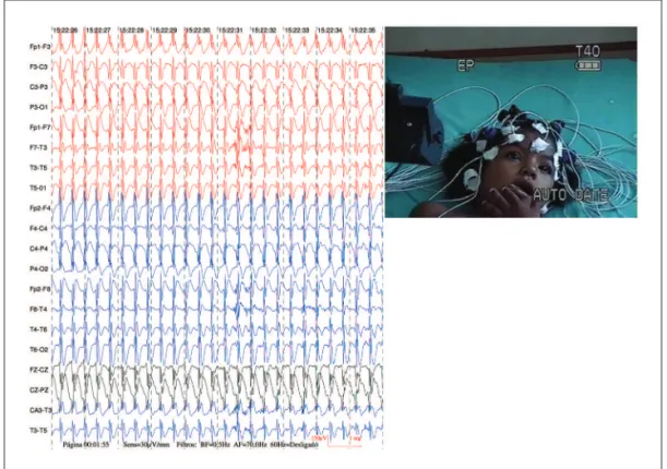
Ictal EEG indings were characteristic of generalized seizures, fulilling the criteria for typical or atypical ab-sence seizures in all patients. In 15 patients (71.4%), video-EEG indings fulilled the criteria for typical absence
Fig 1. Registered ictal video-electroencephalographic showing complex of spike slow waves rhythmic of 3 Hz, high amplitude, with projection widespread, synchronous and symmetrical, with duration of 18 seconds. Clin-ically, seizures of absence with eyelid myoclonia, perioral and language automatism.
Table 2. Results of ictal and interictal video-EEG.
Patient Interictal vídeo-EEG Ictal vídeo-EEG Hiperpnea Light stimulus
Diagnosis before Pann Criteria
Diagnosis after Pann Criteria applyed 1. Irregular slow waves, acute
frontal waves temporal left
Complex spike slow wave 3 Hz ++ – JAE JAE
2. Spike complex and poli spike slow wave frontal and central bilateral
Spike complex, irregular and generalized poli spike slow wave (2.5–3 Hz)
++ – JME JME
3. Intermittent slow wave frontal right
Spike complex and poli spike slow wave intermittent generalized (±4 Hz)
– ++ NC ABEEM
4. Occipital slow wave Complex spike slow wave generalized 3 Hz
++ – EAI NC
5. Diffuse slow track, intermittent slow waves on frontal left
Complex spike slow wave generalized 2 Hz and generalized slow wave acute wave 2 Hz
NA – AA AA
6. CE and poli spike slow wave fronto central
CE and intermittent poli spike slow wave generalized 3–4 Hz
– – CAE Phantom
absences 7. CE and poli spike slow
wave fronto temporal
CE e intermittent poli spike slow wave generalized 3–4 Hz
++ – NC Phantom
absences 8. Irregular slow waves
on temporal left
Complex spike slow wave generalized 1.5–2 Hz
+ – AA NC
9. Irregular acute wave and slow wave
Complex spike slow wave generalized 3–4 Hz
++ – JAE NC
10. CE e poli spike slow wave on frontal left
CE intermittent poli spike slow wave generalized 3–4 Hz
++ ++ JAE AEEM
11. Irregular slow wave generalized
Intermittent complex spike slow wave generalized
– + NC Phantom
absences 12. Diffuse slow track; complex
spike slow wave fronto-central
Complex spike slow wave generalized 2–2.5 Hz
++ – AA NC
13. Complex spike slow wave on frontal right
Intermittent complex spike slow wave generalized 3–4 Hz
– – NC Phantom
absences 14. Irregular slow waves
on parietal right
CE and poli spike slow wave generalized 2.5 Hz
+ + NC NC
15. EF and slow wave/acute wave on frontal and temporal left
CE and intermittent poli spike slow wave generalized
+ + JME NC
16. Irregular slow waves on fronto-temporal; generalized spikes
Generalized spikes and generalized poli spike slow wave
NA – AA NC
17. Fronto temporal spikes on right
Intermittent generalized spike slow wave complex 2 Hz
+ – AA NC
18. Acute and slow waves on fronto-temporal left
CE and generalized poli spike slow wave 3 Hz
NA – NC EAIP
19. Irregular acute and slow wave on temporal left
Generalized spike slow wave complex 3 Hz
– – CAI NC
20. Irregular slow waves on fronto-temporal
generalized spike slow wave complex 1.5–2.5 Hz
+ + AA NC
21. Irregular acute slow wave fronto-central rigth
Generalized spike slow wave complex 3 Hz
+ + JAE JAE
elements with a predominance of morphological charac-teristics similar to focal ictal features in a normal interictal tracing. Of these patients, 4 (80%) had spike complexes and polyspike-slow wave complexes, and 1 patient (20%) had spike-slow wave complex. In one patient (4.76%), interictal recording showed slow wave and intermittent, generalized spike wave activity and in another patient (4.8%), slowing of background activity in the EEG tracings was found in as-sociation with spike-slow wave complex (Table 2).
Ictal recording of 12 patients (57.4%) with spike-slow wave complexes showed regular ictal discharge in 7 (58.3%) and irregular ictal discharge in 5 (41.7%). Of the 12 patients with spike-slow wave complex, frequency was >2.5 Hz in 7 (58.4%) and ≤ 2.5 Hz in 5 (41.7%) (Table 2).
Of the 9 patients (42.8%) in whom ictal recording reg-istered spike complexes and polyspike-slow wave com-plexes, ictal activity was regular in 2 cases (22.2%) and ir-regular in 7 (77.8%). In 3 patients (33.3%), frequency was
>2.5 Hz, while in 1 patient (11.1%), frequency was ≤ 2.5 Hz, and in another (11.1%) frequency varied between >2.5 Hz and ≤ 2.5 Hz during seizures. In 2 patients (22.2%) with spike complexes and polyspike-slow wave complexes
< 2.5 Hz, there was an association of spike-wave and slow-wave paroxysms (Table 2).
Of the 18 patients (85.7%) in whom hyperpnea was successfully accomplished, 13 (72.2%) had abnormalities; 6 (46.2%) had accentuated electroencephalographic
el-ements and seizures were triggered in another 7 (53.8%). Seven patients (33.3%) had an abnormal EEG recording be-cause of photic stimulation, 5 of these patients (71.4%) having accentuated electroencephalographic elements while 2 (28.6%) developed seizures as a result of photo-sensitivity. In three patients (14.3%), the presence of eyelid myoclonus was registered following ocular occlusion in an illuminated environment, with absences in one patient.
Of the 21 patients evaluated, 15 (71.42%) fulilled clin-ical and EEG criteria for typclin-ical absences and 6 patients (28.58%) for atypical absences. Of the 15 patients of the group with clinical and EEG characteristics of typical ab-sences, 9 (60%) were diagnosed with an epileptic syn-drome, while 6 (40%) were not classiied as having any of the syndromes described by the ILAE. Of the 9 patients with an established epileptic syndrome, 5 (55.6%) were classiied as juvenile absence epilepsy, 3 (33.3%) as child-hood absence epilepsy and 1 (11.1%) as juvenile myoclonic epilepsy. After applying Panayiotopoulos’ criteria, 10 pa-tients (66.7%) were able to be diagnosed with speciic syn-dromes, while 5 (33.3%) failed to fulill the criteria for any speciic syndrome. Of the 10 patients with a speciic clas-siication, 2 (20%) retained the diagnosis of juvenile ab-sence epilepsy and 1 (10%) of myoclonic abab-sence epilep-sy, while 4 (40%) were classiied as phantom absences (Fig 3), 2 (20%) as absence epilepsy with eyelid myoclonus and 1 (10%) as early-onset absence epilepsy.
DISCUSSION
The present results show that some absence seizures may include ictal phenomena with both simple and com-plex motor automatisms, confounding diagnosis and lead-ing to inappropriate therapy, as shown by Lombroso, Fer-rie and Holmes, and Brown and Tucker7,8,13. It can justify
why patients with primary generalized seizures were re-ferred to our video-EEG unit.
The rate (10.57%) of patients with drug-resistant epi-lepsy at the HUCFF-UFRJ was lower than igures reported in the literature6,14 and may be attributable to the low rate
of diagnosis or to the age group of the cohorts in those studies. In the present study, this inding may perhaps lect the large number of patients with focal epilepsy re-ferred to epilepsy programs such as that offered at the HUCFF, which receives a greater percentage of patients with supposedly focal seizures.
The age at onset of absence seizures, which ranged from 1 to 39 years, is justiied by the inclusion of patients with different epileptic syndromes in which absence sei-zures are found. Each one of the syndromes is charac-terized by a certain age at onset according to the re-spective age-group. Another factor that may explain this wide range is the inclusion of patients with epileptic syn-dromes involving absence seizures that have not yet been recognized by the ILAE. These syndromes have, howev-er, been the subject of recent studies and their inclusion into the ILAE classiication has been proposed by various investigators1,5,9. One example is the patient in whom
on-set of absence seizures occurred in adulthood (at 39 years of age), a case similar to others previously reported by Trinka10. Another example was the patient with early
on-set at 12 months of age, other investigators also having re-ported similar cases9,10. Despite the disparate variation in
age at onset, the mean age of patients at the onset of ab-sence seizures in the present study was 11.04 years, which is in agreement with data reported from other studies on juvenile absence epilepsy12.
There was a predominance of females in the present study, which is in agreement with indings reported in the literature, principally in studies on generalized epilepsies with absence seizures13. There may be a predominance of
males in cases of epilepsy with myoclonic absences13.
The duration of epilepsy found in this study was lon-ger than expected. This inding is justiied by the fact that patients with drug-resistant epilepsy require referral to specialized treatment centers as early as possible,as em-phasized in the literature14,15. Erroneous initial diagnosis
and the consequent ineficacy of the prescribed antiepi-leptic drugs was the most frequent inding. Syndromes such as atypical absences, which have a natural history of refractoriness to treatment, were another cause of drug-resistant epilepsy16,17.
The present results are in agreement with reports in the literature where in some cases a family history of the condition was common and various different types of sei-zure were found in the same family3,18.
In 14 patients (70%) in this series, an increase was found in the frequency of epileptic seizures that was attributed to the use of antiepileptic drugs that are contraindicated for patients with absence seizures, thereby conirming the importance of the new ILAE classiication19, which now
in-cludes selective response to antiepileptic drugs as part of the classiication criteria2,20-22. Incorrect diagnosis based on
the presence of apparently focal characteristics23,24, as well
as interictal EEG scans with focal abnormalities7, were also
found in our patients and were confounding factors in the differential diagnosis with focal epilepsy. Indeed, differen-tial diagnosis was only possible with the aid of video-EEG. In the present series, four patients had limb and oro-facial (mouth and tongue) automatisms highly suggestive of temporal epilepsy but which occurred during absence seizures and were correlated with EEG showing 3c/s spike-slow wave complex or spike complex and generalized polyspike-slow wave. In another 9 patients, the presence of eyelid myoclonia was confused with facial automatisms or considered to constitute an event unrelated to epilep-sy. In 8 patients, the presence of appendicular myoclonus was confused with temporal lobe automatisms. The di-versity in the types of seizures found in patients with ab-sence seizures in the present study is in agreement with reports in the literature13,23,24. This heterogeneity in the
ab-sence seizures found in patients with generalized epilepsy has been conirmed by other authors6,10,12,13,23,24.
The inding of absence epilepsy in our patients is in agreement with reports in the literature that patients with absence seizures, albeit apparently asymptomatic, may progress to absence epilepsy or generalized tonic-clonic seizures5,25-27.
In the present study, a greater association of atypi-cal absence seizure was found in patients with a histo-ry of function loss following the onset of seizures, men-tal retardation and drug-resistant epilepsy. This inding is in agreement with the literature, conirming that atypical absence seizures are associated with a poorer prognosis with respect to the control of seizures, to impaired cog-nition and neuropsychological and motor development and to a greater variation in the types of seizures13,16,17. In
the present study, patients with atypical absences had sei-zures associated with abnormal muscle tone, either hypo or hypertonia, and astatic seizures were more frequent in this group compared to reports from other studies pub-lished in the literature13,16,17,28.
classiica-tion according to the criteria proposed by Panayiotopou-los et al.1,5 According to Panayiotopoulos2, the
classiica-tion of a speciic syndrome is in agreement with the lit-erature when application of the criteria used results in an increase in that classiication category and a consequent improvement in the therapeutic options available to the respective patients1,5,24,30.
We attribute the large rate of cases of phantom ab-sences in this series to the fact that these patients had sei-zures that were dificult to diagnose, this type of referral being commonplace at specialized centers.
Analysis of this series of patients with absence sei-zures demanded a meticulous review of the currently pro-posed diagnostic criteria and showed that a careful eval-uation of patients with drug-resistant epilepsy may result in a change in the initial diagnosis to a controllable form of epilepsy that is not drug-resistant. In almost 70% of the cases in the present series, modiication and adaptation of the diagnosis of the epileptic syndrome or of the type of seizure altered therapeutical management and the qual-ity of life of these patients and their families, conirming the importance of establishing referral centers for the in-vestigation and treatment of epilepsy.
In conclusion, in patients with epilepsy with typical or atypical absence seizures, ictal phenomenology may in-clude various characteristics, and clinical and EEG evalua-tion conirms that myoclonus, automatisms and autonom-ic disorders are involved and that the consciousness of the patient may be affected to different degrees. Typical absences are more frequent (71.4%) than atypical absenc-es. Cases of juvenile absence epilepsy were the most fre-quent (19%) in this series, followed by childhood absence epilepsy (14.4%) and juvenile myoclonic epilepsy (4.8%). In 14 patients (66.67%), diagnosis was modiied from focal epilepsy to primary generalized epilepsy. AS was success-fully deined in 10 patients following application of Panay-iotopoulos’ criteria. Clinical and EEG diagnosis of absence epilepsy resulted in a dramatic improvement in the con-trol of seizures following modiication of diagnosis and in-dication of an appropriate antiepileptic drug.
REFERENCES
1. Panayiotopoulus CP. Absence epilepsies. In: Engel JR (Ed) Epilepsy: a comprehensive textbook. Philadelphia: Lippincott-Raven Publishers; 1997:2327-2346.
2. Panayiotopoulus CP. Typical absence seizures and their treatment. Arch Dis Child 1999;81:351-355.
3. Engel JJ. Report of the ILAE Classiication Core Group. Epilepsia 2006;
47:1558-1568.
4. Commission on classification and terminology of the International
League Against Epilepsy: proposal for revised classiication of epilep
-sies and epileptic syndromes. Epilepsia 1989;30:389-399.
5. Panayiotopoulus CP. Syndromes of idiophatic generalized epilepsies not recognized by the International League Against Epilepsy. Epilepsia
2005;45(Suppl 9):S57-S66.
6. Duron RM, Medina MT, Martínez-Juárez IE, et al. Seizures of
idiophath-ic generalized epilepsies. Epilepsia 2005;46(Suppl 9):S34-S47. 7. Lombroso CT. Consistent EEG focalities detected in subjects with pri
-mary generalized epilepsies monitored for two decades. Epilepsia
1997;38:797-812.
8. Holmes MD, Brown M, Tucker DM. Are “generalized” seizures truly
generalized? Evidence of localized mesial frontal and frontopolar
dis-charges in absence. Epilepsia 2004;45:1568-1579.
9. Chaix, Y, Daquin, G, Monteiro, F, Villeneuve N, Laguitton V, Genton P. Absence epilepsy with onset before age three years: a heterogeneous and often severe condition. Epilepsia 2003;44:944-949.
10. Trinka E. Absences in adult seizure disordes. Acta Neurol Scand 2005;
112(Suppl 182):S12-S18.
11. TitomanIio L, Romano A, Bellini G, et al. Familial occurrence of early-onset chidhood absence epilepsy. Eur J Paediatr Neurol 2007;11:178-180. 12. Valentin A, Hindocha N, Osei-Lah A, et al. Idiophathic generalized
epilepsy with absences: sindrome classification. Epilepsia 2007;48: 2187-2190.
13. Stefan H, Snead OC, Olofsson O. Typical and atypical absence seizures,
mioclonic absences, and eyelid myovlonia. In: Engel JR J, Pedley TA (Eds). Epilepsy a comprehensive textbook. 2th Ed. Philadelphia: Wolt-ers Kluwer: Lippincott Williams &Wilkins, 2007:572-584.
14. Devinsky O. Current concepts: patients with refractory seizures. N Engl J Med 1999;340:1565-1570.
15. Arroyo S. Evaluatión de La epilepsia farmacorresistente. Rev Neurol 2000;30:881-886.
16. Bare MB, Glauser TA, Strawsburg RH. Need for electroencephalogram video conirmation of atypical absence seizure in children with Lennox-Gastaut syndrome. J Child Neurol 1998;13:498-500.
17. Holmes GL. Generalized seizures. In: Swaikman K, Ashwal S (Eds). Pediatric neurology: principles & practice. 4th Ed. Minnesota: Elservier, 1999:643-645.
18. Winawaer MR, Marini C, Grinton BE, et al. Familial clustering of sei
-zures types within the idiophatic generalized epilepsies. Neurology 2005;65:523-528.
19. Engel JJ. ILAE classiication of epilepsy syndromes. Epilepsy Res 2006; 70(Suppl):S5-S10.
20. Liporace JD, Sperling MR, Dichter MA. Abcence seizures and carbam
-azepine in adults. Epilepsia 1994;35:1026-1028.
21. Genton P. When antiepileptic drugs aggravate epilepsy? Brain Dev 2000;22:75-80.
22. Gelisse P, Genton I, Kuatec C, Pesenti A, Baldy-Moulinier M, Crespel A. Worsening of seizures by oxcabazepine in juvenile idiopathic gen
-eralized epilepsies. Epilepsia 2004;45:1282-1286.
23. Mory SB, Guerreiro CAM, Lim L, et al. Epilepsias generalizadas id -iopáticas diagnosticadas incorretamente como epilepsias parciais. Arq
Neuropsiquiatr 2002;60:788-796.
24. Guilhoto MFF, Manreza ML, Yacubian EM. Syndromic classiication of patients with typical absence seizures. Arq Neuropsiquiatr 2003;61: 580-587.
25. Baykan B, Noachtar S. Perioral myoclonia with absences: an overlooked and misdiagnosed generalized seizure type. Epilepsy Behav 2005;6: 460-462.
26. Shorvon S, Walker M. Status epilepticus in idiopathic generalized
epi-lepsy. Epilepsia 2005;46(Suppl, 9):S73-S79.
27. Genton P, Felarzzo E, Thomas P. Absence status epilepsy: delineation of a distinct idiopathic generalized epilepsy syndrome. Epilepsia 2008;
49:642-648.
28. Niedermeyer E. Epileptic seizure disorders. In: Niedermeyer E, Sil
-va FL (Eds) Electroencephalography: basic principles, clinical appli -cations, and related fields. 4th Ed. Baltimore: Lippincott Willians & Wilkins, 1998:476-585.
29. Commission on classification and terminology of the international league against epilepsy. Proposal for revised clinical and
electroenceph-alographic classiication of epileptic seizures. Epilepsia 1981;22:489-501. 30. Giannakodimos S, Panayiotopulos CP. Eyelid myiclonia with absenc