373 https://doi.org/10.1590/0004-282X20180054
ARTICLE
Neurosyphilis and ocular syphilis clinical and
cerebrospinal fluid characteristics: a case series
Características clínicas e liqúoricas de pacientes com neurossífilis e sífilis ocular: uma
série de casos
Conrado Regis Borges1, Sérgio Monteiro de Almeida1,3, Karen Sue2, Jéssyca Luana Alves Koslyk1, Mario
Teruo Sato2, Naoye Shiokawa2, Hélio Afonso Ghizoni Teive1
Syphilis is a sexually transmitted disease that manifests
itself as primary, secondary and tertiary forms. It affects vari
-ous organ systems, including the central nerv-ous system and
ocular system. Before the antibiotic era, it was one of the
greatest public health concerns worldwide. The availability of penicillin treatment, however, dramatically reduced its preva -lence1,2,3 and epidemiologic importance. Nevertheless, during
the first decade of this century, studies in many populations worldwide have revealed a significant increase in the inci -dence of syphilis and neurosyphilis, especially in some
pop-ulation groups, such as HIV-carriers and men who have sex
with men1,2,3. The aim of this study was to compare clinical
and laboratory characteristics of ocular and central nervous
system syphilis.
1Universidade Federal do Paraná, Hospital de Clínicas, Departamento de Medicina Interna, Serviço de Neurologia, Curitiba PR, Brasil; 2Universidade Federal do Paraná, Hospital de Clínicas, Serviço de Neuro-Oftalmologia, Curitiba PR, Brasil;
3Faculdades Pequeno Príncipe Curitiba, Instituto de Pesquisa Pelé Pequeno Príncipe, Curitiba PR, Brasil.
Correspondence: Conrado Regis Borges; Hospital de Clínicas, Serviço de Neurologia; Rua General Carneiro, 181/4o andar; 80060-900 Curitiba PR, Brasil; E-mail: conradoborges@outlook.com
Conflict of interest: There is no conflict of interest to declare.
Received 16 December 2017; Received in final form 02 March 2018; Accepted 19 March 2018.
ABSTRACT
Background: During the first decade of this century, a significant increase in the incidence of syphilis was documented. Objective: To study clinical and laboratory characteristics of central nervous system and ocular syphilis. Methods: A retrospective case series of 13 patients with a clinical and laboratory diagnosis of neurosyphilis and/or ocular syphilis who had been admitted to the Neurology and Neuro-ophthalmology Service of the Hospital de Clínicas, Federal University of Paraná. Results: Nine patients had a diagnosis of neurosyphilis and two of them also had ocular syphilis. Four patients had a diagnosis of ocular syphilis alone. Among the patients with a diagnosis of neurosyphilis, six had symptomatic syphilitic meningitis, of whom one manifested as cranial nerve palsy alone, one as cranial nerve palsy plus ocular syphilis, two as transverse myelitis (syphilitic meningomyelitis), one as meningitis worsening the patient’s myasthenia gravis symptoms and one as meningitis plus ocular syphilis. Additionally, we diagnosed three patients with meningovascular neurosyphilis. In the univariate analysis, patients without ocular syphilis showed greater levels of total protein and white blood cells in the cerebrospinal fluid than patients with ocular syphilis. Conclusion: This Brazilian case series of patients with neurosyphilis and ocular syphilis highlights the wide variability of this disease. A high degree of diagnostic suspicion is necessary when facing neurological and ocular symptoms for rapid diagnosis and appropriate management of patients.
Keywords: syphilis; neurosyphilis, ocular syphilis.
RESUMO
Introdução: Na primeira década deste século observou-se um aumento significativo da incidência de sífilis no mundo. Objetivo: Estudar características clínicas e laboratoriais da sífilis no Sistema Nervoso Central e da sífilis ocular. Métodos: Estudou-se, retrospectivamente, uma série de treze casos com diagnóstico clínico e laboratorial de neurossífilis e/ou sífilis ocular, admitidos aos Serviços de Neurologia ou Neuroftalmologia do Hospital de Clínicas da Universidade Federal do Paraná. Resultados: Nove pacientes tiveram diagnóstico de neurosífilis e dois destes apresentaram concomitantemente sífilis ocular. Quatro pacientes tiveram somente o diagnóstico de sífilis ocular. Dos pacientes com diagnóstico de neurosífilis, seis apresentaram meningite sifilítica sintomática, dentre os quais um se apresentou com paralisia isolada de par craniano, um com paralisia de par craniano associada sífilis ocular, dois com mielite transversa (manifestação de meningomielite), um com meningite que agravou sintomas de Miastenia Gravis e um com meningite isolada associada a sífilis ocular. Houve 3 casos de neurosífilis meningovascular. Na análise univariada, pacientes sem manifestações oculares de sífilis apresentaram maiores níveis proteína total e leucócitos do que os pacientes com sífilis ocular.
Conclusão: Essa série brasileira de casos de pacientes com neurosífilis e sífilis ocular destaca a alta variabilidade clínica desta doença. Alto grau de suspeição diagnóstica é necessário quando em frente a sintomas neurológicos e oculares para rápido diagnóstico e adequado manejo dos pacientes.
METHODS
We describe a case series of patients admitted between 2013 and 2015 to the Neurology and Neuro-ophthalmology
Services in the Hospital de Clínicas at the Universidade Federal do Paraná (HC-UFPR), which is a public ter
-tiary university hospital situated in southern Brazil. We reviewed retrospectively 13 selected medical records of patients diagnosed and treated as having neurosyphi -lis and/or ocular syphi-lis. The patients were referred to the hospital by the local public health care system after
an initial evaluation by a general practitioner. These cases
were probably not all the neurosyphilis patients who
had been diagnosed in the above-mentioned hospital, because information was retrieved from a paper-based chart system. Care was provided by the neurology and/ or ophthalmology services following the standard proto -cols for neurosyphilis1. This was part of a larger research
study on patients with meningitis, which was approved
by the ethics committee of our institution. All patients had consented to the use of the information obtained
during hospitalization.
Diagnosis of syphilis
As there was clinical suspicion of syphilis, blood samples from the 13 patients were tested for syphilis using the
follow-ing standard laboratory protocol adopted by the HC-UFPR Clinical Pathology Laboratory: a treponemal screening assay (chemiluminescent immunoassay), which, if reactive, was followed by the Venereal Disease Research Laboratory (VDRL) test and fluorescent treponemal antibody absorp
-tion (FTA-Abs) test. Further laboratory tests were ordered by
the physicians who had examined the patients, as required.
The diagnostic criteria for syphilis were reactive treponemal and/or nontreponemal tests and a clinical profile consistent
with the disease.
Diagnosis of ocular syphilis
The diagnostic criteria for ocular syphilis include having a new diagnosis of syphilis (defined as having serologic evi
-dence of syphilis) and evi-dence of syphilitic infection in the eye or documented ocular inflammation related to syphilis
on ocular and imaging examinations4,5.
Diagnosis of neurosyphilis
After serological diagnosis, all patients underwent
lum-bar puncture, and cerebrospinal fluid (CSF) cytology and the VDRL were carried out. Protein and glucose levels were also quantified. The FTA-Abs was only performed on the CSF in
one patient.
Symptomatic neurosyphilis was diagnosed accord-ing to the criteria described by Marra1: the clinical pro
-file had to be consistent with neurosyphilis, with reac
-tive CSF-VDRL and CSF white blood cell count > 5/mm3 or
CSF protein > 45 mg/dL1. No patient with asymptomatic
neu-rosyphilis was included in this series.
Response to treatment
All patients were treated according to the current guide-lines of neurosyphilis and ocular syphilis. Patients were
con-sidered to have responded to syphilis treatment when there was resolution, improvement or stabilization of clinical abnormalities and normalization of CSF findings1,2.
Statistical analysis
For comparative purposes, we divided the patients into two groups: patients with neurosyphilis without ocular syph
-ilis and patients with ocular syph-ilis. Descriptive results were presented as median (IQR) or number (%) as appropriate. Comparative statistics for categorical variables were per
-formed with the chi-square test or, for continuous variables, with the Mann-Whitney test. Statistical significance was obtained with p-values ≤ 0.05.
RESULTS
Thirteen patients fulfilled the criteria for neurosyphilis
and/or ocular syphilis between 2013 and 2015. Demographics,
laboratory characteristics and CSF findings are described in
Table 1. Supplementary information on the patients is shown in Table 2.
Nine patients (69.23%) had a diagnosis of neurosyphilis. Among these, two patients also fulfilled the criteria for ocu
-lar syphilis. Four patients (30.76%) had a diagnosis of ocu-lar
syphilis alone.
Among the nine patients diagnosed with
neurosyphi-lis, six (66.6%) had symptomatic syphilitic meningitis pre
-senting with different clinical manifestations: two (22.2%) with meningomyelitis manifesting as transverse myelitis, one (11.1%) with cranial nerve palsy alone (peripheral facial palsy), one with cranial nerve palsy plus ocular syphilis, one (11.1%) with meningitis that worsened the symptoms of myasthenia gravis and one with meningitis plus ocular syphi
-lis (11.1%). Three (33.3%) patients had meningovascular neu
-rosyphilis. For illustrative purposes, we describe two of the
patients in Figures 1 and 2.
There was a higher proportion of male patients in the ocular syphilis group (83.3%) than the neurosyphilis group (42.86%), although this was not statistically significant (p = 0.26). The neurosyphilis and ocular syphilis patients were
comparable in age, gender, duration of symptoms, frequency
of positivity of serum treponemal and nontreponemal tests
and frequency of HIV infection between groups.
CSF biochemistry and cell characteristics
375
Borges CR et al. Neurosyphilis and ocular syphilis: a case series
than in ocular syphilis (p = 0.03, 0.02 respectively); white
blood cell count and the frequency of patients with pleocy-tosis was higher in those with neurosyphilis than in those with ocular syphilis although it did not reach statistical
sig-nificance (p = 0.073; 0.10) between groups (both were greater in the neurosyphilis group).
Treatment and evolution
All patients were treated with aqueous crystalline
penicillin G for 14 days (nine patients) or for 10 days (one patient), or with ceftriaxone for 14 days (three patients).
The reason that the treatment of one patient was reduced to 10 days was because of the almost asymptomatic clini-cal profile she presented with and the good response to the treatment of the myasthenic crisis. In the follow-up,
three patients (23.1%) showed complete improvement, seven (53.8%) showed partial improvement, one (7.6%) showed no improvement (7.6%) and two were lost to fol
-low-up (15.3%).
DISCUSSION
Neurosyphilis
Demographics and coinfection with HIV
In the first decade of the 2000s, the incidence of syphi-lis increased significantly, especially in certain population groups1,3. The main risk factor associated with this change
was HIV coinfection1,2,3, which affects the severity of syphi
-lis and increases the likelihood of central nervous system involvement6,7. In our case series, the prevalence of comor
-bidities due to HIV was similar to that commonly reported in the literature. Two of the nine patients with
neurosyphi-lis (22.2%) had HIV-positive serology, a prevalence similar to that of the population in the United States of America (22–25%)8. Two of the HIV-positive patients manifested
transverse myelitis, which may be associated with the HIV
infection9, although this is unlikely because of the
dra-matic clinical improvement post-penicillin G observed in
these patients.
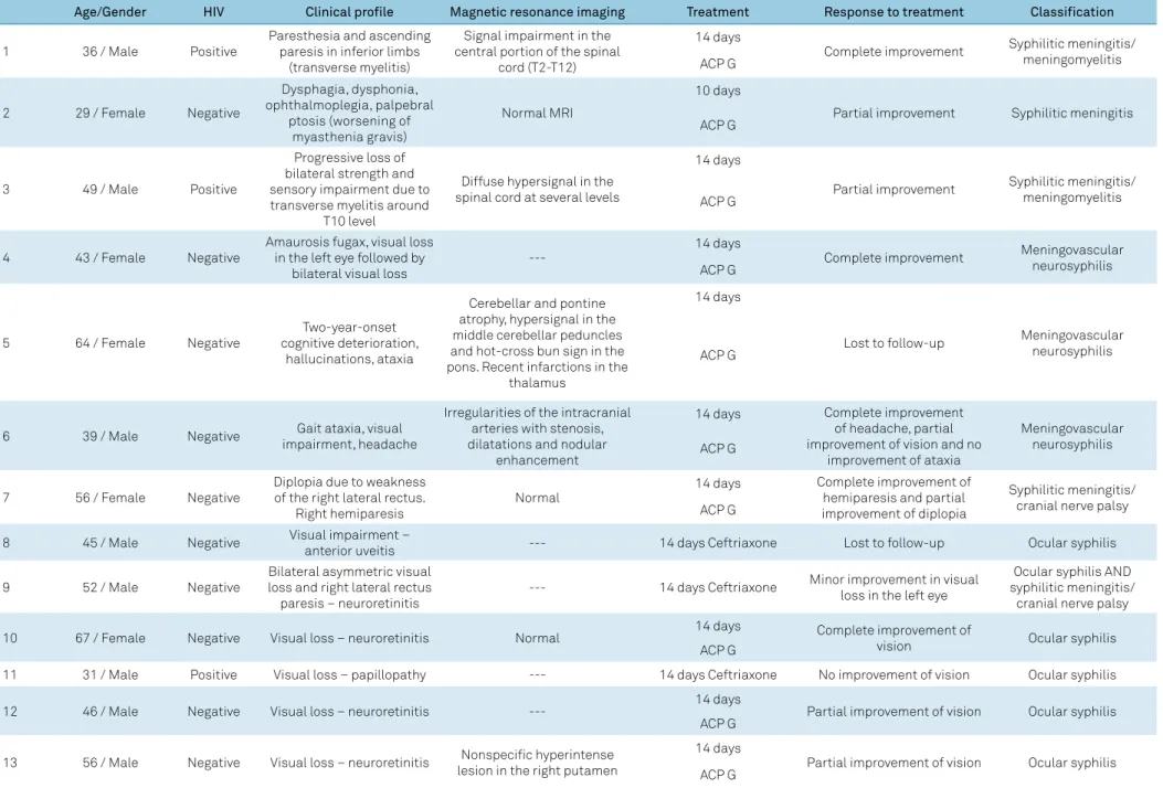
Table 1. Clinical and demographic characteristics of 13 patients with neurosyphilis and ocular syphilis.
Variables Neurosyphilis without
ocular syphilis (n = 7)
Ocular syphilis (total) (n = 6)
Ocular + neurosyphilis
(n = 2) p
Demographics
Age (years) – median (IQR) 43 (37.5;60) 49 (38; 61.5) 59 0.44
Gender (male) – n (%) 3 (42.86%) 5 (83.3%) 1 (50%) 0.26
Symptoms
Symptom duration (days) – median (IQR) 187.5 (10.5; 540) 270 (180; 360) ----
-Serum
HIV+ - n (%) 2 (28.6%) 1 (16.7%) 0
CD4+ – median (IQR) 111.5 (97.2; 125.7) --- ----
CD4+ Nadir– median (IQR) 88 (62;114) --- ----
Viral load – median (IQR) 1.8x10⁵ (1.5 x10⁵; 2.2 x10⁵) --- ----
Reactive CI – n (%) 6 (100%) 6 (100%) 2 (100%)
Reactive VDRL – n (%) 3 (42.8%) 6 (100%) 2 (100%) 0.55
Reactive FTA-Abs – n (%) 4 (80%) 4 (80%) 1 (50%)
CSF
Reactive VDRL – n (%) 2 (28.6%) 1 (20%) 1 (50%)
Reactive FTA-Abs – n (%) --- 1 (100%) 1 (50%)
WBC cell/mm3 – median (IQR) 47 (4;72) 2.5(1;23.5) 0.07
WBC >5 cells/mm³ - n (%) 5 (71.4%) 1 (16.67%) 1 (50%)
Protein mg/dL– median (IQR) 114 (70.70; 407.7) 33,75 (28,05; 59.1) 0.03
Protein> 45mg/dL – n (%) 6 (85.7%) 1 (16.67%) 0 0.02
Glucose mg/dL– median (IQR) 52 (44.50; 114.0) 58 (49.5; 6.5) 0.18
Lactic Acid mmol/L– median (IQR) 2.05 (1.25; 3.2) -
376
Ar
q Neur
opsiquia
tr 2018;
76(6):373-380
Table 2. Neurosyphilis and ocular syphilis – clinical and neuroimaging data and follow-up for 13 patients.
Age/Gender HIV Clinical profile Magnetic resonance imaging Treatment Response to treatment Classification
1 36 / Male Positive
Paresthesia and ascending paresis in inferior limbs
(transverse myelitis)
Signal impairment in the central portion of the spinal
cord (T2-T12)
14 days
Complete improvement Syphilitic meningitis/
meningomyelitis ACP G
2 29 / Female Negative
Dysphagia, dysphonia, ophthalmoplegia, palpebral
ptosis (worsening of myasthenia gravis)
Normal MRI
10 days
Partial improvement Syphilitic meningitis
ACP G
3 49 / Male Positive
Progressive loss of bilateral strength and sensory impairment due to transverse myelitis around
T10 level
Diffuse hypersignal in the spinal cord at several levels
14 days
Partial improvement Syphilitic meningitis/
meningomyelitis ACP G
4 43 / Female Negative
Amaurosis fugax, visual loss in the left eye followed by
bilateral visual loss
---14 days
Complete improvement Meningovascular
neurosyphilis ACP G
5 64 / Female Negative
Two-year-onset cognitive deterioration,
hallucinations, ataxia
Cerebellar and pontine atrophy, hypersignal in the middle cerebellar peduncles and hot-cross bun sign in the pons. Recent infarctions in the
thalamus
14 days
Lost to follow-up Meningovascular
neurosyphilis ACP G
6 39 / Male Negative Gait ataxia, visual
impairment, headache
Irregularities of the intracranial arteries with stenosis, dilatations and nodular
enhancement
14 days Complete improvement
of headache, partial improvement of vision and no
improvement of ataxia
Meningovascular neurosyphilis ACP G
7 56 / Female Negative
Diplopia due to weakness of the right lateral rectus.
Right hemiparesis
Normal
14 days Complete improvement of
hemiparesis and partial improvement of diplopia
Syphilitic meningitis/ cranial nerve palsy ACP G
8 45 / Male Negative Visual impairment –
anterior uveitis --- 14 days Ceftriaxone Lost to follow-up Ocular syphilis
9 52 / Male Negative
Bilateral asymmetric visual loss and right lateral rectus
paresis – neuroretinitis
--- 14 days Ceftriaxone Minor improvement in visual
loss in the left eye
Ocular syphilis AND syphilitic meningitis/
cranial nerve palsy
10 67 / Female Negative Visual loss – neuroretinitis Normal 14 days Complete improvement of
vision Ocular syphilis
ACP G
11 31 / Male Positive Visual loss – papillopathy --- 14 days Ceftriaxone No improvement of vision Ocular syphilis
12 46 / Male Negative Visual loss – neuroretinitis --- 14 days Partial improvement of vision Ocular syphilis
ACP G
13 56 / Male Negative Visual loss – neuroretinitis Nonspecific hyperintense
lesion in the right putamen
14 days
Partial improvement of vision Ocular syphilis
ACP G
377
Borges CR et al. Neurosyphilis and ocular syphilis: a case series
A study, presented at the annual Conference on Retroviruses
and Opportunistic Infections in 201710, observed that some clini
-cal symptoms, such as photophobia, vision loss, gait incoordina
-tion and moderate or severe hearing loss, presenting in positive
HIV patients, could predict the diagnosis of neurosyphilis. One
of the HIV-positive patients showed vision loss, although this patient did not meet the CSF criteria for neurosyphilis.
Another important risk factor for syphilis (and
neuro-syphilis) is male sex and the group of men who have sex with
men1,2,3. However, in our case series we observed a greater fre
-quency of female patients with neurosyphilis (57%). One pos -sible explanation is that most of the patients with HIV and AIDS who are admitted to our hospital are managed solely by the infectious diseases team, and these patients were not included in this series.
Clinical manifestations
In this case series, we observed the characteristic broad clinical spectrum of neurosyphilis. The clinical manifesta
-tions of neurosyphilis are classified according to whether
they are associated with the early or late forms of the disease. Early forms usually occur a few months to a few years after the primary infection and can manifest as
asymptom-atic or symptomasymptom-atic syphilitic meningitis and meningovas -cular neurosyphilis.
Late forms usually occur years to decades after the pri
-mary infection and can affect the brain, causing demen
-tia paralytica (general paresis of the insane), and the spinal
cord, resulting in tabes dorsalis1,2,3,11,12. No cases of paralytic
dementia or tabes dorsalis were observed in our study, in
agreement with reports in the literature that manifestations
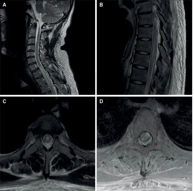
Figure 1. MRI of the vertebral column of a patient with transverse myelitis caused by neurosyphilis. A and B: Sagittal T2-weighted images of cervicothoracic vertebral column showing a hyperintense lesion affecting spinal cord segments from C6-C7 to T11-T12; C: Axial T2-weighted MRI showing hyperintensity involving most of the circumference of the spinal cord; D: Axial T1-weighted post-gadolinium MRI showing contrast enhancement in two different areas of the spinal cord.
A
B
of late neurosyphilis have been observed less frequently since the advent of antibiotics1,2. Instead, most of the
neurosyphi-lis patients studied here showed symptoms associated with syphilitic meningitis. Two patients presented with cranial
nerve palsy, a common manifestation of the early form of syphilitic meningitis that is frequently the first clinical symp -tom of the disease1,2,3,6. Two others had a clinical profile and
MRI consistent with meningomyelitis but responded very well to treatment with penicillin, supporting the diagnosis; HIV serology was positive in both these patients.
While tabes dorsalis used to be the most common spinal cord manifestation of neurosyphilis in the pre-antibiotic era, it has now been superseded by
meningo-myelitis, which develops, on average, six years after infec
-tion, although this can vary between 1-30 years1,2,3,13.
Meningomyelitis is in fact a very rare form6 caused by
spi-nal cord infections secondary to syphilitic meningitis1. Its
onset is insidious and is characterized by weakness and/
or paresthesia starting in the lower limbs, which may be
asymmetric. The symptoms may worsen with time and are associated with important impairments. Neuroaxis
MRI can reveal, as in Figure 2, a high-signal lesion in
T2-weighted images of parenchymal spinal cord confined
to the central portion and extending over multiple levels
as well as in gadolinium-enhanced T1-weighted images.
Unlike tabes dorsalis, the symptoms of this disorder can improve following treatment with penicillin13,14.
We also observed worsening of the clinical picture in the patient with myasthenia gravis as a result of syphilitic menin
-gitis, which improved after treatment. We found only one case
report in the literature describing a similar presentation15.
In our series, the meningovascular form corresponded with
a substantial proportion of the patients studied. Most were
under 50 years of age and, apart from one, who suffered from
amaurosis fugax, all had histories of repeated episodes of
infarc-tion. The MRI angiography revealed characteristic irregularities
of the intracranial arteries with stenosis, dilations and nodular
enhancement in one patient, consistent with meningovascular Figure 2. Workup of a 67-year-old female with neuroretinitis caused by neurosyphilis. A: normal fundoscopy. B: visual field examination showing central scotoma and impairment of the nasal peripheral visual field in the right eye (RE) and mild impairment of the nasal peripheral visual field in the left eye (LE). C: Fluorescein angiography revealing hyperfluorescence in the right superior temporal arch and papilla after four minutes and in the left papilla and macula after nine minutes, suggesting neuroretinitis. D: Fluorescein angiography after treatment showing an improvement in the macular and papillary edemas, as well as a reduction in leakage in the optic disc and retina in both eyes.
RE
RE RE
RE LE
LE LE
LE
2’ 4’ 9’ 18’ 1’ 9’ 11’
1’30” 3’ 9’30” 17’ 1’ 9’ 12’
A
B
379
Borges CR et al. Neurosyphilis and ocular syphilis: a case series
neurosyphilis. Meningovascular neurosyphilis may occur 5-10
years after the initial infection and may manifest as arteritis, resulting in reduced arterial caliber and culminating in throm-bosis followed by ischemic strokes, which may occur repeatedly.
Unlike traditional stroke syndromes, it can present without vas -cular risk factors and at a young age1,2,3. The CSF-VDRL is con
-sidered the gold standard for diagnosis of neurosyphilis but has
low sensitivity (from 30% to 70%)1,2. In our series, there were nine
neurosyphilis patients, of whom only three had reactive CSF-VDRL (33%). The lack of assessment of the IgG index in our lab
-oratory may have contributed to this low sensitivity.
OCULAR SYPHILIS
Demographics, coinfection with HIV and disease manifestations
An American study of demographic characteristics of
ocular syphilis showed similar mean ages (49.4 ± 16.5) and sex (male 74%) to the results presented here16,17.
Because ocular syphilis is frequently found in associa-tion with HIV, serological HIV tests should be performed in all patients with a diagnosis of ocular syphilis. While some
studies report high HIV-positive rates in patients with ocular syphilis (up to 70%)18, we observed a rate of only 16.6% (1/6).
This may be explained by a selection bias, considering that the
patients with ocular syphilis came from an ophthalmology
service that provides care to the general population. Another possible explanation is the difference in the demographic pat
-tern of the distribution of syphilis in developing countries in comparison with developed countries. While the latter show a higher prevalence of patients with syphilis and positive HIV in men who have sex with men, the former exhibit a higher prev -alence of older people without these conditions.
Corroborating the data in the literature, this study found five patients with disorders of the posterior pole ( four with neuroretinitis and one with optic neuritis) and one with anterior uveitis. Currently, the frequency of syphilis patients
diagnosed by ophthalmologists is unknown, but the
poten-tial for successful treatment is very high19.
COMPARISON OF CSF DATA OF PATIENTS WITH NEUROSYPHILIS AND OCULAR SYPHILIS
This study compared the CSF samples of patients with
a diagnosis of neurosyphilis with those with a diagnosis of
ocular syphilis. Only 33% of the patients with ocular syphi
-lis also showed CSF impairments that led to the diagnosis of neurosyphilis, and this differs from the literature about ocular syphilis, which shows 60% of lymphocytic pleocyto
-sis, elevated protein or both in the CSF20. The patients with
neurosyphilis exhibited higher protein levels and a trend to having more white blood cells in the CSF in comparison
with the patients with ocular syphilis. Furthermore, only one
sample showed a positive CSF FTA-Abs and no one exhib
-ited a positive CSF-VDRL result. Despite the fact that ocular
syphilis is not always accompanied by syphilitic meningitis21,
this frequency seems low. However, the small sample in this case series does not allow the generalization of this data and
points to the need for further studies.
All the patients with ocular syphilis had reactive serum VDRL and FTA-Abs, pointing to a diagnosis of syphilis. In four patients, the ophthalmological evaluation was consistent with neuroretinitis, which is strongly suggestive of neurosyphilis22.
The unavailability of the FTA-Abs test in the CSF may have lessened the sensitivity toward the diagnosis of neurosyphi -lis. Although nontreponemal tests are used to screen for
syph-ilis, they have the disadvantage of having low sensitivity and specificity, especially in CSF samples, unlike the FTA-Abs test, which is highly sensitive and, when negative, can exclude a
diagnosis of neurosyphilis19,22. In a 21-patient case series, for
example, 75% of patients with ocular syphilis had a nonreac
-tive VDRL but 100% had a posi-tive serum FTA-Abs23.
However, it is necessary to point out that no laboratory study has proven sufficiently sensitive or specific to serve as a single test for the definitive diagnosis of neurosyphilis. The diagnosis of syphilis has remained more difficult than the diagnosis of most other infections. The standard diagnosis of neurosyphilis is based on an increased CSF white blood cell count and/or a reactive CSF-VDRL test result. The CSF abnormalities include elevated protein levels and pleocyto
-sis, which are found in up to 70% of patients. The FTA-Abs,
despite being a good auxiliary test, has shown a high rate of
false positives and is not recommended by most of the guide -lines for the diagnosis of neurosyphilis1,21.
Although the Centre for Disease Control recommenda -tion from 2015 suggests a lumbar puncture in all patients with ocular syphilis, the literature lacks data describing the frequency of neurosyphilis among the patients diagnosed with ocular syphilis. One explanation for this could be that a lumbar puncture is only indicated in selected cases, such
as syphilis with neurological involvement, patients with a
relapse, before treatment with nonpenicillin regimens and infants with congenital syphilis24.
In conclusion, the case series presented here allowed some important features currently attributed to neurosyphilis and
ocular syphilis to be reviewed. It brings attention to some clin
-ical presentations of neurosyphilis that are different from its
classic manifestations. It also discusses some patients with ocular syphilis, which is a manifestation that is commonly
neglected by the neurologist. The study’s limitations are the few patients studied and its retrospective case series design,
which limits the collection of clinical data and precludes the
description of all patients among the population. Moreover, we did not perform the IgG index of the CSF samples, which could
help to identify intrathecal synthesis of IgG and increase the
Syphilis continues to be an important disease in neu-rology and in neuro-ophthalmology, especially in certain
epidemiological risk groups, such as men who have sex with men and persons living with HIV. In view of the
re-emergence of this disease in the first decade of this cen-tury, neurologists and ophthalmologists should consider
a possible diagnosis of syphilis in many different clinical
contexts. Moreover, it is important to observe the clini -cal and epidemiologi-cal particularities of the disease in the post-antibiotic era. It should be remembered that it was not without reason that Osler called this disease the “Great Imitator”.
References
1. Marra CM. Neurosyphilis. Continuum (Minneap Minn). 2015 Dec;21 6 Neuroinfectious Disease:1714-28.
2. Berger JR, Dean D. Neurosyphilis. Handb Clin Neurol. 2014;121:1461-72. https://doi.org/10.1016/B978-0-7020-4088-7.00098-5
3. Bhai S, Lyons JL. Neurosyphilis update: atypical is the new typical. Curr Infect Dis Rep. 2015 May;17(5):481. https://doi.org/10.1007/s11908-015-0481-x
4. Woolston SL, Dhanireddy S, Marrazzo J. Ocular syphilis: a clinical review. Curr Infect Dis Rep. 2016 Nov;18(11):36. https://doi.org/10.1007/s11908-016-0542-9
5. Davis JL. Ocular syphilis. Curr Opin Ophthalmol. 2014 Nov;25(6):513-8. https://doi.org/10.1097/ICU.0000000000000099
6. Marra CM. Update on neurosyphilis. Curr Infect Dis Rep. 2009 Mar;11(2):127-34. https://doi.org/10.1007/s11908-009-0019-1
7. Gaudio PA. Update on ocular syphilis. Curr Opin Ophthalmol. 2006 Dec;17(6):562-6. https://doi.org/10.1097/ICU.0b013e328010a9b5
8. Chahine LM, Khoriaty RN, Tomford WJ, Hussain MS. The changing face of neurosyphilis. Int J Stroke. 2011 Apr;6(2):136-43. https://doi.org/10.1111/j.1747-4949.2010.00568.x
9. Andrade P, Figueiredo C, Carvalho C, Santos L, Sarmento A. Transverse myelitis and acute HIV infection: a case report. BMC Infect Dis. 2014 Mar;14(1):149. https://doi.org/10.1186/1471-2334-14-149
10. Davis AP, Stern J, Dunaway S, Tantalo L, Sahi S, Crooks A et al. How well do neurologic symptoms identify hiv-infected individuals with neurosyphilis? In: Annals of Conference on Retroviruses ans Oppotunistic Infections; 2017 Feb 13-16; Seattle, USA. Abstract number: 749.
11. Ghanem KG. Neurosyphilis: a historical perspective and review. CNS Neurosci Ther. 2010 Oct;16(5):e157-68. https://doi.org/10.1111/j.1755-5949.2010.00183.x
12. Nitrini R. The history of tabes dorsalis and the impact of observational studies in neurology. Arch Neurol. 2000 Apr;57(4):605-6. https://doi.org/10.1001/archneur.57.4.605
13. Berger JR. Neurosyphilis and the spinal cord: then and now. J Nerv Ment Dis. 2011 Dec;199(12):912-3. https://doi.org/10.1097/NMD.0b013e31823928e8
14. Chilver-Stainer L, Fischer U, Hauf M, Fux CA, Sturzenegger M. Syphilitic myelitis: rare, nonspecific, but treatable. Neurology. 2009 Feb;72(7):673-5. https://doi.org/10.1212/01.wnl.0000342460.07764.5c
15. Wüllenweber M, Schneider U,Hagenah R [Myasthenia gravis in AIDS and neurosyphilis]. Nervenarzt. 1993 Apr;64(4):273-7. German.
16. Moradi A, Salek S, Daniel E, Gangaputra S, Ostheimer TA, Burkholder BM et al. Clinical features and incidence rates of ocular complications in patients with ocular syphilis. Am J Ophthalmol. 2015. Feb;159(2):334-43. https://doi.org/10.1016/j.ajo.2014.10.030
17. Balba GP, Kumar PN, James AN, Malani A, Palestine AG, Welch JN et al. Ocular syphilis in HIV-positive patients receiving highly active antiretroviral therapy. Am J Med. 2006 May;119(5):448.e21-5. https://doi.org/10.1016/j.amjmed.2005.11.016
18. Malerbi FK, Ghanem RC, Chiang J, Takahashi WY. Descolamento de retina exsudativo bilateral associado a alterações de comportamento em paciente com diagnóstico de neurossífilis: relato de caso. Arq Bras Oftalmol. 2006 Jan-Feb;69(1):115-8. https://doi.org/10.1590/S0004-27492006000100022
19. Chao JR, Khurana RN, Fawzi AA, Reddy HS, Rao NA. Syphilis: reemergence of an old adversary. Ophthalmology. 2006
Nov;113(11):2074-9. https://doi.org/10.1016/j.ophtha.2006.05.048
20. Spoor TC, Ramocki JM, Nesi FA, Sorscher M. Prevalence of FTA-ABS reacrivity and cerebrospinal fluid findings. J Clin Neuroophthalmol. 1987;7(4):191-5.
21. Fishman RA. Cerebrospinal fluid in diseases of the nervous system. Philadelphia: Saunders; 1992.
22. Hernández-Bel P, Gómez-Maestra MJ, Torrijos-Aguilar A, López J, Vilata JJ, Alegre V. Ocular syphilis: a rare presentation of secondary syphilis in an immunocompetent patient. Actas Dermosifiliogr. 2010. Mar;101(2):184-6. https://doi.org/10.1016/S1578-2190(10)70611-9
23. Rodrigues RA, Nascimento HM, Muccioli C. Yellowish dots in the retina: a finding of ocular syphilis? Arq Bras Oftalmol. 2014 Oct;77(5):324-6. https://doi.org/10.5935/0004-2749.20140081