Chronic interstitial lung diseases in children*
Doenças pulmonares intersticiais crônicas na criançaMaria Aparecida Soares de Souza Paiva, Sandra Mara Moreira Amaral
Abstract
Interstitial lung diseases (ILDs) in children constitute a heterogeneous group of rare diseases that have been described and classified according to experiences and research in adults. However, pediatric pulmonologists have observed that the clinical spectrum is broader in children than in adults, and that many of these disorders have different courses and treatment responses. In addition, probably due to the various stages of lung development and maturation, new clinical forms have been described, particularly in infants. This has broadened the classification of ILDs in this age bracket. The understanding that neither the usual definition nor the standard classification of these disorders entirely apply to children has prompted multicenter studies designed to increase knowledge of these disorders, as well as to standardize diagnostic and therapeutic strategies. We have reviewed the conceptualization of ILDs in children, taking into consideration the particularities of this group of patients when using the criteria for the classification of these diseases in adults. We have also made a historical review of several multicenter studies in order to further understanding of the problem. We have emphasized the differences in the clinical presentation, in an attempt to highlight knowledge of newly described entities in young children. We underscore the need to standardize management of laboratory and radiological routines, as well as of lung biopsy processing, taking such knowledge into account. It is important to bear in mind that, among the recently described disorders, genetic surfactant dysfunction, which is often classified as an idiopathic disease in adults, should be included in the differential diagnosis of ILDs.
Keywords: Lung diseases, interstitial; Lung diseases, interstitial/diagnosis; Lung diseases, interstitial/therapy;
Child.
Resumo
As doenças pulmonares intersticiais (DPIs) da criança constituem um grupo heterogêneo de doenças raras que têm sido definidas e classificadas de acordo com as experiências e as pesquisas em adultos. Entretanto, os pneumolo-gistas pediátricos vêm observando que o espectro clínico é mais amplo nas crianças, e que muitas destas doenças evoluem e respondem ao tratamento de forma diferente. Além disso, provavelmente devido a estágios diferentes de desenvolvimento e maturação pulmonares, novas formas clínicas têm sido descritas, principalmente em lactentes, ampliando a classificação nessa faixa etária. A compreensão de que nem a definição nem as classificações esta-belecidas se aplicam inteiramente ao grupo pediátrico tem motivado a realização de estudos multicêntricos com o objetivo de estudá-las melhor, unificando as estratégias diagnósticas e terapêuticas. Fizemos a revisão atuali-zando a conceituação das DPIs no grupo pediátrico, considerando as particularidades desse grupo na utilização do esquema de classificação dessas doenças para adultos e revendo o histórico dos esforços para uma melhor compreensão do problema com os estudos multicêntricos. Foram ressaltadas as diferenças na apresentação clínica, procurando realçar os novos conhecimentos sobre as doenças recém descritas nas crianças pequenas. Alertamos também para a necessidade de ser seguida uma rotina padronizada de investigação laboratorial, radiológica e de processamento das biópsias à luz desses conhecimentos. É importante lembrar que, do grupo das novas doenças descritas, as alterações genéticas do surfactante devem constar também do diagnóstico diferencial das DPIs dos adultos, podendo se apresentar nesse grupo como uma das doenças classificadas como idiopáticas.
Descritores: Doenças pulmonares intersticiais; Doenças pulmonares intersticiais/diagnóstico;
Doenças pulmonares intersticiais/terapia; Criança.
* Study carried out at the Rio de Janeiro State Worker’s Hospital, Rio de Janeiro, Brazil.
Correspondence to: Maria Aparecida Soares de Souza Paiva. Av. das Américas, 2300, Casa 37, Barra da Tijuca, CEP 22640-102, Rio de Janeiro, RJ, Brasil.
Tel 55 21 3431-1000. E-mail: [email protected] Financial Support: None.
a period of 3 years (1995-1998) and identi-fied 46 cases of ILD, symptom onset occurring before 1 year of age in 66% of the children. Of the 46 patients, 7 (16%) died, and 9 belonged to one of only four families.
A multicenter study involving 131 children (1995-1997),(8) with questionnaires sent to 187 pulmonology centers in Europe, Australia and South Africa, obtained a response from 20.3% of these centers. In that study, noninvasive tests alone were able to establish the diagnosis in 5 patients (3.8%), and complementary invasive techniques—bronchoalveolar lavage (BAL) and biopsy—allowed the diagnosis of 117 patients (89%). Of these, 64% were males, and mean age ranged from 0.75 to 17.8 years.
Subsequently, a group of pediatric pulmo-nologists from the European Respiratory Society (ERS) formed a work group and sent questionnaires to all pulmonology centers in Europe, evaluating the records of 185 patients (1997-2002).(9) Of these, 58 children were under 2 years of age. The prevalence was higher among males. Approximately 10% of the cases occurred in siblings. The clinical data and the invasive and noninvasive complementary tests allowed the diagnosis of 177 patients (95.6%). In 67 of the patients who underwent biopsy, the diag-nosis reported in the records was not consistent with the classification of ILDs in adults. The authors raised various questions, principally regarding the need for a classification of ILDs in children. Based on the data collected, the patients were divided into four principal groups of diagnosis: 1) diffuse parenchymatous pulmo-nary disease of known causes (hypersensitivity pneumonitis, aspiration pneumonitis, etc.); 2) idiopathic interstitial pneumonia (desqua-mative interstitial pneumonia, lymphocytic interstitial pneumonia, nonspecific interstitial pneumonia [NSIP], etc.); 3) other forms of inter-stitial pneumonia (hemosiderosis, sarcoidosis); and 4) congenital disorders (surfactant dysfunc-tion, lymphangiectasia, etc.). The authors did not clearly suggest a classification.
New forms of ILDs have been described in young children, diagnosed thanks to the advances in immunohistochemistry, genetic tests and electron microscopy (EM): neuroen-docrine cell hyperplasia of infancy,(10) pulmonary interstitial glycogenosis,(11-12) genetic surfactant dysfunction,(2,5) disorders of lung development(2)
Introduction
Interstitial lung diseases (ILDs) constitute a heterogeneous group of diseases, the diagnoses of which are a challenge.(1) The term diffuse parenchymatous pulmonary disease is more appropriate to describe such disorders because, in addition to alterations in the alveolar walls and spaces, the distal airways are commonly affected.(2) The genetic factors are taken into consideration due to various reports of cases in the same family and to new information regarding genetic surfactant dysfunction.(3-5) The prevalence of ILD among children is not well-known; some data suggest a prevalence of 3.6 per million.(3) It is probably underdiagnosed and underreported. However, it is certainly quite rare among children.
There has been a great deal of debate regarding the nomenclature and classification of these disorders in pediatric patients due to the clinical spectrum, which varies with age. Certain clinical situations are unique to chil-dren, principally among those under 2 years of age; others are included in the classification of ILDs in adults. However, ILDs have a less severe course and a better prognosis in children than in adults. In addition, children respond to thera-peutic strategies better than do adults. These differences in presentation and course might be related to the different stages of lung develop-ment and maturation.(6)
Since the 1980s, studies aiming to stand-ardize the diagnosis and treatment of ILDs in children have been conducted. However, the rarity of ILDs among children, the small patient samples and the lack of long-term follow-up, as well as the lack of standardization of the diag-nosis, have been obstacles to such studies.
We believe that it is important to highlight certain multicenter studies or studies that have investigated a greater number of patients in an attempt to gain a deeper understanding of these disorders.
One group of authors investigated 99 children in the USA over a period of 15 years (1980-1994); of the 99 children, 15 died during the study period.(7) Survival rates were 83%, 72% and 64% for 24, 48 and 60 months after the onset of symptoms, respectively, which high-lights the importance of a timely diagnosis.
In order to better define ILDs in children, the work group decided to incorporate clinical data to the definition of the “chILD syndrome” to aid in recognizing these disorders. For the diagnosis of ILDs, it was established that, in the absence of known causes of lung disease, at least 3 of the following criteria had to be present: 1) respiratory symptoms (cough, rapid or difficult breathing, or exercise intolerance); 2) signs (tachypnea at rest, crackles, retractions, digital clubbing, underde-velopment or respiratory failure); 3) hypoxemia; and 4) diffuse alteration on chest X-rays or CT scans. By applying the definition of the chILD syndrome, 3 to 4 criteria were met by 91% of the 218 cases studied in 11 centers in North America (1999-2004).(2,15) The group reviewed the biopsy results of 187 children (1999-2004) and developed a new clinical and histological classification,(2) which principally organized the disorders in children under 2 years of age (Chart 1). This classification is probably not defini-tive. It should be pointed out that the duration should not be added to the definition because the disease progresses rapidly in some neonates. Some of the previous definitions, including that developed by the ERS work group, limited their scope to children presenting disease duration of 3 months.
With regard to older children, we believe that the consensus classification proposed by the American Thoracic Society (ATS) and the ERS(16) is appropriate. However, the experience gained from analyzing this age bracket should be considered. The usual interstitial pneu-monia (UIP) and idiopathic pulmonary fibrosis (IPF) form, for instance, with the character-and chronic pneumonitis of infancy.(13) These
will be discussed further in the present review because we believe in the relevance of furthering the understanding of such entities.
Based on the accumulated experience, there are now certain new concepts related to ILDs in children, principally in those who are under 2 years of age. In 2004, in a meeting regarding ILDs, the US National Institutes of Health founded the Rare Lung Disease Consortium, which also resulted in the establishment of a foundation for the families of children with ILDs. According to specialists,(14) the excellent survey conducted by the European work group should be regarded as the first step in the attempt to standardize the diagnosis and treatment of ILDs in children, so that each center will be have guidelines to follow when diagnosing and treating such disor-ders The data collected will also make it possible to develop protocols and design clinical trials (therapeutic and diagnostic). A work group, which comprised pediatric pulmonologists, pathologists and radiologists from 11 centers with experience in ILDs, was formed. A protocol of investigation, which involved the exchange of imaging test and biopsy results among the largest centers in the world, was proposed.
In addition, this multidisciplinary group, designated Children’s Interstitial Lung Disease (chILD), has turned its attention to the clas-sification and study of ILDs that affect young children, often in the neonatal period, in the belief that the information obtained might increase understanding of these disorders in the other phases of life.(15)
Chart 1 - Most prevalent diffuse lung diseases in patients under two years of age.
Diffuse diseases of development Acinar dysplasia
Congenital alveolar dysplasia
Alveolar capillary dysplasia with misalignment of pulmonary veins
Abnormalities of development Chronic lung disease of infancy
• related to chromosomal alterations • related to congenital heart diseases
Pulmonary hypoplasia
Specific conditions of unknown etiology Neuroendocrine cell hyperplasia of infancy
Pulmonary interstitial glycogenosis
Disorders caused by surfactant dysfunction Mutations in the genes of SP-B, SP-C, ABCA3 and TTF-1
Other histological expressions (PAP, DIP, NSIP, CPI)
under 4 years of age).(20) The clinical data are nonspecific and often less evident than they are in adults. The prognosis also seems to be different, the course of the disease being less favorable. Multiple organ involvement is more common and more severe in younger children. An excellent review of sarcoidosis in children was published in 2005.(21)
Among the group of histologically well-de-fined diseases, we highlight, due to its severity in children, multisystem Langerhans cell histio-cytosis, an immunologic disease that affects the bone marrow, spleen, liver, lymph nodes, thymus, skin, brain, bones, gastrointestinal tract and lungs. Lung disease might occur in isolation, principally in young individuals. It can cause spontaneous pneumothorax. Lung biopsy reveals Langerhans cell infiltrate, interstitial pneumo-nitis and honeycombing with cysts of different dimensions. Lytic lesions in the bones and cysts in the lungs are clues to the diagnosis.(22)
History
The classifications in adult patients have also been evolving.
In 1944, Hamman and Rich reported cases of interstitial pneumonia.(23) However, the study of ILDs began to advance in the 1960s, with the istic fibroblastic foci, is rare among children;
however, a recent study reported that less severe phenotypes of diseases that cause surfactant protein deficiency might progress with various histopathological profiles of ILDs,(17) a case of UIP being reported in a teenager with the ABCA3 mutation.(18) Other forms of presenta-tion, such as respiratory bronchiolitis (related to smoking), have also been excluded from the differential diagnosis in children, although we do not know whether passive smoking might be the cause of a similar problem in children. There have been reports in the literature that hypersen-sitivity pneumonitis is probably underdiagnosed in children, principally in its chronic form. Since hypersensitivity pneumonitis principally results from the exposure to avian antigens and fungi, as well as from methotrexate use, a high degree of suspicion and an excellent history-taking are crucial.(19)
Sarcoidosis, a systemic granulomatous disease, is relatively rare in children, and epide-miological reports in the literature are few. A national registry in Denmark (1979-2004) reported an incidence of 0.29 per 100,000 person-years among those under 15 years of age, an incidence that decreased with age (0.06/100,000 person-years among those
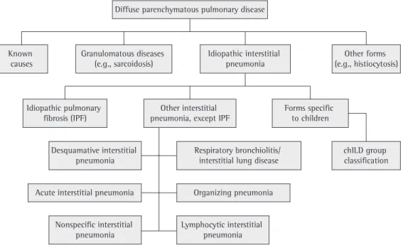
Diffuse parenchymatous pulmonary disease
Known
causes Granulomatous diseases (e.g., sarcoidosis) Idiopathic interstitial pneumonia (e.g., histiocytosis)Other forms
Idiopathic pulmonary fibrosis (IPF)
Other interstitial pneumonia, except IPF
Forms specific to children
chILD group classification Desquamative interstitial
pneumonia Respiratory bronchiolitis/ interstitial lung disease
Acute interstitial pneumonia Organizing pneumonia
Nonspecific interstitial pneumonia
Lymphocytic interstitial pneumonia
Figure 1 - Classification of diffuse lung diseases according to the American Thoracic Society consensus,
tion and ground-glass opacities. Respiratory function tests confirmed obstruction with air trapping. Lung biopsy showed no inflammation or interstitial alterations. The most signifi-cant histological finding was neuroendocrine cell hyperplasia in the distal airways, revealed by bombesin-like immunoreactivity. There was a contrast between the unfavorable clinical picture and the minimal histological alterations observed. Many patients received the standard treatment, and the majority required supple-mental oxygen for months or years. The etiology, pathogenesis and long-term prognosis remain unknown.(1,10) Of the 15 patients, 10 lived at an altitude of 1,600 m, 5 m at sea level, suggesting that hypoxia at elevated altitudes is a relevant factor in the pathogenesis of the disorder.(28)
Neuroendocrine cell hyperplasia or dysfunc-tion might occur in the following situadysfunc-tions: pediatric asthma; cystic fibrosis; bronchopul-monary dysplasia; pulbronchopul-monary hypertension; pneumonia; congenital malformations; sudden death syndrome; congenital central alveolar hypoventilation syndrome; and diffuse idio-pathic neuroendocrine cell hyperplasia in adults. Therefore, the diagnosis of neuroendocrine cell hyperplasia of infancy requires a correlation among clinical, histological and radiological findings.
Pulmonary interstitial glycogenosis
One group of authors(11) reported the cases of 7 children, 6 of whom were male, with nonin-advances in thoracic surgery, making biopsies
more common. Based on histopathological tests, the first classification was established by Liebow in 1975.(24) However, subsequent questions led to revisions, because different terms occasion-ally designated the same disorder. The landmark review conducted by Katzenstein & Myers and published in 1998 divided the cases formerly classified as IPF into four different forms and established consistent histological criteria for each of them, with clinical, therapeutic and prognostic implications.(25)
The need for internationally standardized diagnostic criteria and nomenclature led to a classification proposed by a multidisciplinary group of the ATS/ERS in a consensus concluded in 2001, in which the importance of the clinical, radiological and histopathological interaction in the study of ILDs was reinforced.(16) This classifi-cation, the fruit of the accumulation of a great amount of experience, is still under debate,(26) and it will certainly be modified. However, it organized the information available and allowed a standardized evaluation. Figure 1 shows a flow chart proposed by the ATS/ERS consensus, with the addition of the forms reported in chil-dren under 2 years of age aiming to place such disorders in the context of a classification used routinely, modified based on a study we have published recently.(27) The different types of chronic idiopathic interstitial pneumonia are described in Chart 2, the diagnosis being gener-ally suggested by a detailed history.
It becomes evident that it is not appropriate to classify all pediatric patients according to the classification in adults. The stage of lung devel-opment and maturation should be taken into account for a more appropriate approach.
Clinical forms specific to children
Persistent tachypnea of infancy with
neuroendocrine cell hyperplasia
The neuroendocrine cells are present in the airway mucosa of mammals, in isolation or in combination with neuroepithelial bodies, and produce serotonin, bombesin and calcitonin.(28)
A group of 15 children, of whom 12 were infants, presenting with persistent tachypnea, rales and hypoxemia, was investigated. Chest X-rays and HRCT scans showed
hyperinfla-Chart 2 - Known causes of interstitial lung disease.
Cause Types
Aspiration syndromes GER/swallowing disorders/ malformations
Chronic infections Viral (EBV/CMV/adenovirus)
Bacterial (Chlamydia sp. and
Mycoplasma sp.) Physical and
environmental agents
Hypersensitivity pneumonitis Toxicity from oxygen and other gases
Mineral and organic dust Drugs
Radiation Collagen diseases
and other rheumatic diseases
Interstitial disease caused by genetic
alterations in the surfactant proteins
Surfactant is a phospholipid film that main-tains alveolar stability, preventing alveolar collapse at the end of exhalation. It is composed of lipids (80-90%) and proteins (10-15%). Approximately 2-3% are surfactant proteins (SP-A, -B, -C and -D). Their metabolism involves other molecules, such as ABCA3 and thyroid transcription factor-1 (TTF-1).(31)
For decades, surfactant deficiency has been known to cause hyaline membrane disease in premature neonates. However, only recently has this type of dysfunction been linked to other lung diseases. Mutations in the genes of the surfactant proteins, although rare, are increasingly reported as being the cause of ILDs in children and adults and should therefore be considered in any patient with the chILD syndrome. The form of presenta-tion varies with age. In neonates and infants, the profile is usually severe, and mortality is high; in schoolchildren and adults, the disease is chronic (NSIP, UIP), requiring a high degree of suspicion.(18,30-37)
Mutations in the ABCA3 gene
Mutations in the ABCA3 gene can manifest as fatal lung disease in neonates(33) or as ILDs in older children and adults. The most common symptoms are cough, dyspnea, hypoxemia, digital clubbing and rales.(32,33) In a study of 9 cases,(35) 4 presented pectus excavatum. The HRCT scans show alterations that are commonly observed in ILDs and do not correlate with respi-ratory function tests, hypoxemia or the course of the disease. Lung biopsy might reveal various histological patterns: alveolar proteinosis, desq-uamative interstitial pneumonia, NSIP and chronic interstitial pneumonitis. The case of a 15-year-old teenager with ABCA3 deficiency fectious respiratory symptoms in the neonatal
period. The chest X-rays revealed tachypnea, retractions, hypoxemia, interstitial infiltrate and hyperinflation. Of the 7 children, 4 were prema-ture neonates, the gestational age ranging from 25 to 33 weeks. The age at the onset of the symptoms ranged from 3 h to 4 weeks. Of the 7 children, 5 were submitted to mechanical ventilation, the duration of which ranged from 4 days to 6 months. All children underwent lung biopsy, which revealed uniform interstitial thick-ening, diffuse due to immature cells, which were similar to mesenchymal cells, showing abun-dant cytoplasmic glycogen detected by EM. This unusual finding of EM led the authors to conduct a review of 1,000 cases of lung biop-sies in patients with different pathologies. None of the patients presented glycogen in mesen-chymal cells, regardless of the gestational age. In contrast with other ILDs, there was no inflam-mation or alveolar cell hyperplasia. The course of the disease was favorable, and only one death occurred. Pulmonary interstitial glycogenosis is a rare neonatal disease and is presumably the same pathology that has been designated cellular interstitial pneumonitis.(29) It seems that select interstitial cells are immature, and the etiology and pathogenesis are still unknown.
Chronic pneumonitis of infancy
One group of authors reported, in a samples consisting of 9 individuals (infants and toddlers), a rare interstitial disease that differed from the ILDs previously described. The histopatholog-ical features of this disease, designated chronic pneumonitis of infancy, included septal thick-ening intensified by primitive mesenchymal cells and marked pneumocyte hyperplasia, together with alveolar exudate containing numerous macrophages and eosinophilic cell remnants. Inflammatory cells were scarce. The analysis of these data suggested recurring pneumonia or slow-resolving pneumonia, affecting an immature or malformed lung. Other such cases have been reported. In general, it presents high mortality. The radiological findings are nonspecific.(13) This histological pattern has been observed in certain patients with genetic surfactant dysfunction.(30)
Chart 3 - Severity score.
1- asymptomatic
2- symptomatic without blood gas alterations 3- symptomatic with hypoxemia
(oxygen saturation < 90%) only during sleep or exercise 4- symptomatic with hypoxemia (oxygen saturation < 90%) at rest
is complicated in individuals of such an age) or progress to death.(18,30,38)
The pathophysiology remains unclear. The accumulation of abnormal proteins in the endoplasmic reticulum activates the inflamma-tory cascade, favors apoptosis and interferes with normal protein synthesis. The release of cytokines and the recruitment of T lymphocytes and fibroblasts occur.(31)
Deficiency of thyroid transcription
factor-1
The TTF-1 plays a fundamental role in the formation and development of the lungs and controls the synthesis of SP-B, SP-C and ABCA3. Genetic mutations of the TTF-1 gene cause hypothyroidism, neurological symptoms (hypotonia and chorea) and neonatal respira-tory distress syndrome or chronic lung disease. The BAL reveals a decrease in SP-B associated with an increase in pro-SP-C. The histological findings include enlargement of intercellular junctions, type II pneumocyte hyperplasia and intra-alveolar accumulation of material positive for periodic acid-Schiff staining.(31)
Diagnosis
The clinical profile is variable and nonspe-cific. The onset can be acute or insidious. The most common signs and symptoms are dyspnea, cough, stunted growth (weight and height), crackles, digital clubbing, cyanosis, thoracic deformity and signs of pulmonary hyperten-sion or cor pulmonale. Clinical history-taking should be thorough for pulmonary diseases and systemic diseases.(39) Parents should be ques-tioned about dyspnea and its progression and severity, as well as about weight loss or delayed somatic development of their children. Such complaints are usually not spontaneous and aid in the diagnosis of chronic hypoxemia. In infants, it should be observed whether there is respiratory effort during milk feedings or crying. In term or near-term neonates with persistent course, cough or difficulty in weaning from mechanical ventilation, the possibility of ILD should be considered.(6) Cough might be the only symptom, and it is generally dry.(6) Hemoptysis or bloodstained sputum can be a sign of pulmonary hemosiderosis, other vasculitis or Ehlers-Danlos and pulmonary fibrosis was cited previously in
the present study.(18) Changes in the histological pattern have been observed when the biopsy is repeated. If typical findings are present, EM should be performed.(36)
Deficiency of surfactant protein B
A gene located on chromosome 2 encodes SP-B. The most common mutation, 121ins2, accounts for 70% of the cases of SP-B defi-ciency. Deficiency of SP-B follows an autosomal recessive inheritance pattern, and heterozygous are asymptomatic. The clinical and radiological profiles are similar to those of hyaline membrane disease. Deficiency of SP-B begins before 12 h of life and requires mechanical ventilation in term neonates. The response to exogenous surfactant is minimal or transitory. It does not respond to corticosteroids. Persistent pulmonary hyperten-sion might occur, responsive or not to NO. Most patients with SP-B deficiency evolve to death. Survival rates are low in cases of partial SP-B deficiency.(30,31,37)
The histological examination of the biopsy specimen might reveal alveolar proteinosis or desquamative interstitial pneumonia, or nonspe-cific findings such as interstitial fibrosis and alveolar cell hyperplasia. The use of EM aids in the differential diagnosis.(30)
Deficiency of surfactant protein C
The gene that encodes SP-C (SFTPC) is located on chromosome 8. Approximately 55% of the mutations occur spontaneously, and the remainder follows an autosomal dominant inheritance pattern. The most common muta-tion is I73T.(30)
when necessary. The identification of mutations using blood DNA or DNA from oral swab samples is the definitive diagnostic method. The PCR for specific fragments of ABCA3 proteins and restriction enzyme analysis should be initially performed. If the diagnosis is not established, gene sequencing of the most common muta-tions or complete gene sequencing is required. In order to screen for SP-B, ELISA or Western blot of the tracheal aspirate or of the BAL fluid can be performed as an initial step. An algorithm of diagnosis of these disorders is found in the reference list (30).
Spirometry can show the three types of respi-ratory pattern, although the restrictive pattern is the most common one. The DLCO technique confirms diffusion impairment that translates to hypoxemia. Functional alterations are seen in most children with active connective tissue disease, even in the absence of radiological abnormalities or of symptoms. These patients should be monitored from a functional stand-point.(22) Infants can also be evaluated using various respiratory function tests. These, however, are available in few health care facilities. Some centers take advantage of the sedation used for HRCT to perform such tests.
Hypercapnia appears later and indicates greater severity. However, it appears earlier in dermatomyositis due to respiratory muscle weakness.
Subsequently, we will recommend radio-logical tests and invasive tests such as BAL and biopsy, as well as the tests that should be performed on the material obtained.
Radiology
The conventional radiological test at the time of diagnosis generally shows bilateral inter-stitial infiltrate, as classically reported in the initial forms of ILDs. However, in a time before HRCT, it was reported that 9.6% of a group of 458 adults with histologically diagnosed disease showed normal radiological test results, biopsy being performed due to the results of blood gas analysis and functional tests of diffusion, which revealed significant alterations in gas exchange. Therefore, normal radiological test results do not exclude the diagnosis at the onset of these disorders.(41)
An HRCT scan can diagnose even initial lesions. The experience gained from such tests syndrome.(23,40) In schoolchildren and
adoles-cents, the profile is similar to that in adults. History-taking should also include infections, environmental exposure to mineral or organic dust, ingestion of medications or mineral oil, symptoms suggestive of aspiration syndrome and symptoms related to systemic diseases such as joint, heart, skin, kidney and sinus diseases, as well as neurological diseases. Nonrespiratory symptoms sometimes predominate, and ILD is diagnosed during screening for systemic disease.
Family history is quite important, principally in infants. Mortality at an early age or ill-defined lung disease in adults can be a clue to genetic diseases.
The height-weight curve, dyspnea at rest and on exertion and diffuse crackles, principally in the lung bases, should be evaluated during clinical examination. In a later phase of these disorders, thoracic deformity with flattening of the anteroposterior diameter, pectus exca-vatum and signs of chronic hypoxemia, such as digital clubbing and signs of cor pulmonale, are observed. Persistent pulmonary hyperten-sion might be the only symptom indicative of alveolar capillary dysplasia with misalignment of the pulmonary veins, a disease of develop-ment involving the vasculature and the lobular parenchyma.(6) Cyanosis is a late sign of severity. In sarcoidosis, general symptoms such as weight loss, fever and abdominal discomfort have been reported more often in children than in adults, as have cutaneous sarcoid lesions, erythema nodosum, peripheral adenopathies and symp-toms related to the central nervous system, articular symptoms being less common.(21)
Based on the clinical history and on the physical examination, the following tests should be performed, sequentially, beginning by the noninvasive tests: blood workup; blood gas analysis at rest and, when possible, after exer-tion; radiological test; serology for viruses such as EBV, HIV and cytomegalovirus; serology for mycoplasma and Legionella pneumophila; immunological profile; screening for aspiration syndromes; tests for collagen diseases; precip-itins to organic antigens; the sweat test; tests for sarcoidosis; cardiac evaluation; and respira-tory function tests.
IPF patients, with scores for classifying honey-comb lesions.(49)
A detailed description of how the principal clinical and pathological forms of ILD appear on CT scans is found in the ATS/ERS consensus statement,(16) as well as in excellent reviews conducted recently.(45,46)
Bronchoalveolar lavage
BAL is indicated for screening for etiologic agents and for evaluating cell profiles. Its diag-nostic possibilities, however, are far greater. In adults, BAL has been widely used in ILDs, and the role of differential analysis of cells with diagnostic and prognostic objectives is still controversial in many situations.
An excellent routine recommendation for performing BAL in children and evaluating the material collected has been published.(50) The laboratory must be prepared for processing the test. In the following cases, the first invasive test to be performed should be BAL, which can preclude the need for a biopsy: in suspected cases of aspiration pneumonia caused by mineral oil, hemosiderosis or alveolar proteinosis; when screening for certain infectious agents; and in immunocompromised patients.(51) Depending on the cellularity, BAL results might suggest other diagnoses. In suspected cases of SP-B deficiency, ELISA or Western blot should be performed on the BAL fluid (and on the tracheal aspirate). The detection of SP-B rules out this diagnostic hypothesis, and genetic tests for other surfactant abnormalities are indicated.(30) For the diagnosis of Langerhans cell histiocytosis, the presence of more than 5% of typical cells in the BAL fluid is diagnostic if it is associated with a suggestive clinical profile.
Since children need to be sedated so that BAL can be performed, a biopsy is the method of choice when children present a high degree of hypoxemia, because it provides more objective information (including prognostic information) and it allows screening for fibrosis, which is an irreversible factor of severity.
Biopsy
A biopsy should be performed early in suspected cases of ILD, i.e., as soon as all nonin-vasive tests have been performed, even in severe patients on mechanical ventilation, prior to in adults has been well-documented since their
emergence; however, the rarity of such disorders and the technical difficulties in performing HRCT in children under 5 years of age make it difficult to conduct studies involving that age bracket. One group of authors analyzed the HRCT scans of 20 children (1-16 years of age) with ILDs confirmed by biopsy and classified the images as follows: airway diseases; septal patholo-gies; infiltrative lung disease; airspace disease; and diseases accompanied by cysts.(42) That and other studies showed the limitations of CT scans, when used in isolation, in diagnosing ILDs in children.(43,44) There is a consensus that certain disorders present typical CT characteristics that, in combination with clinical characteristics and less invasive tests, facilitate the diagnosis of such disorders, without the need for a biopsy. Such disorders include alveolar proteinosis, congenital lymphangiectasia and idiopathic hemosiderosis. In adults, the recent advances toward a deeper understanding of the clinical and radiological features of ILDs using a standardized technique have greatly improved the diagnostic accuracy by narrowing the possibilities of differential diagnoses and allowing, in certain situations, a specific diagnosis to be established without the need for a biopsy.(45,46) The standardization of the HRCT techniques in the pediatric radiology routine is essential to make the most of imaging tests in children. A protocol addressing the best technique and emphasizing the dangers of high doses of radiation in children has been suggested to pediatric radiologists.(47) The controlled ventilation technique promises to be useful in pediatric tests in certain situations, reducing the inability of young children to cooperate.(48) The protocols highlight that, in the supine position, opacities in the dependent regions of the lung (small atelectasis) are commonly observed on CT scans, opacities that disappear in the prone posi-tion. The tests performed during inhalation and exhalation increase the sensitivity to aeration disturbances.
A CT scan can aid in defining the best site from which the biopsy specimen is to be taken, sites presenting a ground-glass pattern being generally chosen.
might last months or years.(55) Other measures involve appropriate nutritional support, preven-tion of infecpreven-tions through active or passive immunization against viruses and bacteria and cardiac monitoring. If the cause is identi-fied, specific treatment or removal of triggering factors (environmental or medication-related) should be recommended. Psychological support might be necessary. Most patients require anti-inflammatory or immunosuppressive treat-ment for months or years. There has been little progress in the pharmacological treatment of ILDs in the last decades.
Oral corticosteroid therapy or pulse therapy are the cornerstone of treatment. An initial course of corticosteroids should be administered for 6 to 8 weeks, and the dose should be gradu-ally removed, genergradu-ally over months or years, according to patient response, which varies. If significant collateral effects are observed, or if the response is not satisfactory, other agents such as hydroxychloroquine and immunosuppressants/ cytotoxic agents (azathioprine, cyclophospha-mide, cyclosporine or methotrexate), can be used as corticosteroid-sparing adjuvants or as substi-tutes. Treatment, however, is not standardized, and doubts remain regarding the dose of oral prednisone (1-2 mg/kg/day), the frequency and dose of pulse therapy with i.v. methylprednisolone (10-30 mg/kg/3 days/month), the criteria used for treatment discontinuation and the poten-tial value of inhaled corticosteroids in these disorders.(6) Our experience, with a group of 25 children under prolonged follow-up, was quite satisfactory, and only two deaths occurred.(27) There is no consensus regarding treatment in cases that progress to fibrosis, although various drugs targeted at the activity of cytokines, growth factors and oxidants are being tested, principally in adults.(56-58) The results of a recent meta-analysis of IFN-γ are encouraging.(59) Lung transplants, which are indicated for chil-dren with advanced-stage ILD or early in fatal diseases such as certain mutations of the SPs, are increasing in number. Survival is similar to that of other pathologies.(60)
The course of the chILD syndrome varies according to the cause. It can lead to death still in the neonatal period, or the patient can reach adulthood with minimal or no symptoms. A score for evaluating disease severity has been the onset of pulmonary fibrosis. It should be
performed before anti-inflammatory treatment is initiated. Biopsy is considered the gold standard because it reveals the presence of interstitial inflammation, thickening of the alveolar wall with different patterns of inflammatory cells, alveolar filling and fibrosis. There should be a close understanding among pulmonologists, pathologists and pediatric radiologists in order to make the most of the biopsy evaluation. The use of specific staining methods, immunohisto-chemistry and EM will depend on the clinical hypotheses. The examination of specimens taken from different sites improves the yield of the test in adults,(52) and should be performed whenever the functional conditions allow it. The coopera-tive group formed to study chILDs has proposed a protocol for processing biopsy specimens from young children.(53) In that proposal, the group recommends that, since the tests for DNA anal-ysis take long, a fraction of any biopsy specimen performed in order to evaluate ILDs be saved for a future EM analysis. Since the histological features of the three types of genetic surfactant deficiency might overlap, a biopsy might distin-guish among the three entities.(35)
It has been reported that video-assisted thoracoscopy performed by a trained surgeon decreases morbidity from lung biopsy, allowing more rapid recovery and reducing the number of complications.(54)
Treatment
The physician-patient relationship is central to the monitoring of chronic diseases that gener-ally demand many appointments, frequent tests and prolonged treatment. It is important that the patient remain in the unit, giving care and attention, and help the family understand the disease.
As occurs in adults, due to the lack of rand-omized clinical trials involving large samples, there is no standardization of treatment regi-mens in children. The current therapeutic approach is based on the experience gained from small pediatric groups and on information from studies involving adults. More often than not, these treatment strategies fail, and the reported mortality is still high.
neonatal interstitial lung disease. Am J Respir Crit Care Med. 2002;165(11):1557-65.
12. Onland W, Molenaar JJ, Leguit RJ, van Nierop JC, Noorduyn LA, van Rijn RR, et al. Pulmonary interstitial glycogenosis in identical twins. Pediatr Pulmonol. 2005;40(4):362-6.
13. Katzenstein AL, Gordon LP, Oliphant M, Swender PT. Chronic pneumonitis of infancy. A unique form of interstitial lung disease occurring in early childhood. Am J Surg Pathol. 1995;19(4):439-47.
14. Bush A. Paediatric interstitial lung disease: not just a kid’s stuff. Eur Respir J. 2004; 24(4):521-523. 15. Deterding R. Evaluating infants and children with
interstitial lung disease. Semin Respir Crit Care Med. 2007;28(3):333-41.
16. Demedts M, Costabel U. ATS/ERS international multidisciplinary consensus classification of the idiopathic interstitial pneumonias. Eur Respir J. 2002;19(5):794-6.
17. Stevens PA, Pettenazzo A, Brasch F, Mulugeta S, Baritussio A, Ochs M, et al. Nonspecific interstitial pneumonia, alveolar proteinosis, and abnormal proprotein trafficking resulting from a spontaneous mutation in the surfactant protein C gene. Pediatr Res. 2005;57(1):89-98.
18. Young LR, Nogee LM, Barnett B, Panos RJ, Colby TV, Deutsch GH. Usual interstitial pneumonia in an adolescent with ABCA3 mutations. Chest. 2008;134(1):192-5. 19. Fan LL. Hypersensitivity pneumonitis in children. Curr
Opin Pediatr. 2002;14(3):323-6.
20. Hoffmann AL, Milman N, Byg KE. Childhood sarcoidosis in Denmark 1979-1994: incidence, clinical features and laboratory results at presentation in 48 children. Acta Paediatr. 2004;93(1):30-6.
21. Fauroux B, Clément A. Paediatric sarcoidosis. Paediatr Respir Rev. 2005;6(2):128-33.
22. Dinwiddie R, Sonnappa S. Systemic diseases and the lung. Paediatr Respir Rev. 2005;6(3):181-9.
23. Hamman L, Rich AR. Acute diffuse interstitial fibrosis of the lungs. Bull John Hopkins Hosp. 1944;74:177-212. 24. Liebow AA. Definition and classification of the
interstitial pneumonias in human lung. Prog Respir Res. 1975;8:1-31.
25. Katzenstein AL, Myers JL. Idiopathic pulmonary fibrosis: clinical relevance of pathologic classification. Am J Respir Crit Care Med. 1998;157(4 Pt 1):1301-15. 26. Myers JL, Katzenstein AL. Beyond a consensus
classification for idiopathic interstitial pneumonias: progress and controversies. Histopathology. 2009;54(1):90-103.
27. Paiva MA, Amaral SM. Chronic interstitial lung disease in children. J Pediatr (Rio J). 2007;83(3):233-40. 28. Cutz E, Yeger H, Pan J. Pulmonary neuroendocrine cell
system in pediatric lung disease-recent advances. Pediatr Dev Pathol. 2007;10(6):419-35.
29. Schroeder SA, Shannon DC, Mark EJ. Cellular interstitial pneumonitis in infants. A clinicopathologic study. Chest. 1992;101(4):1065-9.
30. Hamvas A. Inherited surfactant protein-B deficiency and surfactant protein-C associated disease: clinical features and evaluation. Semin Perinatol. 2006;30(6):316-26. 31. Epaud R, Feldmann D, Guillot L, Clément A. Lung
diseases associated with inherited disorders of surfactant metabolism [Article in French]. Arch Pediatr. 2008;15(10):1560-7.
proposed.(7) Higher scores indicate a lower likeli-hood of survival (Chart 3).
Patients who respond well or fairly well to treatment show improved growth (weight and height) and improved psychomotor develop-ment, important parameters in children. There are few data regarding the long-term progres-sion of lung function in children with ILD.(3)
Final considerations
Children present peculiar immunological characteristics of defense and repair. In addi-tion, lung development is still not complete. Therefore, children, principally younger ones, should be distinguished from adults. Because they involve large cohorts, the international collaborative studies conducted recently repre-sent an important step toward the definition and individualization of the various characteristics of ILDs. Such studies focus their recommendations on the diagnosis of and approach to pediatric patients in daily practice and search for new information regarding this group of patients.
References
1. Fan LL, Deterding RR, Langston C. Pediatric interstitial lung disease revisited. Pediatr Pulmonol. 2004;38(5):369-78.
2. Deutsch GH, Young LR, Deterding RR, Fan LL, Dell SD, Bean JA, et al. Diffuse lung disease in young children: application of a novel classification scheme. Am J Respir Crit Care Med. 2007;176(11):1120-8.
3. Dinwiddie R, Sharief N, Crawford O. Idiopathic interstitial pneumonitis in children: a national survey in the United Kingdom and Ireland. Pediatr Pulmonol. 2002;34(1):23-9.
4. Barbato A, Panizzolo C, Cracco A, de Blic J, Dinwiddie R, Zach M. Interstitial lung disease in children: a multicentre survey on diagnostic approach. Eur Respir J. 2000;16(3):509-13.
5. Nogee LM. Genetics of pediatric interstitial lung disease. Curr Opin Pediatr. 2006;18(3):287-92.
6. Clement A, Eber E. Interstitial lung diseases in infants and children. Eur Respir J. 2008;31(3):658-66. 7. Fan LL, Kozinetz CA. Factors influencing survival in
children with chronic interstitial lung disease. Am J Respir Crit Care Med. 1997;156(3 Pt 1):939-42. 8. Barbato A, Panizzolo C. Chronic interstitial lung disease
in children. Paediatr Respir Rev. 2000;1(2):172-8. 9. Clement A; ERS Task Force. Task force on chronic
interstitial lung disease in immunocompetent children. Eur Respir J. 2004;24(4):686-97.
10. Deterding RR, Pye C, Fan LL, Langston C. Persistent tachypnea of infancy is associated with neuroendocrine cell hyperplasia. Pediatr Pulmonol. 2005;40(2):157-65. 11. Canakis AM, Cutz E, Manson D, O’Brodovich H.
47. Owens C. Pearls and pitfalls in HRCT in children. Paediatr Respir Rev. 2006;7 Suppl 1:S44-9.
48. Long FR, Castile RG. Technique and clinical applications of full-inflation and end-exhalation controlled-ventilation chest CT in infants and young children. Pediatr Radiol. 2001;31(6):413-22.
49. Nagao T, Nagai S, Hiramoto Y, Hamada K, Shigematsu M, Hayashi M, et al. Serial evaluation of high-resolution computed tomography findings in patients with idiopathic pulmonary fibrosis in usual interstitial pneumonia. Respiration. 2002;69(5):413-9.
50. de Blic J, Midulla F, Barbato A, Clement A, Dab I, Eber E, et al. Bronchoalveolar lavage in children. ERS Task Force on bronchoalveolar lavage in children. European Respiratory Society. Eur Respir J. 2000;15(1):217-31. 51. Fan LL, Lung MC, Wagener JS. The diagnostic value of
bronchoalveolar lavage in immunocompetent children with chronic diffuse pulmonary infiltrates. Pediatr Pulmonol. 1997;23(1):8-13.
52. Monaghan H, Wells AU, Colby TV, du Bois RM, Hansell DM, Nicholson AG. Prognostic implications of histologic patterns in multiple surgical lung biopsies from patients with idiopathic interstitial pneumonias. Chest. 2004;125(2):522-6.
53. Langston C, Patterson K, Dishop MK; chILD Pathology Co-operative Group:, Askin F, Baker P, et al. A protocol for the handling of tissue obtained by operative lung biopsy: recommendations of the chILD pathology co-operative group. Pediatr Dev Pathol. 2006;9(3):173-80.
54. Fan LL, Kozinetz CA, Wojtczak HA, Chatfield BA, Cohen AH, Rothenberg SS. Diagnostic value of transbronchial, thoracoscopic, and open lung biopsy in immunocompetent children with chronic interstitial lung disease. J Pediatr. 1997;131(4):565-9.
55. Balfour-Lynn IM, Primhak RA, Shaw BN. Home oxygen for children: who, how and when? Thorax. 2005;60(1):76-81.
56. Raghu G, Chang J. Idiopathic pulmonary fibrosis: current trends in management. Clin Chest Med. 2004;25(4):621-36, v.
57. Selman M. From anti-inflammatory drugs through antifibrotic agents to lung transplantation: a long road of research, clinical attempts, and failures in the treatment of idiopathic pulmonary fibrosis. Chest. 2002;122(3):759-61.
58. Azuma A, Nukiwa T, Tsuboi E, Suga M, Abe S, Nakata K, et al. Double-blind, placebo-controlled trial of pirfenidone in patients with idiopathic pulmonary fibrosis. Am J Respir Crit Care Med. 2005;171(9):1040-7.
59. Bajwa EK, Ayas NT, Schulzer M, Mak E, Ryu JH, Malhotra A. Interferon-gamma1b therapy in idiopathic pulmonary fibrosis: a metaanalysis. Chest. 2005;128(1):203-6. 60. Mallory GB, Spray TL. Paediatric lung transplantation.
Eur Respir J. 2004;24(5):839-45. 32. Deterding R, Fan LL. Surfactant dysfunction mutations
in children’s interstitial lung disease and beyond. Am J Respir Crit Care Med. 2005;172(8):940-1.
33. Shulenin S, Nogee LM, Annilo T, Wert SE, Whitsett JA, Dean M. ABCA3 gene mutations in newborns with fatal surfactant deficiency. N Engl J Med. 2004;350(13):1296-303.
34. Bullard JE, Wert SE, Whitsett JA, Dean M, Nogee LM. ABCA3 mutations associated with pediatric interstitial lung disease. Am J Respir Crit Care Med. 2005;172(8):1026-31.
35. Doan ML, Guillerman RP, Dishop MK, Nogee LM, Langston C, Mallory GB, et al. Clinical, radiological and pathological features of ABCA3 mutations in children. Thorax. 2008;63(4):366-73.
36. Somaschini M, Nogee LM, Sassi I, Danhaive O, Presi S, Boldrini R, et al. Unexplained neonatal respiratory distress due to congenital surfactant deficiency. J Pediatr. 2007;150(6):649-53, 653.e1.
37. Hartl D, Griese M. Interstitial lung disease in children -- genetic background and associated phenotypes. Respir Res. 2005;6:32.
38. Cameron HS, Somaschini M, Carrera P, Hamvas A, Whitsett JA, Wert SE, et al. A common mutation in the surfactant protein C gene associated with lung disease. J Pediatr. 2005;146(3):370-5.
39. Hilman BC, Amaro-Galvez R. Diagnosis of interstitial lung disease in children. Paediatr Respir Rev. 2004;5(2):101-7.
40. Susarla SC, Fan LL. Diffuse alveolar hemorrhage syndromes in children. Curr Opin Pediatr. 2007;19(3):314-20. 41. Epler GR, McLoud TC, Gaensler EA, Mikus JP,
Carrington CB. Normal chest roentgenograms in chronic diffuse infiltrative lung disease. N Engl J Med. 1978;298(17):934-9.
42. Lynch DA, Hay T, Newell JD Jr, Divgi VD, Fan LL. Pediatric diffuse lung disease: diagnosis and classification using high-resolution CT. AJR Am J Roentgenol. 1999;173(3):713-8.
43. Vrielynck S, Mamou-Mani T, Emond S, Scheinmann P, Brunelle F, de Blic J. Diagnostic value of high-resolution CT in the evaluation of chronic infiltrative lung disease in children. AJR Am J Roentgenol. 2008;191(3):914-20. 44. Copley SJ, Coren M, Nicholson AG, Rubens MB, Bush A,
Hansell DM. Diagnostic accuracy of thin-section CT and chest radiography of pediatric interstitial lung disease. AJR Am J Roentgenol. 2000;174(2):549-54.
45. Elicker B, Pereira CA, Webb R, Leslie KO. High-resolution computed tomography patterns of diffuse interstitial lung disease with clinical and pathological correlation. J Bras Pneumol. 2008;34(9):715-44.
46. Gotway MB, Freemer MM, King TE Jr. Challenges in pulmonary fibrosis. 1: Use of high resolution CT scanning of the lung for the evaluation of patients with idiopathic interstitial pneumonias. Thorax. 2007;62(6):546-53.
About the authors
Maria Aparecida Soares de Souza Paiva
Pediatric pulmonologist. Rio de Janeiro State Worker’s Hospital, Rio de Janeiro, Brazil.
Sandra Mara Moreira Amaral