Clinicoradiological Session
Case 3/2010 - Two-month-old Male Infant with Fistula from the
Anterior Interventricular Coronary Artery to the Pulmonary Trunk
Edmar Atik, Alexandre S. Cauduro, Marcelo Jatene
Hospital Sírio Libanês, São Paulo, SP - Brazil
Mailing address: Edmar Atik •
Rua Dona Adma Jafet, 74 conj.73 - Bela Vista - 01308-050 - São Paulo, SP - Brazil
E-mail: [email protected], [email protected]
Manuscript received July 30, 2009; revised manuscript received November 12, 2009; accepted November 12, 2009.
Key words
Infant; heart defects, congenital; arteriovenous fistula; myocardial ischemia.
Differential diagnosis
All other acyanotic congenital cardiopathies must be recalled, both with left-right shunt and obstructive ones, as long as they have a mild hemodynamic impact.
Diagnostic confirmation
The clinical elements were decisive for the diagnosis of myocardial ischemic damage, which led to the heart failure condition, represented by the anomalous origin of the left coronary artery. The echocardiogram confirmed the diagnosis, considering the connection between the anterior interventricular artery and the pulmonary trunk. The left cavities were enlarged (Figure 2).
Management
At surgery, a fistula with an external diameter of 1 mm was identified between the anterior interventricular artery and the pulmonary trunk, with a shunt toward the pulmonary artery tree. The fistula was disconnected with suture. The anomalous origin of the coronary artery was not detected. As a result, the systolic murmur disappeared and the electrocardiogram showed a normal ventricular repolarization. The postoperative arterial hypertension was treated with sodium nitroprusside and subsequently, with captopril.
Comments
In spite of the arteriovenous fistula with a left-to-right shunt (increased pulmonary vascularity and left cavities), the patient’s main condition was the myocardial ischemia due to the escape of blood from the coronary circulation (altered electrical repolarization and myocardial dysfunction). Thus, the patient’s condition mimicked an anomalous origin of the left coronary artery.
The surgical anatomical finding (fistula between the anterior interventricular artery and the pulmonary trunk) is in fact characterized as an unusual congenital heart defect in the absence of other abnormalities, such as pulmonary atresia with ventricular septal defect and other situations that accompany pulmonary flow obstruction.
The early surgical correction prevents the unfavorable evolution related to the left ventricular enlargement and dysfunction, in addition to the progressive worsening of the ischemic process.
Clinical data
The patient presented tiredness since birth, which was carried out by Caesarean section at 38 weeks of gestation. The infant had improved slightly after receiving specific medication (digoxin and captopril) about a month before. The assessment was requested after the diagnosis of anomalous origin of the left coronary artery (LCA), with preserved ventricular function (ejection fraction = 53%).
At physical examination, the patient was dyspneic, had normal skin color and normal pulses. The aorta was not palpable at the suprasternal notch.
There were slight precordial impulses at the left sternal border and the ictus cordis was not palpable. The heart sounds were normal and there was a protomesosystolic murmur of +/2 intensity, in the two hemithoraces. The lungs and abdomen were normal.
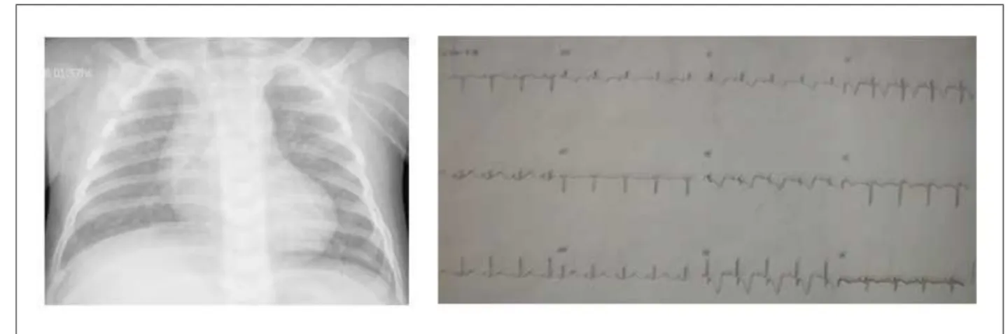
The electrocardiogram (ECG) showed sinus rhythm and signs of electrical ischemia in the anterior wall, with a negative T wave in V1 to V6, accompanied by ST-segment depression and low-voltage QRS complexes (Figure 1).
Radiographic image
Slightly enlarged cardiac silhouette with a round-shaped heart and slightly increased pulmonary vascular network in the pulmonary hila (Figure 1).
Diagnostic impression
Theimage is compatible with the diagnosis of cardiopathy that accompanies intercavitary communication-type pulmonary hyperflow, between the atria or the ventricles.
Clinico Radiological Session
Atik et alArq Bras Cardiol 2010; 94(5) : e57-e58
Figure 1 - The chest X-ray shows a slightly increased cardiac silhouette with a round-shaped heart and slightly increased pulmonary vascularity in the hila. The electrocardiogram discloses signs of ischemia, with a negative T wave in the precordial leads and ST-segment depression in V2 and V3, in addition to low-voltage QRS.
Figure 2 - The echocardiogram shows left ventricular dilation at the four-chamber view in A and the istula between the left coronary artery (LCA) and the pulmonary trunk (PT) in the suprasternal view, in B.
A B
LCA PT