277
Case 1 - 2003 – Pediatric Cardiac Surgery Department – Hospital de Base,
State Medical School, São José do Rio Preto
Correspondence address: Ulisses Alexandre Croti – Hospital de Base – FAMERP – Av. Brigadeiro Faria Lima, 5416 – CEP 15090-000 – São José do Rio Preto – São Paulo – E-mail: [email protected]
Clinical-surgical correlation
Ulisses Alexandre CROTI e Domingo Marcolino BRAILE
Rev Bras Cir Cardiovasc 2003; 18(3): 277
Article received on August, 2003 Article accepted on September, 2003 CLINICAL DATA - A male, white baby of 2900g was born
at full term. After one day of life presented with cyanotic crises on crying. a good general state, flushed, hydrated, tachydyspneic and icteric. Cardiac auscultation evidenced rhythmic, normophonetic sound at two sinus nodes, systolic murmur ++/6+ at the high left sternal margin. Lungs presented with vesicular murmur present and symmetrical without adventitious sounds. The liver was located on the right costal edge. The peripheral pulses were present and symmetrical.
ELECTROCARDIOGRAM - Electrocardiogram demonstrated sinusal rhythm with a frequency of 150 beats per minute, electrical axis + 60º and right atrial and left ventricular overload.
THORACIC RADIOGRAM - Increase in the area of the heart (cardiothoracic index = 0.61) owing to an increase in the right atrium and left ventricle were evidenced by thoracic radiogram with a hollowed mid arch. Pulmonary parenchyma with an increase in the peri-hilar pulmonary vascular system was observed, but it was not visible at the periphery.
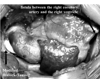
ECHOCARDIOGRAM - Situs solitus at levocardia, normal veno-atrial and atrio-ventricular connections, ventriculo-arterial connection with single aortic outflow, an interatrial communication of 8 mm and an arterial cannel of 4 mm in diameter were seen. Hypoplastic tricuspid valve with a 5-mm annulus and confluent pulmonary branches with diameters of 4 mm were also present. The hypoplastic right ventricle possessed a single inlet and small trabecular portion. Presence of a fistula between the right ventricular chamber and the dilated right coronary artery with retrograde flow during ventricular systole were evidenced, suggesting the presence of sinusoids.
DIFFERENTIAL DIAGNOSIS - Transposition of the great vessels, Fallot’s tetralogy with or without pulmonary atresia, pulmonary stenosis with integral septum, tricuspid atresia,
obstructive-type total anomalous connection of the pulmonary veins and Ebstein’s Disease should all be considered.
DIAGNOSIS - A cineangiocardiographic study was not necessary. An operation confirmed the echocardiographic diagnosis of pulmonary atresia with integral interventricular septum and the presence of a right coronary artery-right ventricle fistula.
OPERATION - A modified Blalock-Taussig operation was chosen by means of median sternotomy with interposition of a 3.5-mm polytetrafluoroethylene tube from the brachiocephalic branch to the right pulmonary artery without the use of cardiopulmonary bypass. During the evolution, the patient presented with bacterial and fungal infections, requiring specific treatment. He was released from hospital on the 22nd post-operative day and attended outpatient
follow-up sessions until the appropriate moment of the second intervention for univentricular correction.
Fig. 1 - Aspect of the heart after the operation demonstrating the position of the modified Blalock-Taussig and the presence of the fistula between the right coronary artery and the right ventricle.
Modified Blalock-Taussig
fistula between the right coronary artery and the right ventricle