C
a s eR
e p o Rt1 2 2 Arq Bras Oftalmol. 2017;80(2):122-4 http://dx.doi.org/10.5935/0004-2749.20170029
Large colloid drusen analyzed with structural
en face
optical coherence tomography
Análise de drusas grandes coloidais através de OCT
en face
estrutural
Nathália Corbelli roberti1, João rafaelde oliveira dias2, eduardo amorim Novais2, Caio saito regatieri2, rubeNs belfort Jr.2
Submitted for publication: October 13, 2016 Accepted for publication: November 7, 2016
1 Department of Ophthalmology, Hospital Brigadeiro, São Paulo, SP, Brazil.
2 Department of Ophthalmology and Visual Sciences, Escola Paulista de Medicina (EPM), Universidade
Federal de São Paulo (UNIFESP), São Paulo, SP, Brazil.
Funding: No specific financial support was available for this study.
Disclosure of potential conflicts of interest: None of the authors have any potential conflict of interest to disclose.
Corresponding author: Nathália Corbelli Robert. Av. Brigadeiro Luís Antônio, 2.651 - São Paulo, SP - 01401-901 - Brazil - E-mail: nathaliaroberti@yahoo.com.br
ABSTRACT
Drusen are extracellular deposits between the basal lamina of the retinal pig-ment epithelium (RPE) and the inner collagenous layer of Bruch’s membrane. Large colloid drusen (LCD) are located below the RPE and are characterized by multiple, large, dome-shaped RPE detachments, with marked attenuation of the ellipsoid zone overlaying the drusen. This report presents the structural en face optical coherence tomography (OCT ) findings of LCD and relates them to findings from fluorescein and indocyanine green angiography. We describe the case of a 55-year-old woman who presented with the chief complaint of a 5-year history of progressively worsening vision. Her best-corrected visual acuities were 20/40 and 20/400 in the right eye and the left eye, respectively. Fundus examination showed large bilateral, symmetrical, sub-retinal, yellowish lesions compatible with LCD. We describe the structural en face OCT characteristics and angiographic findings from this patient.
Keywords: Retinal drusen; Tomography, optical coherence/methods;Fluorescein angiography; Indocyanine green
RESUMO
Drusas são depósitos extracelulares localizados entre a lâmina basal do epitélio pig mentado da retina (RPE) e a camada colágena interna da membrana de Bruch. Drusas grandes coloidais (LCD) estão localizadas abaixo do EPR, e são caracterizadas por múltiplos descolamentos cupuliformes do EPR com atenuação da zona elipsoide sobrejacente às drusas. O objetivo deste relato é apresentar os achados de tomografia de coerência óptica (OCT ) enface estrutural em uma paciente com LCD, bem como correlacioná-los com angiografia fluoresceínica e angiografia com indocianina verde. Descrevemos o caso de uma paciente do sexo feminino, 55 anos, que referiu baixa acuidade visual em ambos os olhos há 5 anos. Sua acuidade visual corrigida era de 20/40 no olho direito e 20/400 no olho esquerdo. Ao exame fundoscópico a paciente apresentava lesões compatíveis com drusas grandes coloidais. As características tomográficas e angiográficas também são descritas neste relato de caso.
Descritores: Drusas retinianas;Tomografia de coerência óptica/métodos; Angiofluo-resceinografia; Verde de indocianina
INTRODUCTION
Drusen are extracellular deposits between the basal lamina of the retinal pigment epithelium (RPE) and the inner collagenous layer of Bruch’s membrane. Although drusen occur more frequently in peo-ple above 50 years of age, some drusen patterns, e.g., cuticular drusen, Malattia Leventinese (ML), and large colloid drusen (LCD) can occur earlier(1,2). LCD are large (200-300 microns) yellowish, bilateral lesions
with hyperpigmented borders scattered throughout the posterior pole. The LCD are found outside of the RPE as is common with conven-tional drusen(3-5). Reticular pseudodrusen also occur in the sub-retinal
rather than in the sub-RPE space(2,3).
Optical coherence tomography (OCT) is an important tool for diffe rentiating between various drusen patterns(6,7). In most cases, LCD
appear on OCT B-scans as multiple convex or dome-shaped structures with medium and homogeneous internal reflectivity and marked attenuation of the ellipsoid zone overlaying the LCD(1,8). These
dru-sen are homogenously hyperfluorescent in late-phase fluorescein an giography images. In late-phase indocyanine green angiography (ICGA) images, LCD are either hyperfluorescent or hypofluorescent and surrounded by a discreet hyperfluorescent halo(8).
This report presents structural en face OCT findings of LCD and cor-relations of these with findings with fluorescein angiography and ICGA.
CASE REPORT
Ro b e Rt i NC, e ta l.
1 2 3 Arq Bras Oftalmol. 2017;80(2):122-4 DISCUSSION
Large colloid drusen develop most often in women without a fa milial history of retinal problems. Drusen do not seem to be related to an increased risk of choroidal neovascularization or significant loss of mean visual acuity(3). The precise incidence and prevalence of
cho-roidal neovascularization in LCD is not well characterized, but most clinicians believe its incidence is significantly lower than in age-re-lated macular degeneration(9).
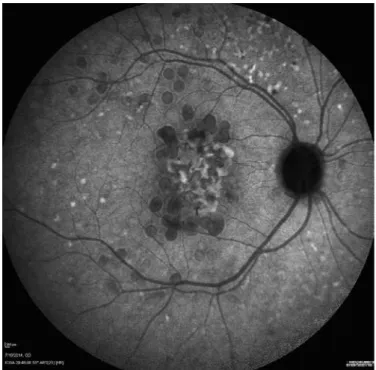
The images obtained from this patient showed features of LCD. In the late-phase ICGA images, the larger drusen appeared
hyperfluores-Figure 1. A fundus photograph showing large, bilateral, yellowish lesions in the macular area and retinal periphery.
A
C
B
D
Figure 2. Optical coherence tomography (OCT) images obtained from a 55-year-old woman with large colloid drusen (LCD). A) Structural en face
OCT with upper segmentation line located at the avascular outer retina and lower segmentation line placed at the sub-retinal pigment epithelium (RPE) space showing a hyper-relective center surrounded by a hypo-relective halo, bordered by hyper-relective and hypo-relective rings, similar to the donut efect. B) Corresponding OCT B-scan of (A) showing the convex contour of LCD with medium and homogeneous internal relectivity under the RPE, as well as marked thinning of the outer nuclear and ellipsoid layers. A small area of RPE atrophy as seen under the horizontal foveal scan, identiied as reverse shadowing (black arrow). No luid accumulation is observed. C) Structural en faceOCT on a choroid slab showing a hy-per-relective center surrounded by a hypo-relective halo. D) Choroidal features on the OCT B-scan were not well deined due to signal blockage caused by the colloid drusen.
cent with a hypofluorescent halo, traditionally described as the donut effect(3) (Figure 3). Drusen are lipid-rich, and the relative
hydropho-bicity of the commonly used angiographic dyes differs. A difference in the lipid composition between the core and the periphery of LCD has been reported, which might be responsible for the typical donut shape observed in the ICGA images(1).
La r g ec o L L o i d d r u s e na n a Ly z e dw i t h s t r u c t u r a Le nf a c eo p t i c a Lc o h e r e n c et o m o g r a p h y
1 2 4 Arq Bras Oftalmol. 2017;80(2):122-4
Figure 3. Large colloid drusen in a late-phase indocyanine green angiography image with a hypoluorescent center surrounded by a hyperluorescent halo. This halo is bordered by a hypoluorescent ring, referred to as the donut efect.
Figure 4. Fluorescein angiography showing early hyperluorescence of large colloid drusen.
and can help differentiate LCD from other early-onset drusen, such as Malattia Leventinese and cuticular drusen. In OCT images, LCD have been described as having a sawtooth pattern, with the height of each LCD approximately equal to its basal diameter. The neurosensory reti-na appears to be spared in the area overlaying the drusen, although the overlaying RPE is much thinner at the apex of each druse than between the drusen. In Malattia Leventinese, confluent sub-RPE accumulation on OCT has been reported. The smaller drusen of this condition have a radial distribution and a confluence of large drusen with sub-retinal fibrous plaque occurs. The typically pale drusen are adjacent to the optic disc(3). Furthermore, OCT allows observation of
focal loss of cellular visibility, which has a mosaic pattern in patients presenting with drusen(6,7). The smallest LCD do not affect the
ellip-soid zone(5). This imaging approach might serve as the foundation for
valuable imaging-based biomarkers for detecting the earliest disease stages, tracking progression, and monitoring treatment response(9).
On fluorescein angiography images, drusen hyperfluorescence increased quickly, especially in the middle periphery. The areas of hy-perfluorescent lesions did not vary among the capillary, venous, and washout angiography phases(4) (Figure 4). One study reported that large
visible drusen in a group of adults with early-onset drusen were con-centric to regions of hyperfluorescence, suggesting that drusen might have clinically detectable, substructural domains(9). Similarly, in another
study, the measurements of the drusen areas on OCT were smaller than the measurements obtained from color fundus images(10).
These results led us to conclude that structural en face OCT is a useful noninvasive tool that facilitates better morphologic evaluation and quantification of structural changes in LCD on high-resolution images. While traditional OCT produces longitudinal cross-sectional images, en face OCT produces transverse images of the retinal and choroidal layers at any specified depth. This provides an extensive overview of the pathological structures in one image. Thus, combi-ning dye-based angiographic images with structural en face OCT may provide a better understanding of this retinal entity.
REFERENCES
1. Guigui B, Querques G, Leveziel N, Bouakkaz H, Massamba N, Coscas G, et al. Spectral domain optical coherence tomography of early onset large colloid drusen. Retina. 2013;33(7):1346-50.
2. De Bats F, Wolff B, Mauget-Faÿsse M, Meunier I, Denis P, Kodjikian L. Association of reticular pseudodrusen and early onset drusen. ISRN Ophthalmol. 2013; 273085. 3. Boon CJ, van de Ven JP, Hoyng CB, den Hollander AI, Klevering BJ. Cuticular drusen:
stars in the sky. Prog Retin Eye Res. 2013;37:90-113.
4. Guigui B, Leveziel N, Martinet V, Massamba N, Sterkers M, Coscas G, et al. Angiography features of early onset drusen. Br J Ophthalmol. 2011;95(2)238-44.
5. Khan KN, Mahroo OA, Khan RS, Mohamed MD, McKibbin M, Bird A, et al. Differentia-ting drusen: drusen and drusen-like appearances associated with ageing, age-related macular degeneration, inherited eye disease and other pathological processes. Prog Retin Eye Res. 2016;53:70-106.
6. Lamory B, Nakashima K, Benchaboune M, Ullern M, Vuaillat E, Sahel JA, et al. In vivo microscopic imaging of drusen using adaptive optics. Invest Ophthalmol Vis Sci. 2011;52(14):4469.
7. Kozak I. Retinal imaging using adaptive optics technology. Saudi J Ophthalmol. 2014; 28(2):117-22.
8. Querques G, Massamba N, Guigui B, Lea Q, Lamory B, Soubrane G, et al. In vivo evalua-tion of photoreceptor mosaic in early onset large colloid drusen using adaptive optics. Acta Ophthalmol. 2012;90(4):e327-8.
9. Russell SR, Gupta RR, Folk JC, Mullins RF, Hageman GS. Comparison of color to fluo-rescein angiographic images from patients with early-adult onset grouped drusen suggests drusen substructure. Am J Ophthalmol. 2004;137(5):924-930.

