325
R
EVIEWA
RTICLEDrusenoid retinal pigment
epithelium detachments
Descolamento do epitélio pigmentar
da retina tipo drusenóide
Miguel Hage Amaro
1,Mario Martins dos Santos Motta
2, Jorge Mitre
3, João Jorge Nassaralla Junior
4, Angelo Leite
5, Teruo
Aihara
61 Instituto de Olhos e Laser de Belém, Belém, PA, Brazil. 2 Universidade do Rio de Janeiro, RJ, Brazil.
3 Faculdade de Medicina do ABC, Santo André, SP, Brazil. 4 Instituto de Olhos de Goiânia, Goiânia, GO, Brazil. 5 Clinica CEOFT, Belém, PA, Brazil.
6Hospital da Irmandade da Santa Casa de Misericórdia de São Paulo, São Paulo, SP, Brazil.
Received for publication 19/05/2014 - Accepted for publication 23/06/2014
The authors declare no conflict of interest
A
BSTRACTThe authors make a review of drusenoid retinal pigment epithelium detachments(DPDs), a form of retinal pigment epithelium detachment(PED) that evolves from confluent and large soft drusen.Drusenoidretinal pigment epithelial detachments are a recognized element of the “dry” AMD. Until now, no treatment is indicated in drusenoid PEDs. The authors describe the clinical characteristics of drusenoid retinal pigment epithelium detachments (DPEDs) and make a review of the DPEDs related in the international literature. We related in this revision paper the multimodal advanced image exams in two cases of dusenoid retinal pigment epithelium detachments (DPEDs) and the general characteristics of thisfinding associated with Dry Macular degeneration.Upon examination of the ocular fundusDPEDs emerge as well-circumscribed yellow or yellow–white elevations of the RPE that are usually found within the macula.They may show scalloped borders and a slightly irregular surface. When visualized using fluorescein angiography (FA),DPEDs are typically described as faint hyper-fluorescent in the early phase followed by a slow increase in fluorescence throughout the transit stage of the study without late leakage. With optical coherence tomography (OCT), drusenoid PEDs usually show a smooth contour of the detached hyperreflective RPE band that may have an undulating appearance.Drusenoid PEDs encompass far above the ground possibility type of “dry” AMD that develops in relationship with large confluent soft drusen.At this point no treatment is utilized in drusenoid retinal pigment epithelium detachment(DPEDs).
Keywords: Macular degeneration; Retinal detachment; Retinal drusen;Fluorescein angiography ; fundus oculi; Aged;Age factors
R
ESUMOOs autores fazem uma revisão do descolamento do epitélio pigmentar tipo drusenoide e apresentam dois casos desta patologiaassociada à degeneração macular relacionadaà idade descrevendo seus achados em avançados exames com imagem da retina.Neste artigo de revisão da literatura sobre os achados característicos dodescolamento do epitélio pigmentar tipodrusenoide e sua evolução descrevemos os achados de dois casosassociados à degeneração macular relacionada à idade, forma seca, utilizando exames como SD-OCT, fundusautofluorescencia e angiografia com indocianinaverde, além de retinografiacolorida e fluoresceínica. Odescolamento do epitélio pigmentar tipo druside evolui á partir de drussas moles confluentes presentes na degeneração macular relacionada à idade e é também associado a outras doenças retinianas. Até este momento não há tratamento para esta forma da doença.
Descritores: Degeneração macular;Descolamento retiniano;Drussasretinianos;Angiofluoresceinografia;Fundo de olho; Idoso;Fatores etários
Rev Bras Oftalmol. 2015; 74 (5): 325-8
326 Amaro MH, Motta MMS, MitreJ, Junior JJN, LeiteA, AiharaT
Rev Bras Oftalmol. 2015; 74 (5): 325-8
I
NTRODUCTIONA
retinal pigment epithelial detachment (PED) isthe separation of theretinal pigment epithelium (RPE) from Bruch’s membrane1,2.Age-related macular degeneration (AMD) is a frequentely association1,2.Drusenoidretinal pigment epithelial detachment
(DPED) is the form of PED that evolves from confluent and large soft drusen2-4. In particular, drusenoid retinal pigment
epithelial detachments (DPEDs) are a recognized element of the “dry” AMD.
Othertypes of PED observed in AMD areserous, vascularized, or mixed categories2-4. Furthermore, it is not unusual
to see more than one type of PED in AMD2-5.In addition
Drusenoid PEDs encompass far above the ground possibility type of “dry”AMD that develops in relationship with large confluent soft drusen.
Drusenoid PEDs may also occur in other retinal disorders including; cuticular drusen6,7, the maculopathy associated with
Type II membranoproliferativecapillary glomerulonephritis8,9,
choroidal nevi10 and mallatialeventinese.11
In a prospective study12,a total of 311 eyes (from 255
participants) with DPEDs were followed for a median of 8 years subsequent to the initial detection of a DPEDs. Of the 282 eyes that did not show advanced AMD at baseline,119eyes(42%) developed advanced AMD within 5 years, with 19% progressing to central geographic atrophy(CGA) and 23% progressing to neovascularage-related macular degeneration(NV-AMD). In theeyes that did not develop advanced AMD, progressive changes occurred in the fundus, including the development of calcified drusen and pigmentary changes. In addition,40% of all eyes showed decreases in visualacuity by >5 letters at 5-years follow-up;overall, mean visual acuity decreased from 76 letters (20/30) baseline to 61 letters (20/60) over 5 years. Five-year decreases in mean visual acuity averaged 26 letters for eyes progressing to advanced AMD and 8 letters for non-progressing eyes.
In a retrospective study13 evaluated the likelihood of
progression to advanced AMD form in 61 eyes with drusenoid PEDs with a mean follow-up of 4.6 years.The outcomes fell into one of three 3 categories: persistent drusenoid PED (38%), the development of geographic atrophy (49%), or the development ofchoroidal neovascularization (CNV) (13%). At 10 years, visu-al outcomes were poor, with progression to geographic atrophy in 75% of eyes and to CNV in 25% of eyes. The authors also noted IDPED size >2 disk diameters(DD) and the presenceof metamorphopsia upon presentation were associated with anincreased likelihood of progression to highly developedAMD. It is not rare to detect thepresence of acompartmentof subretinal fluid in DPEDs, in the absence of CNV;in such cases a analysis with advancedmultimodal imaging, such as indocyanine green angiography (ICGA) can be used to exclude CNV5,14.
Acquired vitelliform lesions isanytimes a finding in DPEDs5.
According any reports, the vitelliform lesions were related withvarious forms of AMD, including: subretinal drusenoid deposits15, cuticular drusen16,largedrusen16,17 and non neovascular
AMD16.Researchers putedthat RPE gradual dysfunction wasthe
mechanism for the development of subretinal fluid or vitelliform lesions in the avascular DPEDs3-5,17-19.
Upon examination of the ocular fundus the researchers
describe1-5 thatDPEDs emerge aswell-circunscribed yellow or
yellow–white elevationsof the RPE that are usually found within the macula.They may show scalloped borders and a slightly irregularsurface. The presence of a speckled or stellate pattern of brown or gray surface pigmentation on their surface is typical.They are often surrounded by large soft drusen and may be undistinguishable from a solitary large drusen in eyes with confluent drusen.
The age-relatedeye disease study defined a large drusen as measuring 125mm or greater and a drusenoid PED as measuring 350 mm or greater12.
When visualized using fluorescein angiography (FA), DPED sare typically described as faint hyperfluorescent in the early phase followed by a slow increase in fluorescence through out the transit stage of the study without late leakage5. Focal
hypofluorescence is often due to the blocking effect of overlying pigment hyperplasia, where as focal hyperfluorescence typically represents window defects caused by RPE atrophy3-5. Authors 3-5 stated that the interpretation of FA findingsfor DPEDs may
be challenging because it is difficult to distinguish characteristic angiographic patterns from those seen with vascularized PED sal though the latter exhibits more intense late staining or obvious leakage. Comparison of FA findings with spectral domain – optical coherence tomography (SD-OCT ) and ICGA5,15analyses may
help differentiate drusenoid from vascularized PEDs.With ICGA using a confocal scanning laser ophthalmoscope (SLO) system, the content of the drusenoid PED will block the fluorescence emitted from the underlying choroidal vasculature and, therefore, the PED will appear as an homogeneous hypofluorescent lesion during the early phase and remain hypofluorescent through out the transit5,20,21. When ICGA is used with a traditional fundus
camera–basedsystem, the DPED may appear isofluorescent or slightly hypofluorescent through out the sequence. Discrepancies may exist in findings obtained using fundus camera-based ICGA versus the confocal SLO-based ICGA because the absorption, diffraction, polarization, and scatter of light are different in the setwo systems5,20,21. One of the primary differences relates to
the confocal aperture of the SLO system that blocks the scattered light and allows transmission only of the images from the focused planes5,20,21. With either system, thereis absence of a
hyperfluorescent hot spot or plaque atthe border or within the drusenoid PED, which helps torule out the presence of associated CNV5,20,21.
Drusenoid PEDs may exhibit decreased fundus autofluorescence (FAF) butthey are isofluorescent or hyperautofluorescen5,22. Insome cases, the degree of FAF may
represent different stages of progression to ward atrophy of the elevatedRPE5. Drusenoid PEDs often show a slight,evenly
distributed increase in the FAF signal surrounded by a welldefined, hypoautofluorescent halo delineating the entire border of the lesion5,22-24. In some cases, areas of increased FAF
can be observed overlying or adjacent todrusenoid PEDs these correspond to focal hyperpigmentationor pigment clumping that can be observed via color photographyandfunduscopic examination5,22-24. Thisfinding may correspond to pigment
hyperplasia or RPEcells or macrophages containinglipofuscin or melanolipofuscinthat have migrated into the retina or subretinal space5. In some eyes with drusenoid PEDs, it is unclear whether
the FAF signal originates from the RPE it self, the material beneath the RPE, or associated vitelliform material in the subretinal space above the PED5,21-23. The spontaneous flattening
327
FAF is a useful indicator of the health of the RPE and may help predict those PEDs evolving toward atrophy5. In the
conventional and enhanced depth imaging optical coherence tomography5drusenoid PEDs usually show a smooth contour of
the detached hyperreflective RPE band that may have an rolling appearance. Pigment clumping lying immediately a top the drusenoid PED is not uncommon and will demonstrate hyperreflective signals with SD-OCT with posterior shadowing5.
Drusenoid PEDs are typically not associated with overlying subretinal orintraretinal fluid5. A retrospective series13 found that
the presence of subretinal or intraretinal fluid and the increase in material hyporeflectivity under the PED were associated with CNV. However, the presence of a hyporeflective area between the neurosensory retina and the elevated RPE line with SD-OCT does not necessarily mean that there is an associated CNV5,13.
Instead, this signal may result from a small pocket of benign subretinal fluid or an acquired vitelliformlesion5. If the
presentation of the drusenoid PED istypical, with the SD-OCT showing homogeneous hyperreflectivecontent of the PED without intraretinal fluid, itis unlikely that ICGA will show CNV. However, if the contentof the drusenoid PED appears heterogeneous or increasinglyhyporeflective on SD-OCT during the follow-up,or if there is evidence of significant subretinal or intraretinal fluid, then it may be helpful to perform ICGA torule out the presence of CNV5.
In some eyes, soft drusen may become confluent,forming RPE detachments in the absence of CNV5. It is hypothesized
that progressive accumulation of lipidswithin Bruch membrane over time may cause it tobecome increasingly hydrophobic5,25,26.
An increasinglystressed RPE pump is unable to adequately move
fluidand debris across Bruch membrane, leading to accumulationof fluid and debris in the sub-RPE space andenlargement of drusen5,26.Authors26analyzed a series of clinical and
clinicopathological cases with drusenand found that “the larger and more fluid the soft drusen has become, the finer the particles into which the original amorphous hard drusen material is found to have disintegrated and the more rapidly the drusen develop and fade, more often leading to geographic atrophy thanchoroidal neovascularization.The conclusions of researchers25,26in this
clinicopathological analyzes suggests that the larger size and more fluidic content of DPEDs may both be predictors of their evolution toward RPE atrophy.
Until now, no treatment is indicated in drusenoid PEDs.
A
CKNOWLEDGMENTSWe would like to express our very great appreciation to Neil Bressler,MD, James Folk,MD and K. Bailey Freund,MD for their valuable and constructive analyses of the cases cited in this review. Theirwillingness to give their time so generously has been very much appreciated.
Figure 1: (A)The color photograph shows a yellow macular lesion surrounded by multiple soft drusen in both eyesand elevated in right eye.The FA shows early hyperfluorescence and late stainning (B)The SD-OCT shows a DPED with a pocket of associated subretinal fluid.
A
B
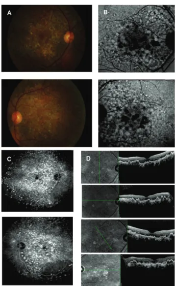
Figure 2: (A)Color photography shows multiple and confluent drusen in both eyes. (B)FAF shows hyperautofluorescence and hypoautofluorescence consistent with soft drusen and early focal atrophy. (C) FA shows late multiple hyperfluoescencedrusen in both eyes.(D)SD-OCT shows multiple DPEDs in both eyes.
Drusenoid retinal pigment epithelium detachments
328
Rev Bras Oftalmol. 2015; 74 (5): 325-8
R
EFERENCES1. Gass JD. Pathogenesis of disciform detachment of the neuroepithe-lium. Am JOphthalmol. 1967;63(3):Suppl:1-139.
2. Gass JD. Drusen and disciform macular detachment and degeneration.ArchOphthalmol. 1973;90(3):206-17.
3. Casswell AG, Kohen D, Bird AC. Retinal pigment epithelial detach-ments in theelderly: classification and outcome. Br J Ophthalmol. 1985;69(6):397-403.
4. Hartnett ME, Weiter JJ, Garsd A, Jalkh AE. Classification of retinal pigmentepithelial detachments associated with drusen. Graefes Arch Clin Exp Ophthalmol. 1992;230(1):11-9.
5. Mrejen S, Sarraf D, Mukkamala SK, Freund KB. Multimodal imaging of pigmentepithelial detachment: a guide to evaluation. Retina. 2013;33(9):1735-62. Review.
6. Querques G, Guigui B, Leveziel N, Querques L, Coscas G, Soubrane G, et al. Insights into pathology of cuticulardrusen from integrated confocal scanninglaser ophthalmoscopy imaging and corresponding spectral domain optical coherence tomography. Graefes Arch Clin Exp Ophthalmol. 2011;249(11):1617-25.
7. Russell SR, Mullins RF, Schneider BL, Hageman GS. Location, sub-structure, and composition of basal laminar drusen compared with drusen associated with agingand age-related macular degeneration. Am J Ophthalmol. 2000;129(2):205-14.
8. Mullins RF, Russell SR, Anderson DH, Hageman GS. Drusen associ-ated with aging and age-relassoci-ated macular degeneration contain pro-teins common to extracellulardeposits associated with atherosclero-sis, elastoatherosclero-sis, amyloidoatherosclero-sis, and densedeposit disease. FASEB J. 2000;14(7):835-46.
9. Ritter M, Bolz M, Haidinger M, Deák G, Sacu S, Säemann M, et al. Functional and morphological macular abnormalities in membranoproliferativeglomerulonephritis type II. Br J Ophthalmol. 2010;94(8):1112-4.
10. Shields CL, Mashayekhi A, Materin MA, Luo CK, Marr BP, Demirci H, et al. Optical coherence tomography of choroidal nevus in 120 patients. Retina. 2005;25(3):243-52.
11. Souied EH, Leveziel N, Letien V, Darmon J, Coscas G, Soubrane G. Opticalcoherent tomography features of malattialeventinese. Am J Ophthalmol. 2006;141(2):404-7.
12. Cukras C, Agrón E, Klein ML, Ferris FL 3rd, Chew EY, Gensler G, Wong WT;Age-Related Eye Disease Study Research Group. Natural history of drusenoidpigment epithelial detachment in age-related macular degeneration: Age-RelatedEye Disease Study Report No. 28.
Ophthalmology. 2010;117(3):489-99.
13. Roquet W, Roudot-Thoraval F, Coscas G, Soubrane G. Clinical features ofdrusenoid pigment epithelial detachment in age related macular de-generation. Br JOphthalmol. 2004;88(5):638-42.
Corresponding author: Miguel Hage Amaro
Address: Trav: Quintino Bocaiúva,516 Bairro Reduto Belém -Pará - Brazil - CEP.: 66053-249
E-mail: [email protected]
14. Yannuzzi LA, Hope-Ross M, Slakter JS, Guyer DR, Sorenson JA, Ho AC, et al. Analysis of vascularized pigment epithelial detachments using indocyanine green videoangiography. Retina. 1994; 14(2):99-113.
15. Zweifel SA, Spaide RF, Yannuzzi LA. Acquired vitelliform detach-ment inpatients with subretinaldrusenoid deposits (reticular pseudodrusen). Retina. 2011;31(2):229-34.
16. Freund KB, Laud K, Lima LH, Spaide RF, Zweifel S, Yannuzzi LA. AcquiredVitelliform Lesions: correlation of clinical findings and mul-tiple imaginganalyses. Retina. 2011;31(1):13-25.
17. Lima LH, Laud K, Freund KB, Yannuzzi LA, Spaide RF. Acquired vitelliformlesion associated with large drusen. Retina. 2012;32(4):647-51.
18. Sarks JP, Sarks SH, Killingsworth MC. Evolution of geographic atro-phy of theretinal pigment epithelium. Eye (Lond). 1988;2 ( Pt 5):552-77.
19. Bressler NM, Silva JC, Bressler SB, Fine SL, Green WR. Clinico patho-logic correlation of drusen and retinal pigment epithelial abnormali-ties in age-related macular degeneration. Retina. 1994;14(2):130-42. 20. Arnold JJ, Quaranta M, Soubrane G, Sarks SH, Coscas G. Indocyanine green angiography of drusen. Am J Ophthalmol. 1997;124(3):344-56. 21. Flower RW, Csaky KG, Murphy RP. Disparity between fundus camera and scanning laser ophthalmoscope indocyanine green imaging of retinal pigment epithelium detachments. Retina. 1998;18(3):260-8. 22. Schmitz-Valckenberg S, Fleckenstein M, Scholl HP, Holz FG. Fundus
autofluorescence and progression of age-related macular degeneration.SurvOphthalmol. 2009;54(1):96-117. Review. 23 .Karadimas P, Bouzas EA. Fundus autofluorescence imaging in serous
anddrusenoid pigment epithelial detachments associated with age-related maculardegeneration. Am J Ophthalmol. 2005;140(6):1163-5.
24. von Rückmann A, Fitzke FW, Bird AC. Fundus autofluorescence in age-relatedmacular disease imaged with a laser scanning ophthalmoscope. Invest OphthalmolVis Sci. 1997;38(2):478-86.
25. Pauleikhoff D, Harper CA, Marshall J, Bird AC. Aging changes in Bruch’smembrane. A histochemical and morphologic study. Ophthal-mology. 1990;97(2):171-8.