O
r i g i n a la
rt i c l e2 4 3 Arq Bras Oftalmol. 2016;79(4):243-6 http://dx.doi.org/10.5935/0004-2749.20160069
INTRODUCTION
Retinal vascular occlusive disorders are the second most com-mon cause of vision loss acom-mong patients with retinal vascular disea-se(1). Macular edema (ME) is a major complication of vein occlusion and the predominant cause of acute vision loss(2). Many factors such as increased hydrostatic pressure in the venous circulation, inlam-mation, endothelial damage, and increased vascular permeability contribute to the development of ME(3). Grid laser photocoagulation(1), antivascular endothelial growth factors (VEGFs)(4-6), and triamcinolone injection(7) have demonstrated eicacy in the treatment of ME caused by retinal vein occlusion (RVO). These treatments aim to reduce exis-ting damage to retinal cells, particularly in the photoreceptor layer, and provide visual improvement by reducing edema, ischemia, and cellular damage. Recently, the intravitreal dexamethasone implant, Ozurdex, has been used as a new treatment modality to reduce ME and increase visual acuity in patients with both types of RVO: branch retinal vein occlusion (BRVO) and central retinal vein occlusion (CRVO)(8).
Clinical assessment of the choroid is typically conducted by indocyanine green angiography (ICGA) or B-scan ultrasonography; however, neither method allows for accurate cross-sectional imaging. Recently, a new approach to optical coherence tomography (OCT), known as enhanced depth imaging (EDI)-OCT, has demonstrated utility in imaging the full thickness of the choroid in a noninvasive, rapid, objective, and reliable manner(9). Choroidal thickness can be afected by ocular pathologies such as age-related macular degene-ration(10), central serous chorioretinopathy(11), Vogt-Koyanagi-Harada disease(12), macular holes(13), high myopia(14), and polypoidal choroidal vasculopathy(15), in addition to systemic diseases such as diabetes. Se-veral previous studies have evaluated choroidal thickness in RVO pa-tients(16-18). However, the results of these studies were contradictory. Du et al.(16) reported no diference in subfoveal choroidal thickness (SFCT) between eyes with longstanding BRVO and normal contrala-teral eyes. In contrast, Lee et al.(17) reported greater choroidal thickness in eyes with BRVO and CRVO compared with normal contralateral
Short-term effects of intravitreal dexamethasone implant (OZURDEX
®) on choroidal
thickness in patients with naive branch retinal vein occlusion
Os efeitos a curto prazo do implante de dexametasona intravítrea (OZURDEX
®) na espessura da coroide
em pacientes com oclusão primária de ramo da veia central da retina
Hasan Basri arifoglu, necati Duru, orHan altunel, BurHan Baskan, BeDirHan alaBay, Mustafa atas
Submitted for publication: January 23, 2016 Accepted for publication: March 6, 2016
Department of Ophthalmology, S.B. Kayseri Education and Research Hospital, Kayseri, Turkey.
Funding: No specific financial support was available for this study.
Disclosure of potential conflicts of interest: None of the authors have any potential conflicts of interest to disclose.
Corresponding author: Hasan Basri Arifoglu. S.B. Kayseri Research and Education Hospital. Department of Ophthalmology- Sanayi Mah. Atatürk Boulevard Hastane Street, 78 - Kayseri 38010 - Turkey - E-mail: [email protected]
Approved by the following research ethics committee: Kayseri Eğitim ve Araştırma Hastanesi (# 16/04/2015-40).
ABSTRACT
Purpose: The objective of this study was to evaluate subfoveal choroidal thickness (SFCT ) using enhanced depth imaging optical coherence tomography (EDI-OCT ) in patients with naïve branch retinal vein occlusion (BRVO) before and after intravitreal dexamethasone implant (Ozurdex®) injection.
Methods: Thirty-nine patients with unilateral BRVO and 35 healthy subjects were included in this prospective study. Choroidal thickness was evaluated by EDI-OCT at baseline and 1 month after dexamethasone implant.
Results: The mean SFCT measured in 39 patients with BRVO was 299.41 ± 55.86 μm, significantly greater than that in contralateral eyes (283.76 ± 57.44 μm; p=0.009) and control eyes (276.14 ± 39.06 μm; p=0.044). The mean SFCT after the treatment was 279.64 ± 50.96 μm, significantly thinner than that before intravitreal dexamethasone therapy (p=0.004).
Conclusions: SFCT in treatment-naive BRVO eyes was significantly greater than that in contralateral eyes and healthy eyes and decreased significantly after in-travitreal dexamethasone implantation.
Keywords: Choroid/pathology; Intravitreal injections; Retinal vein occlusion; To-mography, optical coherence; Macular edema; Dexamethasone/administration and dosage; Visual acuity
RESUMO
Objetivo: O objetivo deste estudo foi avaliar a espessura da coróide (SFCT ) usando imagens de tomografia de coerência óptica com profundidade aprimorada (EDI-OCT ) no tratamento de pacientes com oclusão primária de ramo da veia central da retina (BRVO) antes e após o implante de dexametasona intravítrea (Ozurdex®).
Métodos: Trinta e nove pacientes com BRVO unilateral e 35 indivíduos saudáveis foram incluídos neste estudo prospectivo. Espessura da coróide foi avaliada por EDI-OCT na antes e um mês após o tratamento.
Resultados: A média da SFCT medida em 39 pacientes com BRVO foi 299,41 ± 55,86 µm, o que foi significativamente maior do que a dos olhos contralaterias (283,76 ± 57,44 μm) e dos olhos controle (276,14 ± 39,06 μm) (p=0,009 e p=0,044, respectivamente). A média da SFCT após o tratamento foi 279,64 ± 50,96 μm, o que foi significativamente menor do que antes do mesmo (p=0,004).
Conclusões: A SFCT do tratamento de olhos com BRVO primária foi significativamente maior do que a dos olhos contralaterais e dos olhos saudáveis, e diminuiu significati-vamente após o implante intravítreo de dexametasona.
Sh o rt-t e r m e f f e c t S o f i n t r av i t r e a l d e xa m e t h a S o n e i m p l a n t (oZUrdex®) o n c h o r o i d a l t h i c k n e S S i n pat i e n t S
w i t hn a i v eb r a n c h r e t i n a lv e i n o c c lU S i o n
2 4 4 Arq Bras Oftalmol. 2016;79(4):243-6
eyes. To date, no studies have examined choroidal thickness in treatment-naïve eyes with BRVO before and after Ozurdex injection.
In the present study, EDI-OCT was used to investigate the short-term efects of Ozurdex injection on choroidal thickness in patients with BRVO by comparing choroidal thickness with the eyes of age- and gender-matched healthy control and normal contralateral eyes.
METHODS
S
TUDYPOPULATIONANDDESIGNThis prospective comparative study was performed in the De-partments of Ophthalmology at Kayseri Education and Research Hospital. The present study followed the tenets of the Declaration of Helsinki and was approved by the local ethics committee. All parti-cipants received both oral and written information about the study, and each participant provided written informed consent. Participants were recruited into two groups: the study group consisting of 39 patients with BRVO and the control group consisting of 35 healthy volunteers. Inclusion criteria were as follows: recent-onset (<4 weeks) and treatment-naive unilateral BRVO with ME. The diagnosis of BRVO was made according to the results of clinical examination and supported by fundus luorescein angiography and OCT measurements.
E
XCLUSIONCRITERIAOcular exclusion criteria for the present study were as follows: prior history of signiicant ocular disease, refractive error of either less than -3 D or more than +3 D, amblyopia, intraocular pressure
(IOP) readings greater than 21 mmHg, glaucoma, history of uveitis, retinal disease (except for BRVO), ocular trauma or tumor, poor image quality, and dense media opacities. Patients with a history of previous intraocular laser therapy or the use of any intravitreal injections were also excluded from the present study.
E
XAMINATIONPROTOCOLANDSTUDYMEASUREMENTSAll participants in both groups underwent a complete examina-tion that included Snellen best-corrected visual acuity, biomicroscopy, IOP measured by Goldmann applanation tonometry, and dilated fundus examination with axial length (AL) and OCT measurements. AL measurements were measured with the IOL Master 500 (Carl Zeiss Meditec Inc, Jena, Germany). OCT measurements were retaken 1 month after intravitreal dexamethasone implant injection in the study group. Due to diurnal luctuations, all examinations were per-formed between 9 am and 11 am.
OCT
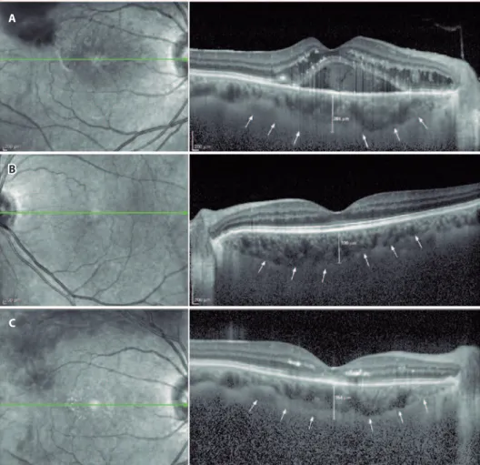
MEASUREMENTSFollowing detailed ophthalmologic examinations, a third-gene-ration Spectralis OCT device (software version 5.6.3.0; Spectralis OCT, Heidelberg Engineering, Dossenheim, Germany) was used for ocular assessments. The method of obtaining EDI-OCT images has been previously reported(9,19). SFCT was determined as the vertical distance from the hyperrelective line of the hyperrelective retinal pigment epithelium to the line of the inner surface of the sclera centered on the fovea, which was taken using a tool with built-in linear measuring. A representative EDI-OCT choroidal image is presented in igure 1.
A
B
C
Ar i f o g l u HB, e t A l.
2 4 5
Arq Bras Oftalmol. 2016;79(4):243-6
Images were captured by one experienced clinician and assessed by another experienced clinician. Group identities remained anonymous to both clinicians.
S
TATISTICALANALYSISAll statistical tests were performed using SPSS (Statistical Package for the Social Sciences) version 20. In the control group, OCT measure-ments from the right eye were used for analyses. For each continuous variable, normality was checked using the Kolmogorov Smirnov test. Diferences in categorical variables between groups were analyzed using the χ2 test. An independent t-test was used to compare
varia-bles between study and control groups. The paired t-test was used for comparisons between BRVO eyes and fellow eyes. The paired t-test was also used for comparisons of preinjection and postinjection measurements. p-Values less than 0.05 were considered statistically signiicant.
RESULTS
Table 1 shows the demographic characteristics of the study and control groups. No statistically signiicant diference in sex or age was observed between the two groups (p=0.929 and p=0.432, respecti-vely). Table 2 shows the results of IOP, AL, and SFCT measurements in the control and study groups. Mean SFCT measurements in eyes with BRVO difered signiicantly from healthy control eyes and fellow eyes (p=0.044 and p=0.009, respectively). However, no statistically signiicant diferences in IOP or AL values were observed between the study and control groups (p>0.05). Table 3 shows the comparison of choroidal thickness between preinjection and postinjection periods.
A statistically signiicant diference in mean SFCT measurements was observed at 1 month after Ozurdex injection in eyes with BRVO (p=0.004).
DISCUSSION
The present study demonstrated that SFCT in recent-onset (<4 weeks) treatment-naive BRVO eyes with ME was greater than that in unafected contralateral and control eyes. Furthermore, choroidal thickness decreased in response to intravitreal dexamethasone im-plant treatment.
RVO is the second most common vascular disease leading to decreased visual acuity, with an annual prevalence 4.42 in 1,000 per-sons(20), slightly lower than diabetic retinopathy. Kolar(21) conducted a meta-analysis to detect risk factors of RVO, identifying advancing age. Other reported risk factors included systemic conditions such as hypertension, arteriosclerosis, diabetes mellitus, hyperlipidemia, vascular cerebral stroke, blood hyperviscosity, and thrombophilia.
Treatment options for ME after the occurrence of BRVO include laser photocoagulation, intravitreal VEGF antagonists, and intravitreal corticosteroids(1,4-8).
The dexamethasone 0.7-mg intravitreal implant (Ozurdex®, Aller-gan, Irvine, CA, USA) is a sustained-biodegradable implant containing the corticosteroid, dexamethasone. Corticosteroids including dexa-methasone are known to have anti-inlammatory and antiangiogenic properties and may inhibit the expression of VEGF and other proin-lammatory cytokines such as IL-6, ICAM-1, and MCP-1(8,22,23).
The choroid is the vascular layer between the retina and the sclera that provides blood supply to the eye and plays an important role in ocular nutrition.
Choroidal blood low is the highest of any tissue in the body and is necessary to satisfy the normal metabolic demands of the outer retina. A structurally and functionally normal choroidal vasculature is essential for retinal function(24), and the choroid itself is important for visual acuity. Eyes with a relatively thicker choroid at baseline may have greater choroidal blood supply and choriocapillaris, which may increase the possibility of a full recovery from ME(17).
Tsuiki et al.(18) posited that VEGF may increase vascular permeability and induce fenestrations of the choriocapillaris, which may in turn increase choroidal thickness. In eyes with RVO, VEGF expression is in creased in retinal endothelial cells, pericytes, RPE, M̈ller cells, gan-glion cells, and astrocytes as a result of hypoxia. Choroidal thickness is also mediated by vascular dilatation induced by nitric oxide (NO) production in response to VEGF expression. Choroidal thickness may increase as a result of either vasodilataton or edema. The indings of the present study support the hypothesis of Tsuiki et al.
Table 1. Patient demographics and characteristics
Study group Control group p-value
Number of eyes/patients 39/39 35/35
-Sex
Female 23 (59%) 21 (60%) 0.929*
Male 16 (41%) 14 (40%)
Age (years)
Mean ± SD 64.71 ± 10.99 62.91 ± 8.24 0.432**
Range 41-89 46-83
*= Chi-Square test; **= independent-sample t test.
Table 2. Comparison of choroidal thickness and other clinical measurements between study group and control group
Study group
Control group
p-values
Eyes with BRVO Fellow eyes BRVO versus control* BRVO versus fellow** Fellow versus control*
SFCT (µm)
Mean ± SD 299.41 ± 55.86 283.76 ± 57.44 276.14 ± 39.06 0.044 0.009 0.511
Range 188-425 164-390 190-378
AL (mm)
Mean ± SD 022.82 ± 00.64 023.04 ± 00.55 022.98 ± 00.69 0.307 0.112 0.667
Range 21.24-23.98 21.98-24.11 21.87-24.02
IOP (mmHg)
Mean ± SD 014.69 ± 02.86 014.89 ± 02.41 015.42 ± 02.42 0.240 0.570 0.349
Range 9-20 10-20 10-21
Sh o rt-t e r m e f f e c t S o f i n t r av i t r e a l d e xa m e t h a S o n e i m p l a n t (oZUrdex®) o n c h o r o i d a l t h i c k n e S S i n pat i e n t S
w i t hn a i v eb r a n c h r e t i n a lv e i n o c c lU S i o n
2 4 6 Arq Bras Oftalmol. 2016;79(4):243-6
Since Spaide et al.(9) developed a method called EDI-OCT, which enables both in vivo cross-sectional imaging of the choroid and the measurement of choroidal thickness, an increasing number of studies have reported the choroidal thickness in eyes with various diseases(10-18).
In a population-based cross-sectional study, Du et al.(16) reported no diference in SFCT between eyes with RVO and normal contralate-ral eyes. In contrast, Tsuki et al.(18) reported signiicantly greater SFCT in CRVO eyes compared with normal contralateral eyes. In addition, Lee et al.(17) recently reported signiicantly greater SFCT in eyes with CRVO and BRVO compared with normal contralateral eyes. These inconsis-tencies may be attributable to diferences in patient characteristics. In contrast to the present study, Du et al. included subjects with longstanding RVOs without marked cystoid ME. The present indings were similar to the indings of Tsuki and Lee’s studies. The present study included recent onset (<4 weeks) and treatment-naïve BRVO patients, which allowed the evaluation of early choroidal changes in BRVO without any efect of treatment modalities such as anti-VEGF agents or laser treatments.
Tsuiki et al.(18) reported decreased SFCT 1 month after intravitreal bevacizumab treatment in patients with CRVO. Lee et al.(17) also repor-ted that increased SFCT in CRVO and BRVO patients decreased after injection of the dexamethasone implant. The present study found that the efects of the dexamethasone implant on choroidal thickness were similar to those reported by previous studies. However, the present study assessed only the efects of Ozurdex on choroidal thickness in patients with recent-onset BRVO.
Corticosteroids inhibit the synthesis of NO and decrease tissue edema. Studies have shown that corticosteroids downregulate VEGF expression and other pro-inlammatory cytokines(22,25). Accordingly, the dexamethasone implant may reduce choroidal thickness via this mechanism.
The present study had several limitations. First, choroidal vessels were not evaluated by ICGA. If ICGA had been performed, it may have been possible to compare angiographic changes on OCT. Second, the present study sample size was somewhat small, and the follow-up period was relatively short, both of which decrease statistical gene-ralizability. Third, SFCT measurements were obtained manually. Auto-mated software may allow more objective evaluation by erasing any potential bias. Future studies with larger numbers of subjects, longer follow-up durations, and the use of automated software are required to fully validate the results of the present study.
In conclusion, SFCT in recent-onset, treatment naïve BRVO eyes is signiicantly greater in both normal contralateral eyes and age- and sex-matched healthy control eyes. Choroidal thickness is signiicantly decreased 1 month after the injection of the dexamethasone implant. SFCT may have utility in assessing the efects of antiedematous treatment on choroidal vascular structures by measuring choroidal thickness noninvasively with EDI OCT, thereby allowing patients to be observed more reliably. Further studies are required to elucidate the role of the choroid in the pathogenesis of BRVO.
ACKNOWLEDGMENT
Special thanks to Halime YILDIZ, our experienced OCT technician.
REFERENCES
1 Argon laser photocoagulation for macular edema in branch vein occlusion. The Branch Vein Occlusion Study Group. Am J Ophthalmol. 1984;98(3):271-82.
2. Mitchell P, Smith W, Chang A. Prevalence and associations of retinal vein occlusion in Australia: the blue mountains eye study. Arch Ophthalmol. 1996;114(10):1243-7. 3. Rehak J, Rehak M. Branch retinal vein occlusion: pathogenesis, visual prognosis, and
treatment modalities. Curr Eye Res. 2008;33(2):111-131.
4. Gerding, H, Monés J, Tadayoni R, Boscia F, Pearce I, Priglinger S. Ranibizumab in retinal vein occlusion: treatment recommendations by an expert panel. Br J Ophthalmol. 2015; 99(3):297-304.
5. Hoeh AE, Ach T, Schaal KB, Scheuerle AF, Dithmar S. Long-term follow-up of OCT-guided bevacizumab treatment of macular edema due to retinal vein occlusion. Graefes Arch Clin Exp Ophthalmol. 2009;247(12):1635-41.
6. Campochiaro PA, Clark WL, Boyer DS, Heier JS, Brown DM, Vittir R, et al. Intravitreal alibercept for macular edema following branch retinal vein occlusion: the 24-week results of the VIBRANT study. Ophthalmology. 2015;122(3):538-44. Comment in: Oph-thal mology. 2015;122(3):443-4.
7. Scott IU, Ip MS, VanVeldhuisen PC. Oden NL, Blodi BA, Fischer M, Chan CK, Gonzalez VH, Singerman LJ, Toletino M; SCORE Study Research Group. A randomized trial comparing the eicacy and safety of intravitreal triamcinolone with standard care to treat vision loss associated with macular edema secondary to branch retinal vein occlusion: the Standard Care vs Corticosteroid for Retinal Vein Occlusion (SCORE) study report 6. Arch Ophthalmol. 2009;127(9):1115-28. Erratum in: Arch Ophthalmol. 2009;127(12):1655. Comment in: Arch Ophthalmol. 2009;127(9):1203-4.
8. Haller JA, Bandello F, Belfort R Jr, Blumenkranza MS, Gillies M, Heier J, Loewenstein A, Yoon YH, Jiao Li, Li XY, Whitcp SM; Ozudex GENEVA Study Group, Li J. Dexa methasone intravitreal implant in patients with macular edema related to branch or central retinal vein occlusion twelve-month study results. Ophthalmology. 2011;118(12):2453-60. Comment in: Ophthalmology. 2012;119(12):2654-5.e.1; author reply 2655. 9. Spaide RF, Koizumi H, Pozzoni MC. Enhanced depth imaging spectral-domain optical
coherence tomography. Am J Ophthalmol. 2008;146(4):496-500. Erratum in Am J Ophthalmol. 2009;148(2):325. Pozonni, Maria C [correctd to Pozonni, Maria C]. 10. Spaide RF. Enhanced depth imaging optical coherence tomography of retinal pigment
epithelial detachment in age-related macular degeneration. Am J Ophthalmol. 2009; 147(4):644-52. Comment in: Retina. 2010;30(8):1320-1; author reply 1321-2. 11. Imamura Y, Fujiwara T, Margolis R, Spaide RF. Enhanced depth imaging optical
coheren-ce tomography of the choroid in coheren-central serous chorioretinopathy. Retina. 2009;29(10): 1469-73.
12. Maruko I, Iida T, Sugano Y, Oyamada H, Sekiryu T, Fujiwara T, Spaide RF. Subfoveal choroidal thickness after treatment of Vogt-Koyanagi-Harada disease. Retina. 2011;31(3):510-7. 13. Reibaldi M, Boscia F, Avitabile T, Uva MG, Russo V, Zagari M, Boniglio V, et al. Enhanced
depth imaging optical coherence tomography of the choroid in idiopathic macular hole: a cross-sectional prospective study. Am J Ophthalmol. 2011;151(1):112-7. Comment in: Am J Ophthalmol. 2011;151(3):560-1; author reply 561.
14. Fujiwara T, Imamura Y, Margolis R, Slakter JS, Spaide RF. Enhanced depth imaging optical coherence tomography of the choroid in highly myopic eyes. Am J Ophthalmol. 2009; 148(3):445-50.
15. Chung SE, Kang SW, Lee JH, Kim YT. Choroidal thickness in polypoidal choroidal vas-culopathy and exudative age-related macular degeneration. Ophthalmology. 2011; 118(5):840-5.
16. Du KF, Xu L, Shao L, Chen CX, Zhou JQ, Wang YX, et al. Subfoveal choroidal thickness in retinal vein occlusion. Ophthalmology. 2013;120(12):2749-50.
17. Lee EK, Han JM, Hyon JY, Yu HG. Changes in choroidal thickness after intravitreal dexame-thasone implant injection in retinal vein occlusion. Br J Ophthalmol. 2015;99(11):1543-9. 18. Tsuiki E, Suzuma K, Ueki R, Maekawa Y, Kitaoka T. Enhanced depth imaging optical
coherencetomography of the choroid in central retinal vein occlusion. Am J Ophthal-mol. 2013;156(3):543-7.
19. Margolis R, Spaide RF. A pilot study of enhanced depth imaging optical coherence tomography of the choroid in normal eyes. Am J Ophthalmol. 2009;147(5):811-5. 20. Rogers S, McIntosh RL, Cheung N, Lim L, Wang JJ, Mitchell P, Kowalski JW, Nguyen H,
Wong TY; International Eye Disease Consortium. The prevalence of retinal vein occlusion: pooled data from population studies from the United States, Europe, Asia, and Australia. Ophthalmology. 2010;117(2):313-9.
21. Kolar P. Risk factors for central and branch retinal vein occlusion: a meta-analysis of published clinical data. J Ophthalmol. 2014:2014:724780.
22. McAllister IL, Vijayasekaran S, Chen SD, Yu DY. Efect of triamcinolone acetonide on vascular endothelial growth factor and occludin levels in branch retinal vein occlusion. Am J Ophthalmol. 2009;147(5):838-46.
23. Pister M, Rothweiler F, Michaelis M, Cinatl J Jr, Schubert R, Koch FH, et al. Correlation of inlammatory and proangiogenic cytokines from undiluted vitreous samples with spectral domain OCT scans, in untreated branch retinal vein occlusion. Clin Ophthalmol. 2013;7:1061-7.
24. Ryan SJ. Retina. 4th ed. Philadelphia, PA: Elsevier Mosby; 2006.
25. Liu Y, Mladinov D, Pietrusz JL, Usa K, Liang M. Glucocorticoid response elements and 11 beta-hydroxysteroid dehydrogenases in the regulation of endothelial nitric oxide synthase expression. Cardiovasc Res. 2009;81(1):140-7.
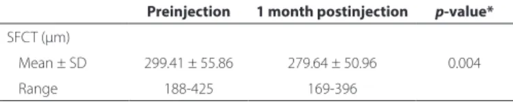
Table 3. Comprasion of choroidal thickness between preinjection and postinjection
Preinjection 1 month postinjection p-value* SFCT (µm)
Mean ± SD 299.41 ± 55.86 279.64 ± 50.96 0.004
Range 188-425 169-396