258
ment and are indicated for more aggressive therapies, such as myotomy-myectomy-septal resection, dual-chamber pacemaker implantation, alcohol injection in the septal artery, defibrillator implantation (used in cases at high risk for sudden death), and even heart transplantation 3,4.
Mitral stenosis is usually of rheumatic origin, occurring mainly among women (approximately 66% of the cases), but in rare cases, it may be congenital 7. Pure or predominant mitral stenosis occurs in approximately 40% of all patients with rheumatic heart disease 7. The clinical findings are extremely varied and have a significant relation with the degree of valvular impairment. These findings may vary from the mere presence of some clinical signs without symptoms until the presence of pulmonary edema, atrial arrhythmias, hemoptysis, and right ventricular failure 7.
Until the present time, however, from the pathophysiological point of view, no cases of an association of myotomy-myectomy-septal resection and the appearance of long-term mitral stenosis have been reported in the scientific literature.
We report the case of a 67-year-old female, who, 18 years after undergoing myotomy-myectomy-septal resection for correction of asymmetric septal hypertrophic cardiomyopathy, had symptoms of severe mitral stenosis. The patient presented to the Instituto do Coração (InCor) of the Hospital das Clínicas of the FMUSP for appropriate clinical assessment, being then referred for surgery.
Case report
We report the case of a 67-year-old female of mixed heritage, born in the city of Montes Claros, in the state of Minas Gerais, and coming from the city of Belo Horizonte, in the state of Minas Gerais, who complained of progressive dyspnea for 1 year, which was lately triggered by minimum exertion (New York Heart Asso-ciation functional class III). The patient reported, as personal ante-cedents, systemic arterial hypertension and dyslipidemia, in addition to ischemic stroke in 1995, with no sequelae.
In 1985, after undergoing complementary examinations, asym-metric septal hypertrophic cardiomyopathy and pulmonary hyper-tension were diagnosed. On that occasion, the patient had pro-gressive dyspnea and dizziness but denied any history of rheumatic disease. Myotomy-myectomy-septal resection was indicated and performed through an aortotomy. The anatomicopathological study of the left ventricular fragments removed during the surgery showed hypertrophic cardiomyocytes and areas of disarrangement and
Case Report
Severe Mitral Stenosis in the Long-term Evolution
of Myotomy-Myectomy-Septal Resection
Alexandre de Matos Soeiro, Juliana Ascenção de Souza, Carlos Vicente Serrano Júnior,
Luiz Alberto Benvenuti, Reynaldo Castro Miranda, José Carlos Nicolau,
José Antônio F. Ramires, Sérgio Almeida de Oliveira
São Paulo, SP - Brazil
InCor of the Hospital das Clínicas of the FMUSP
Mailing address: Alexandre de Matos Soeiro - Rua Clemente Ferreira, 122/ 22 - 09530-440 - São Caetano do Sul, SP, Brazil
e-mail: [email protected] Received for publication: 9/5/03 Accepted for publication: 2/4/04 English version by Stela Maris Costalonga
The patient is a 67-year-old female with severe nonrheumatic mitral stenosis occurring 18 years after myotomy-myectomy-septal resection for correction of asymmetric septal hypertrophic cardio-myopathy. The stenotic mitral valve was replaced by a mechanical prosthesis. The combination of 2 extremely rare syndromes with a poor prognosis made this patient a unique case.
Hypertrophic cardiomyopathy, also known as idiopathic hyper-trophic subaortic stenosis or hyperhyper-trophic obstructive cardiomyo-pathy, is a disease classically characterized by nondilated left ventricular hypertrophy in the absence of any other cause of increase in myocardial mass1-4. Its incidence in the general population ranges from 0.02% to 5% 1,5. In approximately half of the cases, hyper-trophic cardiomyopathy is a genetic disease, with a dominant autosomal transmission and variable expression; in the remaining cases, it may result from new mutations or recessive autosomal transmission with reduced penetrance of the gene1,4,6.
The pattern of myocardial hypertrophy is usually heterogeneous, and contiguous segments of the left ventricle may greatly differ in their thickness. More frequently, the hypertrophy involves the interventricular septum, whose thickness is disproportionately increased in regard to the free left ventricular wall, characterizing asymmetric septal hypertrophic cardiomyopathy2,4-6. Independently of its morphological pattern, asymmetric septal hypertrophic car-diomyopathy has a bizarre and disorganized arrangement of the muscle cells in the septum, with alteration in the myofibrillar architecture, regardless of the presence or absence of an intra-ventricular systolic pressure gradient. In addition, variable myo-cardial fibrosis and thickening of the small intramural coronary arteries may be present. The pathophysiological abnormality pre-sent is diastolic, characterized by greater stiffness of the hyper-trophied muscle, which primarily results from an abnormality in the management of intracellular calcium, causing an increase in diastolic filling pressures 4, 6.
treat-259
fibrosis. In the postoperative period, the patient had atrial fibrillation and complete atrioventricular block, which required the implan-tation of a definitive pacemaker. Postoperative transthoracic echo-cardiography showed residual myocardial hypertrophy (interven-tricular septum thickness of 1.8 cm), preserved left ven(interven-tricular function, and no signs of subaortic obstruction. Calcification of the mitral valve ring was also observed, but neither valvular insuf-ficiency nor stenosis was identified.
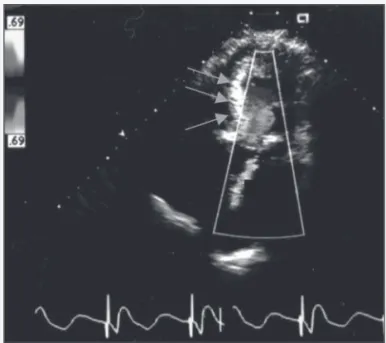
Both clinical and echocardiographic control during the following years showed a progressive enlargement of the left atrium, without worsening of the myocardial hypertrophy, but with evident mitral valvular impairment due to stenosis. In 2002, progressive dyspnea on exertion appeared, as well as nocturnal paroxysmal dyspnea, but with no other cardiovascular symptoms. The patient sought medical care on that occasion, when transthoracic echocardio-graphy was performed (fig. 1), revealing significant enlargement of the left atrium (5.7 cm of diameter), a mean mitral transvalvular gradient of 18 mmHg, and a peak gradient of 31 mmHg, with a reduction in the valvular area (1.15 cm²). The diagnosis of mode-rate to severe mitral stenosis was established, and the existence of pulmonary hypertension (pulmonary artery systolic pressure of 64 mmHg) was evidenced. In addition, a relatively hyperechoic morphologically altered interventricular septum was observed, with thinning in its basal, middle, and apical portions, and asynchronous motion. The overall systolic function was preserved with an ejection fraction of 80%.
Despite the medicamentous treatment with amiodarone (200 mg/day), furosemide (40 mg/day), phenytoin (100 mg/day), verapamil (120 mg/day), and warfarin (5 mg/day), the patient showed no clinical improvement. Thus, in April 2003, the patient was hospitalized, so that her clinical status could be better assessed through an adequate diagnostic investigation.
On physical examination, the patient was in regular general condition, acyanotic, anicteric, hydrated, eupneic, and had healthy coloring. Her blood pressure was 130/70 mmHg. Jugular venous distension was observed at 45º, crepitant rales were heard on both pulmonary bases, and the liver was tender and palpated
4 cm from the costal margin in the right midclavicular line. On cardiac auscultation, her heart rate was 70 bpm, her rhythm was regular, the intensity of the mitral (M1) and pulmonary (P2) components of the first and second cardiac sounds, respectively, was increased, and a diastolic murmur could be heard on the aortic and mitral positions. The electrocardiogram showed a base-line rhythm of atrial fibrillation, with a pacemaker functioning at a heart rate of 70 bpm. The chest X-ray in the posteroanterior and left lateral projections (fig. 2) showed signs suggestive of mitral stenosis and pulmonary hypertension. Transesophageal echo-cardiography (fig. 3) showed significant stenosis of the mitral valve (area of 1cm²), mild mitral insufficiency, and no intracavitary thrombi. Cinecoronary angiography and ventriculography showed the presence of normal coronary arteries, a calcified mitral valve, and preserved LV function.
Because of the clinical findings of severe mitral stenosis, surgi-cal treatment for the mitral valve was chosen, with replacement
Fig. 1 - Two-dimensional transthoracic echocardiography with color-flow mapping showing a relatively hyperechoic interventricular septum, which is morphologically altered with thinning in its basal, middle, and apical portions (arrows).
Fig. 2 - Chest X-rays in the posteroanterior (A) and left lateral (B) projections indicating the presence of cardiomegaly, upper bulging of the middle arch, increased pulmonary vascularization with the cephalization pattern, image of double cardiac contour in the right border, and increased carinal angle. The presence of a pacemaker is noted.
260
Severe Mitral Stenosis in the Long-term Evolution of Myotomy-Myectomy-Septal Resection
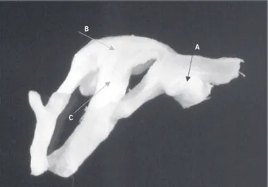
of the stenotic valve by a St. Jude 25-mm mechanical prosthesis. On surgical field inspection, the mitral valve was intensely thicke-ned and stenotic, with extensive calcification of its ring, which extended from the valve to the left ventricular apex. Still during surgery, a 2.3 x 2.0 x 0.8 cm fragment of the mitral valve was removed, and its anatomicopathological study showed thickening of the leaflet and chordae tendineae, with focal areas of calcifica-tion of the leaflet and fusion of the chordae tendineae (fig. 4). On histological examination (fig. 5), fibrosis, mucoid degeneration, calcification, and focal areas of neovascularization were observed. Calcification was also observed to occur mainly close to the inser-tion border of the leaflet (valvular ring).
In the postoperative period, the patient had complications, such as a delay in extubation due to pulmonary congestion and the need for vasoactive and inotropic drugs (noradrenaline, dobu-tamine, and milrinone). The patient evolved with respiratory infec-tion and acute renal failure, with complete reversion of the findings after adequate therapy was instituted.
On postoperative echocardiography, the presence of a normo-functioning mechanical prosthesis was observed in the mitral posi-tion, with a valvular area of 2.7 cm². In addiposi-tion, maintenance of pulmonary hypertension (pulmonary artery systolic pressure of 61 mmHg) and preserved left ventricular function were observed.
The patient was discharged from the hospital after 46 days of hospitalization, in good general condition and with a significant improvement in functional class (New York Heart Association func-tional class I). On hospital discharge, the following drugs were prescribed: warfarin (5 mg/day); losartan (50 mg/day); amiodarone (200 mg/day); and hydrochlorothiazide (25 mg/day).
Discussion
Asymmetric septal hypertrophic cardiomyopathy is usually greatly heterogeneous in its morphology, its clinical features, and its evolution 1-4. In a considerable number of cases, asymmetric septal hypertrophic cardiomyopathy has been reported in asso-ciation with valvular changes, mostly mitral insufficiency, which usually resolves after myotomy-myectomy-septal resection is perfor-med 2-5,8. More commonly, the mitral insufficiency occurring in asymmetric septal hypertrophic cardiomyopathy follows the obstruction caused by the systolic anterior motion of the mitral valve 2-4, 8.
A study by la Morena et al 2 assessed, by means of echocar-diography, the characteristics of a population of 72 patients with hypertrophic cardiomyopathy, 31 of whom had asymmetric septal hypertrophic cardiomyopathy. Of their patients with hypertrophic cardiomyopathy, 44 had mitral insufficiency, which was mild in 25 patients, moderate in 15, and severe in 4. This shows the close relation between hypertrophic cardiomyopathy and that valvu-lar problem 2. In addition, in 33 patients with systolic anterior motion of the mitral valve, the occurrence of mitral insufficiency was 78.7%, while in those with no systolic anterior motion of the mitral valve, that index was 46.1% 2.
Currently, 5% to 10% of the patients are refractory to clinical treatment, and more aggressive therapies should be indicated in these patients. Myotomy-myectomy-septal resection continues to be an important alternative to the treatment of asymmetric septal hypertrophic cardiomyopathy, being, up to the present moment, the preferred choice in very symptomatic patients, who do not respond to clinical treatment, or in cases with a baseline pressure gradient ≥ 30 mmHg 3,4,8,9. In many cases, myotomy-myectomy-septal resection is performed in association with reconstruction of
Fig. 3 - Transesophageal echocardiography showing the following: valvular thi-ckening and calcification, and reduction in the valvular opening, characterizing mitral valvular stenosis.
Fig. 4 - Gross analysis of the mitral valvular leaflet removed during surgery. Evident areas of calcification close to the leaflet insertion (A arrow), and thickening of the valvular leaflet (B arrow) and of the chordae tendineae (C arrow) may be seen.
A B
C
261
the mitral ring 3,8,9. However, replacement of the mitral valve should be the exception and performed only in cases of great hypertrophy with severe mitral insufficiency 9.
Fighali et al 3 studied 36 patients with hypertrophic cardiopathy undergoing surgical procedures, 6 of whom underwent myo-tomy-myectomy-septal resection, and 9 underwent that procedure in association with repair of the mitral valve. In the first group, 1 patient was not diagnosed with mitral insufficiency, 4 had mild mitral insufficiency, and 1 had moderate mitral insufficiency. In the second group, mitral insufficiency was mild in 3 patients, moderate in 2, and severe in 4. After surgery, a significant improvement in mitral insufficiency was observed in both groups, and only 1 patient remained with mild valvular insufficiency in each group 3.
Therefore, myotomy-myectomy-septal resection improves asym-metric septal hypertrophic cardiomyopathy in association with mitral insufficiency. In addition, the appearance of mitral insuffi-ciency after myotomy-myectomy-septal resection is not frequent. Current survival for this type of surgery is around 80% in 10 years, with good immediate and late results in regard to symptom improvement. In the immediate postoperative period, approximately 70% of the cases already have expressive improvement in symp-toms. In-hospital mortality, however, ranges from approximately 0% to 17.6%, depending mainly on the age group affected, and intraoperative mortality is close to 5%. The major complications of this procedure reported so far are as follows: complete atrio-ventricular block (incidence between 5% and 7%), interatrio-ventricular septal defect, endocarditis, and thromboembolism 3,4,8,9.
Cases of mitral stenosis in association with hypertrophic cardio-myopathy or with myotomy-myectomy-septal resection are rare; when present, this association occurs in patients with previous rheumatic disease 5,10. Therefore, no correlation exists between these 2 diseases. Up to the present time, no case has been reported in the scientific literature showing, from the pathophy-siological point of view, a relation between asymmetric septal hypertrophic cardiomyopathy or myotomy-myectomy-septal resec-tion and the appearance of mitral stenosis.
We report the evolution of a female patient with asymmetric septal hypertrophic cardiomyopathy, with no previous history of rheumatic disease, who developed mitral stenosis after undergoing myotomy-myectomy-septal resection.
In the anatomicopathological analysis of the valve performed in the leaflet fragment removed during the last surgery, we observed characteristics that could be present in degenerative and rheumatic processes, or in a possible extension of the healing process due to resection of the basal region of the interventricular septum perfor-med during myotomy-myectomy-septal resection 18 years before. The gross and microscopic characteristics reported, especially those concerning neovascularization, could be found in the rheuma-tic process, due to its inflammatory character 6,7. However, the presence of the rheumatic process does not justify the morpholo-gical aspect found in the interventricular septum of this patient 6. The surgical and echocardiographic findings of intense fibrosis,
thinning, and hypokinesia of the interventricular septum are not observed in rheumatic disease, and this hypothesis does not also explain the continuity of the process to the region of the mitral valve 6. In addition, the patient did not have a previous history of rheumatic disease. It would be expected that the signs and symp-toms of rheumatic disease would manifest until the fourth decade of life. Characteristically, rheumatic disease has its first manifes-tation before the age of 20 years, and, on average, 20 years later, symptomatic mitral stenosis may appear as a long-term compli-cation 7. In the present case, the patient had no alterations in the mitral valve that could indicate a possible impairment due to the rheumatic process on the occasion of myotomy-myectomy-septal resection at the age of 49 years. If rheumatic disease existed at that age, the patient might already have severe impairment of the valve, and this impairment might have been diagnosed on the preoperative examinations, or the alterations in the valvular appa-ratus might have been observed during the surgical procedure performed on the occasion. Therefore, when the characteristics of the present case are seen as a whole, the chance that mitral stenosis is related to rheumatic disease is considered minimal.
In regard to the possibility of the presence of a valvular dege-nerative process in the present case, the anatomicopathological analysis is very suggestive. The findings of extensive calcification and thickening of the mitral valve are characteristic of a degene-rative process 6, which had already been suggested in 1985 on echocardiography, and confirmed in 2003, with removal of the mitral valve. Degeneration of the mitral valve ring could, by itself, cause mitral stenosis, mainly in the elderly, although this is seen only in extremely rare cases. Characteristically, mitral insufficiency is the most common occurrence 6. Moreover, characteristics, such as thickening of the leaflet with focal areas of calcification, thickening, and fusion of the chordae tendineae, septal morpho-logical alteration, and, mainly, areas of neovascularization, are not present in the isolated degenerative process. Although, in our case, the presence of degeneration of the valvular ring continued to be considered, the direct association of this process with the occurrence of mitral stenosis was not performed.
Therefore, based on the surgical, echocardiographic, and ana-tomicopathological observations, the mitral stenosis of the present case was concluded to result from an extension of the healing process, which occurred primarily in the interventricular septum after the myotomy-myectomy-septal resection performed in 1985. This healing process is supposed to have also affected the conti-guous mitral valve. This extension of the healing process (charac-teristically inflammatory) justifies the presence of the intense fibro-sis in the interventricular septum not restricted to its basal region, later reaching the valvular leaflet.
262
Severe Mitral Stenosis in the Long-term Evolution of Myotomy-Myectomy-Septal Resection
1. Almeida DR, Diniz RVZ, Carvalho AS, et al. Incidence of the hypertrophic cardio-myopathy. Rev Soc Cardiol Est São Paulo 2000; 4: 431-4.
2. De la Morena G, Ruiperez JA, Garcia Garcia J, Salas J, Jean E, Lopez Candel J. Hypertrophic myocardiopathy (I). The clinical and echocardiographic characteris-tics of a population of 72 patients. Rev Esp Cardiol 1991; 44: 11-7.
3. Fighali S, Krajcer Z, Leachman RD. Septal myomectomy and mitral valve replace-ment for idiopathic subaortic stenosis: short and long-term follow-up. J Am Coll Cardiol 1984; 3: 1127-34.
4. Wynne J, Braunwald E. Miocardiopatias e miocardites. In: Fauci AS, Braunwald E, Isselbacher KJ, Wilson JD, et al. Harrison’s Principles of Internal Medicine. 14th ed. Rio de Janeiro: McGraw-Hill Interamericana do Brasil; 1998: c1417-23. 5. Shapiro LM, Howat AP, Crean PA, Westgate CJ. An echocardiographic study of
localized subaortic hypertrophy. Eur Heart J 1986; 7: 127-32.
References
6. Schoen FJ. The heart. In: Cotran RS, Kumar V, Collins T. Robbins Pathologic Basis of Disease. 6th ed. Rio de Janeiro: Guanabara Koogan; 2000: 486-539. 7. Braunwald E. Valvopatias cardíacas. In: Fauci AS, Braunwald E, Isselbacher KJ,
Wilson JD, et al. Harrison’s Principles of Internal Medicine. 14th ed. Rio de Janeiro: McGraw-Hill Interamericana do Brasil Ltda; 1998: 1400-13.
8. Krajcer Z, Lufschanowski R, Angelini P, Leachman RD, Cookey DA. Septal myomec-tomy and mitral valve replacement for idiopathic hypertrophic subaortic stenosis. An echocardiographic and hemodynamic study. Circulation 1980; 62 (2 Pt 2): I158-64.
9. Pomerantzeff PMA, Brandão CMA, Stolf NAG, Oliveira SA. Surgical treatment of hypertrophic cardiomyopathy. Rev Soc Cardiol Est São Paulo 2000; 4: 487-91. 10. Gash AK, Ferri DP, Kolff J, Carabello BA, Spann JF. Coexisting mitral stenosis and