AR
TIGO ORIGINAL / ORIGINAL AR
TICLE
INTRODUCTION
The gastroesophageal relux disease (GERD) is a condition that develops when gastric content passes to the esophagus and causes the onset of annoying symp-toms and/or complications(17). It is a disease of great medical and social importance due to its increasing incidence and long-lasting symptoms, which hinder patients quality of life(5, 20).
The oral lesions resulting from GERD are not usually noticed by patients, physicians or dentists until they cause signiicant damage. They may range from pruritus and burning on the oral mucosa, tooth sensitivity, aphthae, sour taste, decrease in the vertical dimension of occlusion to irreversible damage such as dental erosion, which can be increased by friction or abrasion(19).
SALIVARY PARAMETERS AND
TEETH EROSIONS IN PATIENTS WITH
GASTROESOPHAGEAL REFLUX DISEASE
Maria Carolina Canteras Scarillo Falotico
CORRÊA
1, Mauro Masson
LERCO
2,
Maria de Lourdes Ribeiro de Sousa da
CUNHA
3and
Maria Aparecida Coelho de Arruda
HENRY
4ABSTRACT – Context - In the gastroesophageal relux disease (GERD), a highly prevalent digestive disorder, gastric content may return to the esophagus and reach the mouth, thus leading to a small number of carious lesions and high incidence of dental ero-sion. Since saliva plays a major role in oral homeostasis, evaluating salivary parameters is necessary in attempting to explain such outcome. Objectives - This study aimed at analyzing salivary parameters (salivary low, pH and buffering capacity), bacterial count, caries index and dental erosion in patients with GERD. Materials - Sixty patients were studied, and of these, 30 had GERD (group 1), and 30 were controls (group 2). Gastroesophageal relux disease diagnosis conirmation was achieved by means of endoscopy, manometry and pH metric esophageal monitoring. The above mentioned salivary parameters were evaluated in patients from groups 1 and 2. Results - The number of erosions in patients with GERD (group 1) was larger than in controls (P<0.001). The number of carious teeth was smaller in group 1 than in group 2 (P<0.001). Salivary low (non-stimulated and stimulated) and pH did not show differences between the 2 groups (P = 0.49; P = 0.80 and P = 0.85, respectively). Salivary buffering capacity in patients with GERD showed lower values in controls (P = 0.018). The number of bacteria (Lactobacilli and Streptococci) was smaller in patients with gastroesophageal relux disease than in controls (P = 0.0067 and P = 0.0017, respectively). Conclusion - It was concluded that the large number of erosions must be a result of GERD patients reduced salivary buffering capacity. The reduced number of caries of patients in group 1 can be explained by the low prevalence of bacteria (Lactobacilli and Streptococci), observed in the saliva of patients with chronic relux.
HEADINGS – Saliva. Tooth erosion. Dental caries. Gastroesophageal relux.
Declared conflict of interest of all authors: none.
1 General Surgery Bases in the Department of Surgery and Orthopedy, Botucatu School of Medicine, State University of São Paulo (UNESP) 2 Department of Surgery and
Orthopedy, Botucatu School of Medicine, UNESP; 3 Department of Microbiology, Biosciences Institute, UNESP; 4 Department of Surgery and Orthopedy, Botucatu School
of Medicine, UNESP, Botucatu, SP, Brazil.
Correspondence: Dr. Maria Carolina C.S.F. Corrêa - Departamento de Cirurgia e Ortopedia da Faculdade de Medicina de Botucatu, Distrito de Rubião Jr. s/n - 18618-970 – Botucatu, SP, Brazil. E-mail: [email protected] or [email protected]
Studies on the oral health of patients with chronic relux have shown a prevalence of dental erosion and a small number of caries in individuals with GERD as compared to control groups(3, 4, 10).
Silva et al.(23) studied patients with GERD and reported that saliva is an important dental erosion modifier. However, they did not relate the reflux disease to the presence of carious lesions, but only to microscopic alterations on the palatal mucosa.
ulcers and the feeling of mucosal burning, candidiasis, caries and susceptibility to dental erosion. The buffering capacity of saliva keeps the mouth’s pH so as to ensure the integrity of tooth structure and inhibit the acid from bacterial plaque. Salivary low reduction will cause the loss of certain salivary functions, allowing for the development of bacterial plaque, high caries activity and smaller salivary buffering capacity.
The dissolution of dental structure in caries and dental erosion occurs when saliva pH reaches a lower value than 5.5. This is “critical oral pH”, under which dental tissue demineralization initiates(15).
Dental caries is an infectious, contagious, and incurable disease that affects 95% of the world population in diffe-rent ways for each individual. Caries initiation requires the interaction of several factors, such as the host, microlora, cariogenic substrate and time for the disease’s development. According to Krasse(13), caries activity is inversely propor-tional to saliva buffering capacity and directly proporpropor-tional to the presence of Streptococcusmutans. Krasse(13) deined dental caries as a destruction located on teeth. The destruc-tion of dental enamel, which consists of 95% hydroxyapatite, is mainly caused by the organic acids produced by the action of microorganisms that ferment carbohydrates, particularly sugars. In the dentin, such process is followed by the diges-tion of organic structure. The three main factors for caries development are the host, the microbiota and diet. Some microorganisms are more important in caries pathogeny, such as Streptococcusmutans, which are related to the initial phase, and Lactobacillus, whose growth produces acid and reduces oral pH to levels below critical pH when microorganisms have adhered to the tooth. Reduction in saliva pH and alterations in its buffering capacity and viscosity directly inluence the severity of erosive lesions.
This study aimed at evaluating the results of saliva tests (non-stimulated salivary low, stimulated salivary low, pH, salivary buffering capacity), caries and erosion index and bacterial count (Streptococcus mutans and Lactobacillus)
in individuals with GERD.
METHODS
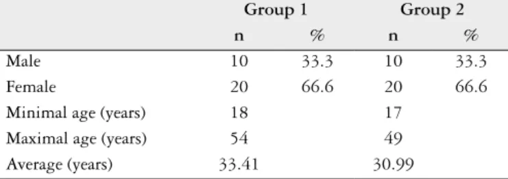
In this prospective study, 60 patients cared by Department of Surgery Orthopedy and were clinicaly evaluated. Of these, 20 were males (33.3%) and 40 were females (66.6%) with ages raging from 17 to 60 years, who were divided into two groups. The demographic aspects are listed in Table 1.
Group 1: 30 individuals with GERD in pre operative period of laparoscopic fundoplication, cared for by the Discipline of Surgical Gastroenterology at the Department of Surgery and Orthopedy of the Botucatu School of Medi-cine – UNESP, Botucatu, SP, Brazil. The diagnosis of GERD was conirmed by the clinical, endoscopic, manometric and pHmetric indings. All patients presented tipical symptoms for GERD.
Group 2: 30 patients without clinical evidence of GERD, located in the inirmary in the pre operative period of ingui-nal hernia.
Inclusion criteria: males and females individuals aged 17 to 60 years, with at least 20 teeth. Systemic medications were systemically recorded in the questionnaire for later correlation with salivary parameters after agreement in par-ticipation in this study. The informed consent was obtained in all patients. The protocol was approved by the Ethical Committee of School of Medicine of Botucatu-UNESP (OF340-2007). Exclusion criteria: patients under 20 years old and over 60 years old; pregnant women and individuals with systemic diseases affecting esophageal motor activity and salivary low.
The patients in group 1, 30 patients were submitted to upper digestive endoscopy, manometry and esophageal pH-metry for GERD diagnosis conirmation, in addition to a questionnaire containing questions concerning risk factors for the relux disease (pirosis, regurgitation and alimentary customs), a detailed clinical oral examination, salivary tests and saliva bacterium count. The individuals in group 2 were submitted to clinical oral examination and salivary tests and answered a questionnaire. The endoscopic, manometric and pH-metric esophageal tests were not performed for this group as recommended by the Ethics Committee.
After a 12-hour fasting period, the patients were en-doscopically evaluated by using an endoscopic video (GIF-Olympus). This test enabled the analysis of the esophageal mucosa, cardia competence and presence of hiatal hernia. The endoscopic appoints informations con-cerning the cardia capacity, by the retroversion maneuner. The Savary-Miller and Los Angeles classiications have the impropriety of not considering the light disorders (edema, eritema and disappearance of the gastroesophageal junction).
After the performance of esophageal manometry, the methodology previously standardized in the laboratory was used, according the pull through technique with evaluation of the following attributes: pressure amplitude in the lower and upper esophageal sphincters and motor activity of the esophageal body. For esophageal manometry, a light lumen low compliance infused system with computerized data ac-quisition and analysis was employed. We consider normal values when LES pressure were above 10.0 mm Hg.
Esophageal pHmetry was performed in all group 1 patients. The disposable catheter in our laboratory at that time had one sensor. The pHmetric study was performed according the technique standardized in our laboratory(12), considering the relux episode when the esophageal pH was lower than 4 units(12).
TABLE 1. Demographic aspects presented by patients with GERD (group 1) and controls (group 2)
Group 1 Group 2
n % n %
Male 10 33.3 10 33.3
Female 20 66.6 20 66.6
This test enables the analysis of various parameters, of which the most important are the percentage of time (of the 24 hours) in which the esophagus showed values below 4 units and De Meester score.
All the individuals (groups 1 and 2) were initially submit-ted to a detailed questionnaire with questions concerning risk factors for GERD, diet, medication and oral hygiene. The clinical oral evaluation performed using the clinical mirror and convenient catheter made it possible to evaluate the dental caries index and erosion(4). Next, the gingiva was probed by a millimeter probe recommended by the Health World Organization, for analysing the depth of the gingiva lesions, and the erosions were evaluated according to Eccles and Jenkins classiication(7, 8). This exam was performed by a dental surgeon, without knowledge of patients with GERD.
Total saliva was collected for 5 minutes for stimulated by the chewing of a small fragment (1 cm) of a strangling-stick, and non-stimulated, at rest with the half open mouth allow-ing the salivary low. The values were evaluated by the gravi-metric method proposed by Lussi(16). The stimuled low was obtained after chewing a strangling-stick during 5 minutes.
Salivary pH and buffering capacity were measured by a portable pHmeter.
Buffering capacity is the salivary capacity to neutralize weak acids produced in saliva and on bacterial plaque, which lead to the dissolution of dental mineral tissues, that is, resistance against pH alterations. The method used is that preconized by Lussi(16) .
The saliva samples were taken to the laboratory and see-ded for at least 1 hour after collection and properly protected in order to prevent bacterial proliferation. After homogeniza-tion and diluhomogeniza-tion in autoclaved yeast extract, 100 microliters were seeded on each dish containing selective medium for S. mutans and Lactobacillus(1).
Statistical analysis
The Chi-square or Fisher’s exact tests were used for group comparison. Student’s t test for independent populations was utilized for the quantitative variables. The level of signiicance adopted was 5%.
RESULTS
Of the 30 patients in group 1, 20 (66%) showed non-ero-sive GERD, and 10 patients showed level-1 and level-2 relux esophagitis according to the Savary and Miller classiica-tion(22). Hiatal hernia was found in 17 (56.6%), with a mean size of 3.7 cm. By the retroversion maneuver, cardia incom-petence was observed in 28 patients in this group (93.3%). The diagnosis of GERD was performed after the analysis of the clinical, endoscopic, manometric and pHmetric indings. The mean pressure in the lower esophageal sphincter was of 10.75 ± 2.42 mm Hg, and in the upper sphincter, it was 75.24 ± 28.08 mm Hg. No disorder was observed for esophageal body motor activity in any of the patients with GERD (group 1).
Twenty-four-hour esophageal pH-metry enabled the analysis of several parameters, of which the most signiicant were the percentage of time during which esophageal pH was below 4 units, with a mean of 7.03 ± 5.49% and the DeMeester score, with a mean of 27.55 ± 18.98. In seven (23%) of the patients in group 1, such score was normal, and the diagnosis of GERD was conirmed by the clinical, endoscopic and manometric indings. The mean number of teeth with dental erosion for patients with GERD was of 5.22 ± 2.52, and in controls, it was of 0.06 ± 0.36 (P<0.001) (Table 2).
Forty-one carious teeth were observed in patients in group 1 (mean of 3.63 ± 0.70) and 156 for controls, with a mean of 5.77 ± 4.39 (P<0.001).
The mean non-stimulated salivary low for group 1 was of 0.26 ± 0.18 mL/min, and for group 2, it was of 0.23 ± 0.13 mL/min (P = 0.49). As regards the stimulated salivary low, the mean value for group 1 was 0.75 ± 0.29 mL/min and 0.78 ± 0.52 mL/min for group 2 (P = 0.80). Saliva pH in both groups did not show signiicant difference (P = 0.85). Buffering capacity was smaller in group 1, than in group 2 (3.77 ± 0.9 X 3.21 ± 0,7; P = 0.018).
As concerns the number of bacterial colonies found in saliva, lower values were observed in patients with re-flux (group 1) than in controls, both for the evaluation
TABLE 2. Mean and standard desviation for the analyzed variables according to groups
Group
G1 G2 P value
Variables Mean SD Mean SD
Number of level-1 erosions 3.76 2.13 0.13 0.51 <0.001
Number of level-2 erosions 0.83 1.58 0.00 0.00 0.005 Number of level-3 erosions 0.10 0.55 0.00 0.00 0.32
Total number of erosions 4.70 2.52 0.06 0.36 <0.001
Number of carious teeth 1.3 2.5 5.2 1.3 <0.001
Non-stimulated salivary low mL/min 0.26 0.18 0.23 0.13 0.49
Stimulated salivary low mL/min 0.75 0.29 0.78 0.52 0.80 pH salivary
buffering capacity
7.1 3,2
0.4 0,7
7.0 3,7
0.4 0,9
of Lactobacilli (P = 0.0067) and for that of Streptococci (P = 0.0017) (Table 3).
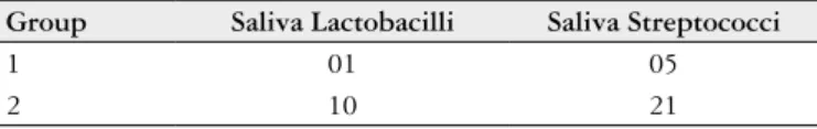
TABLE 3. Conluent type count (Petri dish with more than 300 colonies) of Streptococci and Lactobacilli in saliva
Group Saliva Lactobacilli Saliva Streptococci
1 01 05
2 10 21
P = 0.0067 P = 0.0017
DISCUSSION
Among the individuals with relux (group 1), 66.6% were females, which is in agreement with previously published articles, as a higher incidence of GERD in women has been reported by many authors(17). In group 2 (control), the distribution of patients in relation to gender was similar to that in group 1, with a higher percentage of females (66.6%).
The mean age of patients in group 1 was 33.41 years, which was lower than that reported by Mota et al.(18). The patient’s young age can be explained by the fact that one of factors for their inclusion in this study was the presence of teeth in reasonably good conditions, a fact that was not observed in older individuals.
Among the oral lesions observed in individuals with GERD, the most evident was dental erosion, with a higher incidence in group 1 than in controls (P<0.001), a similar result to that observed by various authors(3, 4).
The classiication used for the patients in this study was that proposed by Eccles and Jenkins(7). The most frequently found erosions were levels 1 and 2, which were more numerous in individuals with GERD than in controls (P<0.001) (Table 1).
In this study, it was observed that the number of ca-ries was smaller in patients with GERD than in controls (P<0.001- Table 1). Our results are in agreement with those reported by Cazzonato et al.(3), Corrêa et al.(4) and Ersin et al.(10), who reported an inverse relation between caries and gastroesophageal relux. However, Linnett et al.(14) report that individuals with relux have more erosions and dental caries than the control group. Silva et al.(23) did not ind a relation between GERD and the presence of caries.
In patients in group 2 (control), non-stimulated salivary low showed a mean value of 0,23 ± 0.13 mL/min, which was considered to be normal(16, 21). In this group, the stimulated sa-livary low observed was of 0.78 ± 0.52 mL/min, characterized as a small salivary low, according Lussi(16) and Raphael et al.(21).
In individuals with pathological gastroesophageal relux (group 1), non-stimulated salivary low showed a mean value of 0.26 ± 0.18 mL/min, and stimulated low showed a mean of 0.75 ± 0.29 mL/min.
Statistical analysis (Table 2) showed that there was no dif-ference between the two groups as regards the non-stimulated (P = 0.49) or stimulated (P = 0.80) salivary low.
This result does not explain the caries found in controls or their relative absence in individuals with relux (P<0.001 - Table 2), in addition to being in disagreement with several authors(2, 3, 6).
In patients with GERD (Group 1), salivary pH ranged from 6.45 to 7.86 units. In the controls, the extreme values observed were 6.47 and 8.69 units (mean of 7.0 ± 0.4). The statistical analysis did not show difference between the two groups (P = 0.85) (Table 2).
The result observed in our study disagrees with that
reported by Ecley and Costa(9). Those authors reported a
positive association between GERD and salivary pH altera-tions, thus suggesting that its measurement can be used for diagnosing acid laryngopharyngeal relux. Such test can be easily performed and could replace prolonged esophageal pHmetry using a pH microelectrode.
Salivary buffering capacity was evaluated in 60 patients in this study. In the patients with relux, salivary buffering capacity ranged from 2.42 to 4.27 units, with a mean of 3.2 ± 0.7 units, and in controls, the mean ranged was 3.7 ± 0.9 units, with extreme values of 2.18 and 6.40. As concerns the means representing low buffering capacity(16), the statistical analysis showed that, in patients in group 1 (GERD), salivary buffering capacity was smaller than in controls (P = 0.018). This result can explain the high rate of dental erosions in this group as compared to controls (P<0.001).
In this study, it was observed that the number of caries was much smaller in patients with GERD than in controls (P<0.001), thus confirming the findings of our previous study(4), a fact that disagrees with the study by Ersin et al.(10), who reported that children with GERD had a signiicantly higher rate of caries and salivary microorganisms than con-trols. After seeding the saliva in appropriate culture media, it was observed that the conluent type count (more than 300 bacterial colonies) was of one Lactobacilli count in patients in group 1 and of 10 in controls, thus showing a reduced number of Lactobacilli in patients with GERD (P = 0.0067). As con-cerns Streptococci, a signiicant difference was also observed (5 x 21) (P = 0.0017), with a smaller number of conluent colonies in patients with relux than in controls (Table 2).
The results above are in agreement with the indings by Gábris et al.(11), who reported that the larger the salivary microbiota, the larger the number of caries.
The results in this study lead to the following conclusions: 1. salivary flow (non-stimulated and stimulated) and
salivary pH in patients with GERD do not differ from those observed in controls;
2. salivary buffering capacity is more reduced in indivi-duals with GERD than in controls;
3. bacterial count (Lactobacilli and Streptococci) was more reduced in patients with GERD than in controls.
ACKNOWLEDGEMENT
To Fundação para o Desenvolvimento da UNESP - FUNDUNESP, for the grant provided.
To CAPES for the fellowship provided during the re-search.
REFERENCES
1. Almeida RVD, Padilha WWN, Pereira MSV, Sampaio TPD. Avaliação de teste salivar microbiológico colorimétrico no risco à cárie dentária. Rev Bras Ciênc Saúde. 2002;6:259-68.
2. Campisi G, Lo Russo L, Di Liberto C, Di Nicola F, Butera D, Vigneri S, Com-pilato D, Lo Muzio L, Di Fede O. Saliva variations in gastro-oesophageal relux disease. J Dent. 2008;36:268-71.
3. Cazzonatto Jr H, Bernasconi GCR, Pedrazzolli JR. Gastroesophageal relux and oral lesions: is the acid that bad? GED Gastroenterol Endosc Dig. 2003;22:42-6. 4. Corrêa MC, Lerco MM, Henry MA. [Study in oral cavity alterations in patients
with gastroesophageal relux disease]. Arq Gastroenterol. 2008;45:132-6. 5. de Oliveira SS, dos Santos Ida S, da Silva JF, Machado EC. [Gastroesophageal relux
disease: prevalence and associated factors]. Arq Gastroenterol. 2005;42:116-21. 6. Di Fede O, Di Liberto C, Occhipinti G, Vigneri S, Russo LL, Fedele S, Lo Muzio
L, Campisi G. Oral manifestations in patients with gastro-oesophageal relux disease: a single-center case-control study. J Oral Pathol Med. 2008;37:336-40. 7. Eccles JD, Jenkins WG. Dental erosion and diet. J Dent. 1974;2:153-9. 8. Eccles JD. Erosion of teeth by gastric contents. Lancet. 1978;2:479.
9. Eckley CA, Costa HO. Estudo comparativo do pH e do volume salivar em indivíduos com laringofaringite crônica por doença do reluxo gastroesofágico antes e após tratamento. Rev Bras Otorrinolaringol. 2006;72:55-60.
10. Ersin NK, Onça˘g O, Tümgör G, Aydo˘g du S, Hilmio˘g lu S. Oral and dental mani-festations of gastroesophageal relux disease in children: a preliminary study. Pediatr Dent. 2006;28:279-84.
11. Gábris K, Nagy G, Madléna M, Dénes Z, Márton S, Keszthelyi G, Bánóczy J. Associations between microbiological and salivary caries activity tests and caries experience in Hungarian adolescents. Caries Res. 1999;33:191-5.
12. Henry MA. [Continuous evaluation of esophageal pH for 24 hours and its use in the diagnosis of gastroesophageal relux]. Rev Hosp Clin Fac Med S Paulo. 1984;39:203-7.
13. Krasse BO. Risco de cáries: um guia prático para avaliação e controle. São Paulo: Quintessence; 1986. 113p.
14. Linnett V, Seow WK, Connor F, Shepherd R. Oral health of children with gastro esophageal relux disease: a controlled study. Aust Dent J. 2002;47:156-62. 15. Loesche WJ. Role of Streptococcus mutans in human dental decay. Microbiol
Rev. 1986;50:353-80.
16. Lussi A. Dental erosion clinical diagnosis and case history taking. part:2. Eur J Oral Sci. 1996;104:191-8.
17. Moraes-Filho J, Cecconello I, Gama-Rodrigues J, Castro L, Henry MA, Me-neghelli UG, Quigley E; Brazilian Consensus Group. Brazilian consensus on gastroesophageal relux disease: proposals for assessment, classiication, and management. Am J Gastroenterol. 2002;97:241-8.
18. Mota LAA, Santos ACO, Melo Jr BC, Travassos RO, Melo MSI. Videolarin-goscopia e atividade da pepsina na saliva em voluntários com sintomas sugestivos de reluxo laringofaríngeo. Arq Int Otorrinolaringol. 2008;12:89-94.
19. Myklebust S, Espelid I, Svalestad S, Tveit AB. Dental health behavior, gastro-esophageal disorders and dietary habits among Norwegian recruits in 1990 and 1999. Acta Odontol Scand. 2003;61:100-4.
20. Nasi A, Michelsohn NH. Avaliação funcional do esôfago: manometria e pH metria esofágicas. São Paulo: Roca; 2001. p.39-251.
21. Raphael Jr A, Bacaltchuck BB, Jacobs ACP. Avaliação do luxo salivar e da capacidade tampão em pacientes da clínica integrada. Rev Assoc Paul Cir Dent. 2005;59:108-12.
22. Savary M, Miller G. L’esophage. Manuel et atlas d’endoscopie. Solothurn: Gassmann; 1977.
23. Silva MAGS, Damante JH, Stipp ACM, Tolentino MM, Carlotto PR, Fleury RN. Gastroesophageal relux disease: new oral indings. Oral Surg Oral Med Oral Pathol Oral Radiol Endod. 2001;91:301-10.
Received 10/11/2011. Accepted 19/6/2012. Corrêa MCCSF, Lerco MM, Cunha MLRS, Henry MACA. Parâmetros salivares e na erosão dentária na doença do reluxo gastroesofágico. Arq
Gastro-enterol. 2012;49(3):214-8.
RESUMO - Contexto - Na doença do reluxo gastroesofágico, afecção digestiva de elevada prevalência, o conteúdo gástrico pode retornar ao esôfago e atingir a cavidade oral, acarretando pequeno número de lesões cariosas e elevada incidência de erosões dentais. Sendo a saliva a principal responsável pela homeostase oral, a avaliação dos parâmetros salivares é imperiosa, numa tentativa de explicar este resultado. Objetivo - O objetivo deste trabalho foi analisar os parâmetros salivares (luxo, pH e capacidade tampão da saliva), contagem de bactérias, índice de cárie e erosão dental em pacientes com a doença do reluxo gastroesofágico. Métodos - Foram estudados 60 pacientes, sendo 30 com a doença do reluxo gastroesofágico (Grupo 1) e 30 controles (Grupo 2). A conirmação do diagnóstico da doença do reluxo gastroesofágico foi realizada através de exames endoscópico, manométrico e pHmétrico do esôfago. Os parâmetros salivares assinalados foram realizados nos pacientes dos grupos 1 e 2. Resultados - O número de erosões nos pacientes com a doença do reluxo gastroesofágico (grupo 1) foi mais elevado que nos controles (P<0,001). O número de dentes cariados foi menor no grupo 1 do que no grupo 2 (P<0,001). O luxo salivar (não estimulado e estimulado) e o pH não apresentaram diferença nos dois grupos estudados (P = 0,49; P = 0,80 e P = 0,85, respectivamente). A capacidade de tampão salivar dos pacientes com a doença do reluxo gastroesofágico apresentou valores mais baixos que nos controles (P = 0,018). O número de bactérias (lactobacilos e estreptococos) foi menor nos pacientes com a doença do reluxo gastroesofágico do que nos controles (P = 0,0067 e P = 0,0017, respectivamente). Conclusão - Os autores concluem que o elevado número de erosões deve ser decorrente da reduzida capacidade tampão salivar dos pacientes com doença do reluxo gastroesofágico. O reduzido número de cáries dos pacientes do grupo 1 pode ser explicado pela pequena prevalência de bactérias (lactobacilos e estreptococos), observada na saliva dos pacientes reluidores crônicos.