Márcio Bruno Figueiredo Amaral
Tratamento do fenômeno de extravasamento/retenção de muco
pela técnica de micro-marsupialização modificada associado
ou não ao uso do laser de baixa intensidade: ensaio clínico
controlado e randomizado
Universidade Federal de Minas Gerais Faculdade de Odontologia
Belo Horizonte – MG
Márcio Bruno Figueiredo Amaral
Tratamento do fenômeno de extravasamento/retenção de muco
pela técnica de micro-marsupialização modificada associado
ou não ao uso do laser de baixa intensidade: ensaio clínico
controlado e randomizado
Dissertação apresentada ao Colegiado do Programa de Pós-graduação da Faculdade de Odontologia da
Universidade Federal de Minas Gerais, como requisito parcial para obtenção do grau de Mestre em Odontologia - área de concentração em Estomatologia
Orientador: Prof. Dr. Ricardo Alves Mesquita
Universidade Federal de Minas Gerais Faculdade de Odontologia
Belo Horizonte – MG
A485t Amaral, Márcio Bruno Figueiredo
2011 Tratamento do fenômeno de extravasamento/retenção de muco pela
T técnica da micro-marsupialização modificada associado ou não ao uso do
laser de baixa intensidade: ensaio clínico controlado e randomizado /
Márcio Bruno Figueiredo Amaral. 2011. 73 f.: il.
Orientador: Ricardo Alves de Mesquita
Dissertação (Mestrado)- Universidade Federal de Minas Gerais,
Faculdade de Odontologia.
1. Glândulas salivares – patologia – Teses. 2. Rânula – cirurgia –
Teses. 3. Terapia a laser de baixa intensidade – Teses. I. Mesquita, Ricardo
Alves de. II. Universidade Federal de Minas Gerais.
Faculdade de Odontologia. III. Título.
A Deus que me deu saúde e possibilitou a realização de mais este trabalho.
AGRADECIMENTOS
Primeiramente a Deus, por me guiar, dar sabedoria e discernimento
durante minhas escolhas até este momento.
Aos meus pais Geraldo e Elziron, por sempre abdicarem de muitas
coisas na vida para darem o melhor aos seus filhos. Amo muito vocês.
Ao meu irmão, Juliano, pela amizade e companheirismo durante toda a
minha vida.
A Paulinha, pelo carinho e apoio durante estes quase quatro anos!
Ao meu orientador Professor Doutor Ricardo Alves Mesquita, pelos
ensinamentos e dedicação para que este trabalho fosse realizado. Tenho
grande orgulho de sua orientação e amizade.
Ao Cristian, pela amizade e parceria profissional durante todos estes
anos!
Ao Professor João Batista de Freitas, pela amizade e ensinamentos
durante minha vida pessoal e profissional.
Aos professores Belini Freire-Maia e Vasco Araújo, pela amizade e
ensinamentos durante minha vida profissional.
À acadêmica Isabel Zanforlin Freitas, que não mediu esforços em
auxiliar na pesquisa durante a aplicação do laser. Muito obrigado.
Aos professores e alunos do curso de especialização em Cirurgia e
Aos colegas de mestrado, Fabrício, Frederico, Silvia, Telma e Vladmir,
pela ajuda e momentos compartilhados.
Aos professores do Departamento de Clínica, Patologia e Cirurgia da
FO-UFMG, Maria Cássia Aguiar, Maria Auxiliadora do Carmo, Tarcília
Aparecida da Silva e Ricardo Gomez, com os quais muito aprendi durante o mestrado.
Agradeço a todos que de alguma forma contribuíram para a realização
deste trabalho.
A humildade exprime, ao contrário, uma das raras certezas de que estou
certo: a de que ninguém é superior a ninguém.
LISTA DE SÍMBOLOS E SIGLAS
AsGaAl Arseanato de Gálio- Alumínio
COEP Comitê de Ética e Pesquisa
GaAlAs Gallium Aluminum Arsenide
LBI Laser de Baixa Intensidade
LLLT Low Level Laser Therapy
mW miliwatts
RESUMO
Objetivo: Avaliar o efeito do laser do laser de baixa intensidade (LBI) em reduzir recorrências e aliviar a dor tratamento do fenômeno de extravasamento/retenção de muco pela técnica de micro-marsupialização modificada.
Materiais e Métodos: Seis pacientes com rânulas orais e mucoceles selecionadas foram submetidos ao tratamento de micro-marsupialização (grupo controle) e onze pacientes foram submetidos ao tratamento de micro-marsupialização associado ao LBI (grupo Laser). Os pacientes do grupo laser foram irradiados utilizando um laser de Arseanato de Gálio- Alumínio (AsGaAl) em um comprimento de onda de 660 nm em modo contínuo, com uma potência de 100 mW. A aplicação do laser foi realizada imediatamente após o tratamento com marsupialização e 24, 48 e 72 horas após a micro-marsupialização. Uma escala visual numérica e uma escala visual análoga de faces foram usadas para avaliar a dor. A relação entre os grupos tratados e dor pós-operatória foi avaliada através do teste de Mann-Whitney.
Resultado: Todos os pacientes com rânulas orais e mucoceles selecionadas tiveram cura clínica das lesões sem evidência de recorrência durante um acompanhamento médio de 10.88 meses. Pode ser observado que os pacientes do grupo laser relataram menos dor em comparação com os pacientes do grupo controle.
Conclusão: O LBI é uma boa alternativa para reduzir a dor pós-operatória causada pelo tratamento de micro-marsupialização em pacientes com fenômeno de extravasamento/retenção de muco.
ABSTRACT
Objective: To investigate the ability of low-level-laser therapy (LLLT) to reduce recurrence rates and alleviate pain caused by the modified
micro-marsupialization treatment of mucus extravasation or retention phenomena.
Patients and Methods: Six patients with oral ranulas and selected mucoceles were submitted to micro-marsupialization treatment (control group) and eleven
were submitted to micro-marsupialization treatment associated with LLLT (LLLT
group). Laser irradiation of patients with the LLLT group was performed using a
100 mW 660 nm continuous wave Gallium Aluminum Arsenide (GaAlAs) laser.
Irradiation was carried out immediately following the micro-marsupialization
treatment and at 24, 48 and 72 hours post-micro-marsupialization treatment. A
numeral rating scale and a visual analogue scale of faces were used to assess
the pain. The relationship between the treated groups and pain was assessed
using the Mann-Whitney test.
Results: All treated oral ranulas and selected mucoceles had clinical healing and no evidence of recurrence during the mean 10.88-month follow-up period. It
could be observed that the LLLT group reported less pain than did control
group.
Conclusion: LLLT is a good alternative to reduce post-surgical pain caused by micro-marsupialization treatment of mucus extravasation or retention
phenomena.
SUMÁRIO
1 INTRODUÇÃO 11
2 SÍNTESE BIBLIOGRÁFICA 12
2.1 Etiopatogenia dos fenômenos de extravasamento/retenção de muco 12 2.2 Características das mucoceles e rânulas 13
2.3 Formas de tratamento 14
2.4 Laser de baixa intensidade 16
3 OBJETIVOS 17
3.1 Objetivo geral 17
3.2 Objetivos específicos 17
4 MATERIAIS E MÉTODOS 18
4.1 Pacientes 18
4.2 Técnica de micro-marsupialização modificada 21
4.3 Protocolo do Laser de Baixa Intensidade 21
4.4 Universo 22
4.5 Critérios de inclusão e exclusão 23
4.6 Plano amostral 23
4.7 Análise estatística 24
5 RESULTADOS 24
5.1 Artigo 1 24
5.2 Artigo 2 40
6 CONSIDERAÇÕES FINAIS 59
7 CONCLUSÕES 61
8 REFERÊNCIAS 63
9 ANEXOS 69
ANEXO A – Aprovação do Comitê de Ética em Pesquisa da UFMG 69
ANEXO B – Termo de Consentimento Livre e Esclarecido 70
ANEXO C – Ficha Clínica (Micro-marsupialização) 72
1. INTRODUÇÃO
Rânulas e mucoceles são fenômenos de retenção de muco, que se
desenvolvem pelo extravasamento de saliva após trauma no sistema de ductos
das glândulas sublinguais ou glândulas salivares menores (Yoshimura et al.,
1995).
Rânula é o termo utilizado para as mucoceles que se localizam no
soalho bucal. O nome deriva do latim “rana”, que significa rã, pois o aspecto
clínico da lesão lembra o ventre translucente do anfíbio. Existem três tipos de
rânula: rânula oral, rânula mergulhante e rânula mista (Zhao et al., 2004).
O cisto de retenção de muco é um cisto verdadeiro devido ao
revestimento epitelial, que se origina dos tecidos da glândula salivar
(Baurmash, 2003).
O tratamento destas lesões tem sido controverso, devido à diversidade
terapêutica, principalmente para a abordagem de rânulas. A técnica de
micro-marsupialização, assim denominada pela primeira vez por Cardoso em 1974,
utilizada para tratamento de mucoceles por Delbem et al. em 2000, e
modificada por Sandrini et al. em 2007, mostra-se um método eficaz para
tratamento de rânulas e mucoceles, por ser uma técnica de fácil execução,
minimamente invasiva, baixa ou nenhuma dor pós-operatória, baixos custos e
com baixo índice de recorrência (Delbem et al., 2000; Sandrini et al., 2007).
O laser de baixa intensidade (LBI) com suas propriedades bem descritas
na literatura de analgesia, antiinflamatório e bioestimulante (Walsh et al., 1997;
especificamente associado ao tratamento do fenômeno de
extravasamento/retenção de muco.
De acordo com as características da técnica de micro-marsupialização e
das propriedades do LBI descritas, este trabalho teve como objetivo a
realização de um ensaio clínico randomizado, duplo cego, para o tratamento da
mucocele e rânula oral utilizando a micro-marsupialização modificada
associado ou não ao LBI. Verificou-se e comparou-se o desempenho destas
duas modalidades de tratamento através da 1) cura clínica ou não da lesão, 2)
avaliação de edema, 3) avaliação de dor pós-operatória e 4) avaliação de
recorrência após 6 meses de acompanhamento.
2. SÍNTESE BIBLIOGRÁFICA
2.1 Etiopatogenia dos fenômenos de extravasamento/retenção de muco
Mucocele é uma lesão comum da mucosa oral, que se origina a partir da
ruptura de um ducto de glândula salivar e consequente acúmulo de mucina
para o interior dos tecidos moles adjacentes (Delbem et al., 2000).
Rânula é um fenômeno de extravasamento de saliva após trauma ou
retenção de muco pela obstrução dos ductos da glândula sublingual e/ou
Fenômenos de retenção de muco denominados de cisto de retenção de
muco ou cisto do ducto salivar são raros. É um cisto verdadeiro causado pelo
estreitamento da abertura do ducto salivar que não pode acomodar
adequadamente a saída da saliva produzida, levando a dilatação do ducto e
aumento de volume na superfície. Clinicamente são semelhantes às
mucoceles, sendo sua diferenciação realizada apenas histologicamente
(Baurmash, 2003).
2.2 Características clínicas das mucoceles e rânulas
Mucoceles podem ser classificadas como um fenômeno extravasamento de
muco, proveniente de uma glândula salivar menor. A localização preferencial
destas lesões é o lábio inferior, mas também podem ocorrer no ventre lingual,
palato e mucosa jugal (Oliveira et al., 1993).
Estas lesões podem ser de base séssil ou pediculada, são de
consistência flácida ou fibrosa, podendo medir mais de 1,0 cm de diâmetro,
sendo lesões maiores que 1,5cm raras. As mucoceles que se localizam
superficialmente podem apresentar uma coloração azulada devido a presença
de capilares superficiais sobre a lesão. Quando localizada profundamente nos
tecidos sua coloração é semelhante a mucosa normal (Delbem et al., 2000;
Baurmash, 2003).
As mucoceles que ocorrem no soalho bucal são denominadas rânulas
(Baurmash, 2003). Segundo Zhao et al., (2004) são três as formas de rânulas,
1) rânula oral ou superficial: que se apresenta como um aumento de
volume em forma de cúpula, flutuante, no soalho da boca de coloração
variando conforme a profundidade da lesão. Ocorre mais comumente na e 2°
década de vida, com discreta predileção pelo sexo feminino.
2) rânula mergulhante: também denominada de rânula cervical, ocorre
quando há aumento de volume somente extra-oral pela dissecção da mucina
pelo músculo milohióideo, se acumulando na região submandibular e/ou
sublingual, podendo ser confundida com cisto do ducto tireoglosso, cisto
dermóide ou epidermóide, má formação vascular ou sialoadenite de glândula
submandibular.
3) rânula mista: ocorre quando há aumento de volume tanto intral-oral
quanto extra-oral, sendo na verdade a manifestação simultânea da rânula oral
e da rânula mergulhante.
2.3 Formas de tratamento
As mucoceles que ocorrem no lábio inferior são tratadas com sucesso através
da remoção cirúrgica da lesão e das glândulas salivares adjacentes (McGurk et
al., 2008).
Entretanto, o soalho bucal é o segundo local mais comum das
mucoceles de extravasamento, sendo o tratamento diversificado, e nem
sempre com sucesso (McGurk, 2007).
Segundo Zhao et al., (2004); e Yoshimura et al., (1995) são várias as
marsupialização com ou sem cauterização das margens da lesão, 4) excisão
da lesão e da glândula sublingual por acesso intra-oral, 5) excisão intra-oral da
glândula sublingual e drenagem da lesão, 6) excisão da lesão por acesso
cervical algumas vezes combinada com excisão da glândula sublingual, 7)
excisão e vaporização com laser de alta intensidade e 8)
micro-marsupialização.
A técnica amplamente utilizada na literatura para tratamento de rânula
tanto oral quanto mergulhante é remoção da lesão associada à retirada da
glândula sublingual ipsilateral, devido ao baixo índice de recidiva das lesões
com esta forma de tratamento (Yoshimura et al., 1995; Zhao et al., 2004 e
2005). No entanto há a necessidade de um procedimento cirúrgico mais
invasivo, envolvendo maiores riscos como lesões a estruturas nobres,
principalmente o nervo lingual e o ducto da glândula submandibular ou ambos
(Baurmash, 2001).
Técnicas menos invasivas podem ser utilizadas como tratamento de
rânulas orais e mucoceles, algumas com bons resultados. A transfixação da
lesão com fio cirúrgico em seda foi denominada micro-marsupialização por
Cardoso em 1974 e considerada como uma escolha para tratamento de
mucoceles. Morton e Bartley (1995) declararam que rânulas poderiam ser
tratadas com uma sutura em seda colocada dentro da cúpula do cisto.
A técnica de micro-marsupialização modificada por Sandrini et al. (2007),
tem se mostrado uma boa alternativa para tratamento de mucoceles e rânulas
orais, principalmente em crianças, por ser de fácil execução, utilizando
várias suturas na lesão, com o objetivo de se formar “novos ductos” para a
drenagem de mucina. A sutura é mantida por um período de 30 dias que,
segundo Selvig et al. (1998), é o tempo suficiente para epitelização da mucosa
bucal ao redor do fio de sutura.
2.4 Laser de baixa intensidade
A palavra LASER é o acrônimo Light Amplification by Stimulated Emission of
Radiation (Amplificação da Luz por Emissão Estimulada de Radiação). O laser
é uma forma de radiação que se transforma em energia luminosa, visível ou
não, dependendo da matéria que produz esse tipo de radiação (Genovese,
2000; Coluzzi, 2004). A radiação a laser pode ser classificada em alta, média e
baixa intensidade.
O laser de alta intensidade por suas propriedades de corte, vaporização,
coagulação e esterilização é utilizado em odontologia para cirurgia de tecidos
moles da cavidade oral, inclusive sendo seu uso descrito para tratamento
cirúrgico de rânulas orais (Lai & Poon, 2009). Entretanto apesar do laser de alta
intensidade ser utilizado em odontologia deste a década de 1960, não tem sido
difundido em países como o Brasil, provavelmente pelo alto custo dos
aparelhos.
O laser de baixa intensidade é um aparelho que emite radiação de baixa
potência, sem potencial destrutivo. Segundo Walsh (1997) a maior absorção
das estruturas no comprimento de onda vermelho visível e infravermelho
proteínas. Entretanto a identificação dos fotorreceptores responsáveis pelos
efeitos biológicos do laser de baixa intensidade não são bem definidos.
Segundo Sun e Tunér (2004) a teoria fotoquímica é a mais aceita para
explicar os efeitos e mecanismos do LBI. De acordo com esta teoria a luz é
absorvida por certas moléculas, seguido por uma cascata de efeitos biológicos
(Ewemeka, 1999). Sugere também que os fotorreceptores são porfirinas
endógenas e moléculas da cadeia respiratória, como o citocromo c-oxidase,
levando ao aumento da produção de ATP (adenosine tri-phosphate) (Lubart et
al., 1992; Karu, 2003).
O LBI tem ações analgésica, antiinflamatória e de bioestimulação bem
descritas na literatura, com grande aplicabilidade em quase todas as
especialidades odontológicas (Sun & Tunér, 2004).
Especificamente sobre mucoceles e rânulas orais a literatura não traz
nenhum relato sobre os efeitos do LBI nas diversas formas de tratamento
destas lesões.
3 OBJETIVOS
3.1 Objetivo geral
O objetivo geral deste estudo foi avaliar a eficácia do LBI associado a técnica
de micro-marsupialização modificada, para o tratamento do fenômeno de
3.2 Objetivos específicos
1- Avaliou-se a eficácia do LBI associado a técnica de micro-marsupialização
modificada no tratamento de mucoceles e rânulas orais, quanto à cura clínica
ou não das lesões;
2- Avaliou-se a eficácia do LBI associado a técnica de micro-marsupialização
modificada no tratamento de mucoceles e rânulas orais, quanto ao edema;
3- Avaliou-se a eficácia do LBI associado a técnica de micro-marsupialização
modificada no tratamento de mucoceles e rânulas orais, quanto à dor
pós-operatória;
4- Avaliou-se a eficácia do LBI associado a técnica de micro-marsupialização
modificada no tratamento de mucoceles e rânulas orais, quanto à recorrência
ou não das lesões após seis meses de acompanhamento.
4 MATERIAIS E MÉTODOS
4.1 Pacientes
Foi realizado um ensaio clínico randomizado, duplo cego, onde dezessete
pacientes com lesões de diagnóstico clínico de mucocele ou rânula oral foram
tratados pela técnica de micro-marsupialização modificada. No grupo controle
foi aplicada apenas a técnica de micro-marsupialização modificada. No grupo
a aplicação do LBI. Ambos os grupos foram acompanhados por quatro
semanas quando a sutura foi removida (Sandrini et al., 2007).
As duas modalidades de tratamento foram avaliadas através de: 1) cura
clínica ou não da lesão após quatro semanas de acompanhamento dos
pacientes, 2) avaliação do edema no momento do 7° dia pós-operatório por
questionamento, 3) avaliação da dor no momento do 7° dia pós-operatório, 4)
avaliação de recorrência ou não da lesão após 6 meses de seguimento. A dor
pós-operatória foi avaliada através de escalas específicas para dor (Figuras 1 e
2). As escalas utilizadas foram uma escala análoga visual de faces e uma
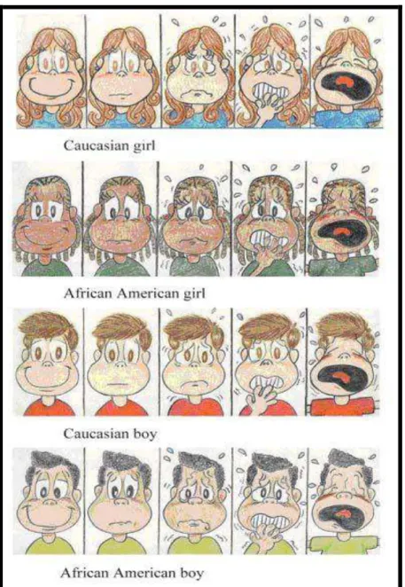
escala análoga visual numérica. A escala visual análoga de faces proposta por
Barrêtto et al. (2004) foi utilizada para pacientes com até seis anos de idade, e
uma escala visual numérica de zero a dez foi utilizada para pacientes com mais
de 6 anos de idade (Mannion et al., 2007).
Os dados foram coletados e anotados em ficha clínica específica para
Figura 2 - Escala análoga visual numérica proposta por Mannion et al. (2007), utilizada para pacientes acima de seis anos de idade.
4.2 Técnica de micro-marsupialização modificada
A técnica foi semelhante à utilizada por Sandrini et al. (2007), com algumas
modificações que incluíram sutura isolada utilizando fio de seda 3.0 ao longo
da lesão, com uma distância de 3 a 5mm entre cada entrada e a saída da
agulha, associado ao alargamento mecânico dos orifícios utilizando o próprio
fio. As suturas foram mantidas por um período de 30 dias. Segundo Selvig et
al., 1998 tempo suficiente para epitelização ao redor do fio. Permitindo assim
formação de vários “ductos” para drenagem do conteúdo da lesão.
4.3 Protocolo de aplicação do laser de baixa intensidade
Foi aplicado nos pacientes do grupo intervenção em quatro sessões, iniciando
no pós-operatório imediato, e sessões subseqüentes com intervalo de 24, 48 e
72 horas. A densidade energética foi de 4 J/ cm² e a forma de aplicação foi
pontual em cada ponto de transfixação do fio de sutura. Foi utilizado o laser de
AsGaAl no comprimento de onda de 660 nm, cor vermelha (visível) (Genovese,
2000).
Cada paciente incluído na pesquisa foi locado no grupo intervenção ou
controle através da técnica de amostragem aleatória simples.
Nos pacientes do grupo controle o LBI foi posicionado na cavidade bucal
da mesma forma que no grupo intervenção. Entretanto o aparelho não foi
acionado. Um pesquisador independente foi responsável ou não pelo
descrita. Nem o paciente nem o pesquisador que realizou a técnica cirúrgica e
avaliou o pós-operatório tiveram conhecimento da aplicação ou não do LBI.
4.4 Universo
Dezessete indivíduos independente de sexo e idade, com diagnóstico clínico
de mucocele ou rânula oral, encaminhados a clínica de estomatologia da
faculdade de odontologia da UFMG.
Os pacientes encaminhados a clínica de estomatologia da faculdade de
odontologia da UFMG foram esclarecidos da não maleficência e da
beneficência do tratamento e puderam negar a participação na pesquisa,
porém continuaram a receber o tratamento proposto. Aqueles que se
interessaram em participar da pesquisa leram e assinaram o Termo de
Consentimento Livre e Esclarecido (ANEXO B). A confidencialidade da
identidade dos participantes foi resguardada.
4.5 Critérios de inclusão e exclusão
Foram incluídos no estudo todos os indivíduos com diagnóstico clínico de
mucocele ou rânula oral. O diagnóstico foi confirmado durante a aplicação da
técnica de micro-marsupialização modificada, onde necessariamente houve
drenagem de muco após passagem da agulha de sutura (Sandrini et al., 2007).
As lesões com diagnóstico de mucocele possuíam pelo menos 10 mm
Foram incluídas no estudo lesões localizadas apenas no lábio inferior,
ventre lingual e soalho bucal (rânula oral).
Lesões menores que 10 mm, de base pediculada, consistência fibrosa e
localizadas na mucosa jugal ou palato foram excluídos do estudo.
Todos os casos encaminhados com diagnóstico diferente de mucocele e
rânula oral, ou que não se encaixaram nos critérios descritos, foram
submetidos à biópsia incisional ou excisional para confirmação diagnóstica e
tratados adequadamente.
4.6 Plano Amostral
Foi realizada uma série de casos com dezessete pacientes encaminhados a
clínica de estomatologia no período de agosto de 2009 a dezembro de 2010,
onde todos os indivíduos foram submetidos ao tratamento proposto, sendo
onze casos aplicados o LBI, e os resultados analisados posteriormente.
4.7 Análise estatística
A relação entre os grupos tratados e a dor pós-operatória foi avaliada
pelo teste de significância Mann-Whitney. As análises estatísticas foram
realizadas utilizando o software SPSS 17.0 (SPSS Inc., Chicago, IL, EUA). Um
valor de p ≤ 0,05 foi considerado estatisticamente significativo.
Os resultados foram apresentados na forma de artigos científicos submetidos
aos periódicos International Journal of Oral and Maxillofacial Surgery and
Lasers in Medical Science.
Ms. Ref. No.: IJOMS-D-11-00374
Title: Upgrading of the micro-marsupialization technique for the management of
mucus extravasation or retention phenomena
International Journal of Oral & Maxillofacial Surgery
Dear Prof. Amaral,
Your submission entitled "Upgrading of the micro-marsupialization technique for
the management of mucus extravasation or retention phenomena" has been
assigned the following manuscript number: IJOMS-D-11-00374.
You may check on the progress of your paper by logging on to the Elsevier
Editorial System as an author. The URL is http://ees.elsevier.com/ijoms/. Your username is Your username is: Márcio Amaral.
If you need to retrieve password details,
please go to: http://ees.elsevier.com/ijoms/automail_query.asp
Thank you for submitting your work to this journal.
Kind regards,
Jacqui Merrison
Editorial Office
Title: Upgrading of the micro-marsupialization technique for the management of
mucus extravasation or retention phenomena
Abstract
The present study evaluated the performance of an upgrading of the
micro-marsupialization technique for the management of mucus extravasation or
retention phenomena. This study presents a prospective case series of
management of ranulas and mucoceles, with a follow-up ranging from 6 to 18
months. Data included the age and gender of patients, as well as the type, size,
and site of lesions, and number of transections. The treatment performance was
evaluated according to: 1) postoperative pain, 2) edema, 3) secondary infection,
4) clinical healing, 5) retreatment, and 6) recurrence of the lesions. All patients
showed a clinical healing of the lesions within 30 days after having applied the
micro-marsupialization technique. None of patients presented a recurrence or
retreatment cases, no edema and no infection. No pain or mild pain were
reported by the majority of the patients (58.81%). Micro-marsupialization proved
to be a simple, low cost, relatively non-invasive, painless, effective, and low
recurrence technique to treat mucus extravasation or retention phenomena. The
micro-marsupialization can be recommended primarily to treat oral ranulas and
Introduction
Ranulas and mucoceles are mucus extravasation phenomena which develop by
means of ruptures within the salivary duct, with mucin spillage onto the
sublingual glands or minor salivary glands, respectively1. Mucus retention
phenomena, also referred to as salivary duct cyst occurs quite infrequently and
exhibits a true cystic epithelial lining due to a ductal dilatation as a result of
saliva produced that cannot be adequately accommodated2. The salivary duct
cyst cannot be clinically differentiated from oral mucoceles and ranulas1.
There are various treatments for mucus extravasation or retention
phenomena, especially for ranulas. Ranulas are treated by the removal of the
sublingual gland, the removal of the sublingual gland and ranula, the removal of
only the ranula, marsupialization, micro-marsupialization, incision and drainage,
the injection of OK-432, homeopathy, or no treatment3. Ranulas may be
classified as oral (simple) ranula or plunging (cervical) ranula4,5. Morton and
Bartley6 stated that ranula can be treated by placing a silk suture in the dome of
the cyst, known as the micro-marsupialization technique, a simple technique
used in the management of mucoceles7, which was later modified by Sandrini et
al.8. According to the pathophysiology of oral ranulas and mucoceles, the
present study aims to illustrate an upgrading of the micro-marsupialization
technique. Therefore, the goal of this study is to present a prospective case
series of ranulas and mucoceles that were managed by means of an upgrading
of this technique.
Patients
This prospective case series study consists of 17 patients with a clinical
diagnosis of mucocele and oral ranula. The patients were referred to the Oral
Medicine Services of the School of Dentistry at Universidade Federal de Minas
Gerais (UFMG) in Belo Horizonte, Brazil, from August 2009 to December 2010.
Informed written consent forms were obtained from all patients. The criteria to
selected mucoceles were performed according to that set forth by Delbem et
al.7. These criteria included: patients who presented mucoceles, located on the
lower lip or ventral surface of the tongue with a smooth surface; on the thin
mucosa, with a bluish or mucosa-like color; on a sessile base; and with a flaccid
consistency appearing to contain a quantity of mucus (Figure 1A). For ranulas,
only oral (simple) ranulas with a bluish or mucosa-like color, close to the
mucosa surface which appear to contain a quantity of mucus, were included in
this study. To both lesions, the minimal size considered was 10mm (Figures 2A
and 2D). All lesions diagnosed as plunging ranulas were excluded. Patients
currently using anti-inflammatory or analgesic medications were also excluded
from this study.
Upgraded micro-marsupialization technique
All patients were submitted to the micro-marsupialization technique as proposed
by Morton and Bartely6, and modified by Sandrini et al.8, with some current
modifications. Topical anesthesia (Emla® AstraZeneca do Brasil LTDA, São
Paulo, Brazil or Benzocaine 20%, DFL Indústria e Comércio S.A., Rio de
current modifications of the micro-marsupialization technique were: 1) the use
of a 3.0 silk suture (Shalon®, Shalon Fios Cirúrgicos, Goiás, Brazil) with a
round cross-section needle; 2) the mechanical enlargement of the pathways
performed by a to and fro movement using silk sutures (Figure 1C); and 3) the
clearance of total mucus by conventional suction, together with local manual
discrete pressure on the inside of the lesion (Figure 1D and 1E). The sutures
were placed both in the mucosa and in the dome of the lesion at a distance of 3
to 5mm from each other (Figures 1E, 2B and 2E). The sutures were maintained
for 30 days8. All patients underwent a follow-up period of at least 6 months. The
performance of the upgraded micro-marsupialization technique was evaluated
according to: 1) postoperative pain, 2) edema, 3) secondary infection, 4) clinical
healing, 5) retreatment, and 6) recurrence of the lesion.
Measure of postoperative pain, edema, and secondary infection
The measure of postoperative pain and edema was performed subjectively one
week after the micro-marsupialization technique had been applied. The patients
were asked, in terms that could be easily understood, if the pain they felt was 1)
no pain; 2) mild pain; 3) moderate pain; 4) severe pain; or 5) unbearable pain.
The patients were also asked whether or not an edema was present. Secondary
infection was investigated by the presence or absence of local exudation and
fever.
All patients underwent special oral hygiene care, especially as regards hot,
hard, and acidy foods, during the post-micro-marsupialization period. In
addition, edemas and pain associated with micro-marsupialization were also
observed. Patients were instructed not to ingest any form of analgesic during
the post-micro-marsupialization period, except in case of the unbearable pain.
Results
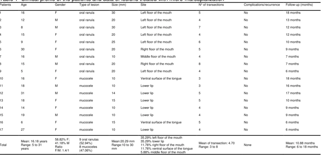
The clinical profile of the patients and data concerning the lesions treated with
the upgraded micro-marsupialization technique are presented in table 1.
Patient ages ranged from 5 years to 31 years of age (mean of 16.18 ±
7.56). The study sample consisted of 10 females (58.82%) and 7 males
(41.18%). Nine (52.94%) patients presented a clinical diagnosis of oral ranula,
while 8 (47.06%) presented a diagnosis of mucocele. Six patients (35.29%) with
oral ranula presented the lesion on the left side of the floor of mouth, 2 (11.76%)
on the right side, and 1 (5.88%) in the middle of the floor of the mouth. Six
patients (35.29%) with mucocele presented lesions on the lower lip and 2
(11.76%) on the ventral surface of the tongue. The size of the lesions ranged
from 10 to 30mm (mean of 20.29mm). Lesion transections ranged from 3 to 8
sutures, with a mean of 4.70. The distance between each transection was
3.6mm (Figure 1E). The patients classified the intensity of pain as no pain in
47.05% of the cases, mild in 11.76%, moderate in 11.76%, and severe in
29.41% of cases. No patient reported any unbearable pain. Moreover, no
patients reported any sign of edemas, and no infections could be observed. All
patients presented a clinical healing of oral ranulas and mucoceles within the
ranulas or mucoceles could be identified in any patient (Figures 1F, 2C, and
2F). New ducts of minor salivary glands created in the places of sutures were
visualized in all cases (Figure 1F).
Discussion
The upgrading of the the micro-marsupialization technique was performed
based on the pathophysiology of the lesions. The drainage of total mucus
eliminates the local foreign body and inflammatory reaction9. In addition, the
drainage of mucus provides the patient with greater comfort and decreases the
possibility of retreatment and recurrence. The increase in suture thickness and
the mechanical enlargement of the pathways facilitate the permanent
epithelization of the mucosa around the suture, in turn leading to several new
path tract formations. Moreover, Sandrini et al.8 established the number of
lesion transections with the use of as many sutures as possible. The present
study verified that the average distance between sutures was 3.6mm. In
additional, the maintenance of sutures for 30 days is related to the physiological
characteristics of the oral mucosa, considering that the occurrence of
reepithelialization is quite likely10.
English-language literature describes a number of techniques that can
used to manage oral ranulas and mucoceles. Some authors advocate more
invasive techniques, mainly associated with ranulas, such as the removal of the
sublingual gland5,11,12. However, other authors prefer to follow a more
conservative manner in which to treat oral ranulas,2,6,13. In addition, pediatric
period of 5 months. If the lesion does not recede within this period, surgical
management is recommended4,11. This time frame was maintained for patients
3, 5, and 9 (table 1); however, the lesions did not recede, and the
micro-marsupialization technique was performed.
The excision of the sublingual gland showed a satisfactory outcome with
relatively few recurrences5. However, this technique can injure the lingual nerve
and the submandibular gland duct, and therefore should only be used to treat
plunging ranulas and recurrence cases2. The excision of the ranula and classic
marsupialization proved to lead to unacceptable results with high recurrence
rates, 57.69% and 66.67%, respectively5. Mucoceles is classically treated by
removing the lesion and minor salivary glands associated with low recurrence
rates2; however, this procedure is more invasive than micro-marsupialization,
mainly in children. The present study demonstrated a satisfactory outcome
when applying the upgraded micro-marsupialization technique, with no surgical
complications, no postoperative infection, and no recurrence rates in the
management of oral ranulas and selected mucoceles. Additionally, the current
study represents the largest case series of oral ranulas and mucoceles
managed by means of the micro-marsupialization technique.
Postoperative follow-up proved to be uneventful, with no edemas and
with either no pain or mild pain reported by the majority of the patients
(58.81%). A mild to moderate inflammation of the mucosa could be observed
after the withdrawal of the sutures due to plaque accumulation, which
disappeared completely within 7 days after the removal of the sutures. Bacterial
observed in any of the 17 patients. Additionally, new ducts of minor salivary
glands were created in the places of sutures.
The micro-marsupialization technique can be executed simply, with
topical anesthesia, rendering it less traumatic and more easily tolerated by the
patients, thus representing a good alternative for the management of oral
ranulas and mucoceles7,8. The micro-marsupialization technique is not
recommended as treatment for mucoceles located in the palate and buccal
mucosa, given that benign or malignant salivary gland tumors are more
common in these areas15, which can lead to a clinical misdiagnosis. In a recent
review published by Harrison3, all cases reported in the English-language
literature that were treated with the micro-marsupialization technique proved to
be successful. Delbem et al.7, managed 14 cases, with only 2 (14.28%) cases
of retreatment, whereas Sandrini et al.8 managed 7 cases with 3 (42.85%)
cases retreatment. The present study presented healing in all cases, with no
retreatment and no recurrence.
Randomized clinical trials may be performed to address more information
concerning the management of oral ranulas and mucoceles by comparing the
main techniques described in the literature with micro-marsupialization. In
addition, resorbable sutures, including synthetic materials such as Polyglactin
910, should be investigated as an alternative to silk sutures, which can easily
sustain more bacteria, in turn causing infection14. However, infection was not
observed in the present case series. Moreover, resorbable sutures serve to
pediatric patients. However, the cost of resorbable sutures may be higher than
that of silk sutures.
The micro-marsupialization technique proved to be a simple, low cost,
relatively non-invasive, painless, and effective, with no need for retreatment or
recurrence in the treatment of mucus extravasation or retention phenomena. It
can therefore be concluded that micro-marsupialization can be recommended
primarily for the treatment of oral ranulas and selected mucoceles.
Acknowledgments
We would like to thank the Conselho Nacional de Desenvolvimento Científico e
Tecnológico (CNPq, #309209/2010-2). RA Mesquita is research fellow of
CNPq.
Funding: Conselho Nacional de Desenvolvimento Científico e Tecnológico
(CNPq, #309209/2010-2).
Competing interests: None declared
Ethical Approval: Approved by the Ethics Committee of Universidade Federal
References
1- Chi AC, Lambert PR 3rd, Richardson MS, Neville BW. Oral Mucoceles A
clinicopathologic review of 1,824 cases, including unusual variants. J Oral
Maxillofac Surg 2011: 69: 1086-1093.
2- Baurmash HD. Mucoceles and ranulas. J Oral Maxillofac Surg 2003: 61:
369-378.
3- Harrison JD. Modern management and pathophysiology of ranula:
literature review. Head Neck 2010: 32 1310-1320.
4- Haberal I, Göçmen H, Samim E. Surgical management of pediatric ranula.
Int J Pediatr Otorhinolaryngol 2004: 68: 161-163.
5- Zhao YF, Jia Y, Chen XM, Zhang WF. Clinical review of 580 ranulas. Oral
Surg Oral Med Oral Pathol Oral Radiol Endod 2004: 98: 281-287.
6- Morton RP, Bartley JR. Simple sublingual ranulas: pathogenesis and
management. J Otolaryngol 1995: 24: 253-254.
7- Delbem AC, Cunha RF, Vieira AE, Ribeiro LL. Treatment of mucus
retention phenomena in children by the micro-marsupialization technique:
case reports. Pediatr Dent 2000: 22: 155-158.
8- Sandrini FA, Sant’ana-Filho M, Rados PV. Ranula management:
suggested modifications in the micro-marsupialization technique. J Oral
Maxillofac Surg 2007: 65: 1436-1438.
9- Serhan CN, Brain SD, Buckley CD, Gilroy DW, Haslett C, O'Neill LA, et al.
Resolution of inflammation: state of the art, definitions and terms. FASEB
10- Miloro M, Ghali GE, Larsen PE, Waite PD. Peterson´s principles of oral
and maxillofacial surgery. In: Shetty V, Bertolami CN, ed.: Wound healing.
Hamilton-London: BC Decker Inc, 2004: 3.
11- Pandit RT, Park AH. Management of pediatric ranula. Otolaryngol Head
Neck Surg 2002: 127: 115-118.
12- Yoshimura Y, Obara S, Kondoh T, Naitoh S. A comparison of three
methods used for treatment of ranula. J Oral Maxillofac Surg 1995:53:
280-283.
13- Baurmash HD. Marsupialization for treatment of oral ranula: a second look
at the procedure. J Oral Maxillofac Surg 1992: 50: 1274-1279.
14- Selvig KA, Biagiotti GR, Leknes KN, Wikesjö UM. Oral tissue reactions to
suture materials. Int J Periodontics Restorative Dent 1998: 18: 474-487.
15- Waldron CA, el-Mofty SK, Gnepp DR. Tumors of the intraoral minor
salivary glands: a demographic and histologic study of 426 cases. Oral
Tables
Table 1- Clinical profile of the patients and data of lesions treated with micro-marsupialization
Patients Age Gender Type of lesion Size (mm) Site N° of transections Complications/recurrence Follow-up (months)
1 16 F oral ranula 30 Left floor of the mouth 5 No 18 months
2 12 M oral ranula 20 Left floor of the mouth 4 No 13 months
3 8 M oral ranula 30 Left floor of the mouth 7 No 12 months
4 15 F oral ranula 20 Left floor of the mouth 4 No 12 months
5 9 F oral ranula 25 Left floor of the mouth 6 No 10 months
6 30 F oral ranula 20 Right floor of the mouth 5 No 9 months
7 16 M oral ranula 10 Middle floor of the mouth 4 No 7 months
8 15 M oral ranula 20 Right floor of the mouth 8 No 7 months
9 5 F oral ranula 20 Left floor of the mouth 4 No 6 months
10 16 F mucocele 10 Ventral surface of the tongue 3 No 18 months
11 18 M mucocele 10 Lower lip 3 No 16 months
12 31 M mucocele 14 Lower lip 5 No 17 months
13 18 F mucocele 15 Lower lip 5 No 10 months
14 14 F mucocele 10 Lower lip 4 No 9 months
15 19 M mucocele 10 Lower lip 4 No 9 months
16 6 F mucocele 15 Ventral surface of the tongue 5 No 6 months
17 27 F mucocele 10 Lower lip 4 No 6 months
Total Mean: 16.18 years Range: 5 to 31 years
58.82% F; 41.18% M Ratio F/M: 1.4/1
9 oral ranulas (52.94%) 8 mucoceles (47.06%)
Mean:20.29 mm Range:10 to 30 mm
35.29% left floor of the mouth 35.29% lower lip
11.76% right floor of the mouth 11.76% ventral surface of the tongue 5.88% middle floor of the mouth
Mean of transection: 4.70
Range: 3 to 8 None Mean: 10.88 months Range: 6 to 18 months
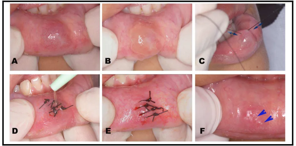
Figure 1 - Patient #14. Clinical aspect of selected mucocele and upgraded micro-marsupialization technique, step by step. A – Sessil nodular lesion with a smooth
surface, a bluish color, 10mm in size on the right lower lip; B – Topical anesthesia; C – To and fro movement (yellow arrows) with silk suture; D – Press and aspiration of
all mucus from inside the lesion; E – Decrease in the size of the lesion after suction, and transections using a 3.0 silk suture at a distance of 3.6mm between each other;
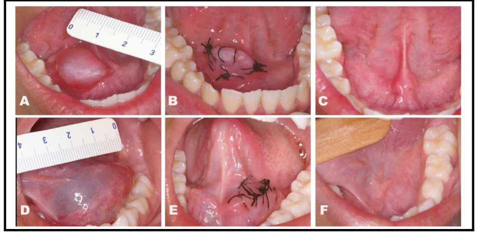
Figure 2 - Patients #3 and #8. Clinical aspects of oral ranulas and upgraded micro-marsupialization technique. A and D – Sessil nodular lesions with smooth surface,
mucosa-like color (A) and bluish color (D), 20mm in size (A), and 30mm (D) on the right floor of the mouth (A) and on the left floor of the mouth (D); B and E – Decrease
in the size of the lesion after suction, and transections using a 3.0 silk suture at a distance of 4mm between each other; C and F – Clinical healing and no signs of
De:Lasers in Medical Science [email protected]
Para: Márcio Bruno Amaral [email protected]
Enviadas: Quinta-feira, 29 de Setembro de 2011 2:03 Assunto: Acknowledgement of Receipt
Dear Prof. Márcio Bruno Amaral:
Thank you for submitting your manuscript, "A randomized double-blind clinical
trial of low-level laser effect after micro-marsupialization technique in treating
ranulas and mucoceles: a preliminary results", to Lasers in Medical Science.
During the review process, you can keep track of the status of your manuscript
by accessing the following web site:
http://lims.edmgr.com/
Your username is: Márcio Amaral
With kind regards,
The Editorial Office
Title: A randomized double-blind clinical trial of low-level laser effect after
micro-marsupialization technique in treating ranulas and mucoceles: a preliminary
results
Abstract
The aim of this preliminary study was to investigate the low level laser therapy
(LLLT) on alleviate pain caused by the micro-marsupialization when treating
oral ranulas and selected mucoceles. LLLT can enhance wound healing and
alleviate pain on injured tissues, and is used on treating of oral lesions.
However, there are not reports on the LLLT management of oral ranulas and
selected mucoceles. Six patients with oral ranulas or selected mucoceles
underwent micro-marsupialization treatment (control group), while eleven
underwent micro-marsupialization treatment associated with LLLT (LLLT
group). The LLLT group were irradiated with a 660nm continuous wave from a
InGaAsP diode laser, at 100 mW, with a spot size on the tissue surface of
0.0283 cm2 (irradiance=3.53 w/cm2). Each needle transaction area was
irradiated in contact mode for 40 s per point (4.0J at 141J/cm2 per point).
Irradiation was carried out immediately following micro-marsupialization
treatment, as well as at 24, 48, and 72 hours post-micro-marsupialization. The
relationship between the groups and pain was assessed using the
Mann-Whitney test. All treated oral ranulas and selected mucoceles presented clinical
healing. No evidence of recurrence could be identified during a mean
10.88-month follow-up period. It could be observed that the LLLT group reported less
pain than did control group. InGaAsP diode laser within the parameters tested
post-micro-marsupialization of oral ranulas and selected mucoceles. However, it seems not
affect the healing of the lesions after the treatment.
Key words: LLLT, micro-marsupialization, ranula, mucocele
Introduction
Ranulas and mucoceles are saliva extravasation phenomena which can affect
both the major and minor salivary glands [1]. These lesions are mainly located
on the lower lip, ventral surface of the tongue or floor of the mouth and
presented with a smooth surface, a thin mucosa of either bluish or mucosa-like
color, a sessile base, and a flaccid consistency [1]. Several conducts reported
to treat ranulas and mucoceles include the excision of the lesion associated
with the affected salivary glands, laser ablation, marsupialization treatment for
ranulas, and micro-marsupialization [2].
Micro-marsupialization uses silk sutures in the dome of a cyst to permit
new epithelialized drainage pathways [3, 4]. This technique is simpler, less
traumatic, and well-tolerated by the patients [5]. Micro-marsupialization can be
recommended for patients with a primarily clinical diagnosis of oral ranulas or
mucoceles, especially in children. Prior studies have demonstrated the success
of the micro-marsupialization technique in treating oral ranulas or mucoceles [4,
5]. By contrast, similar studies have reported nonhealing cases, which in turn
lead to a necessary repetition of the technique.
In this light, the use of low level laser therapy (LLLT) can aid in repairing
the mucosa and controlling the pain. Studies in the literature have demonstrated
post-surgical pain [7-9]. LLLT is commonly recommended to heal wounds and to
alleviate pain due to its well-known biological effects found in the interaction
between laser energy and injured tissues [7, 8].
Considering the efficiency of micro-marsupialization in treating oral
ranulas or mucoceles and the acceleration of the repair process and the
anti-inflammatory/analgesic properties that LLLT provides, the present preliminary
study aimed to investigate the ability of LLLT to alleviate the pain caused by the
micro-marsupialization in treating oral ranulas or selected mucoceles, as well as
to verify the possibility of reducing the incidence of nonhealing cases.
Material and Methods Patients
This study was approved by the Ethics Committee of Universidade Federal de
Minas Gerais (#0311.0.203.000-10) and informed written consent forms were
obtained from all participants. Seventeen patients were recruited from the Oral
Medicine Clinic, School of Dentistry, Universidade Federal de Minas Gerais,
Belo Horizonte, Brazil, from August 2009 to December 2010. The selection of
the cases included patients who presented mucoceles or oral ranulas located
on the lower lip, ventral surface of the tongue or floor of the mouth with a
smooth surface, a thin mucosa of either bluish or mucosa-like color, a sessile
base, and a flaccid consistency (Figures 1a and 1d). Only lesions of at least
10mm were included. Patients currently using anti-inflammatory or analgesic
medications were excluded. Patients were randomly assigned to one of two
groups: control and LLLT. The control group (n= 9) underwent
micro-marsupialization associated with LLLT. Three patients from the control group
removed the sutures after having undergone micro-marsupialization treatment
and were therefore excluded from the study (patient’s lost). Micro
-marsupialization was performed by one calibrated, trained oral surgeon (MBA).
An oral medicine professional performed the LLLT (RAM). Clinical evaluation
during each visit was performed by MBA, who was blinded to the LLLT status.
All patients (n=17) underwent the micro-marsupialization with design of LLLT
treatment; however, in the control group, the laser apparatus was not turn on.
Micro-marsupialization technique
Following topical anesthesia (Eutectic Mixture of Lidocaine and Prilocaine 5%,
AstraZeneca do Brasil LTDA, São Paulo, Brazil or Benzocaine 20%, DFL
Indústria e Comércio S.A., Rio de Janeiro, Brazil), the micro-marsupialization
technique was carried out on all patients. This technique was performed using
3.0 silk sutures by introducing a round cross-section needle into the dome of the
cyst. Mechanical enlargement of the pathways was also performed using the
suture line. During the procedure, all saliva was removed from within the lesion
by conventional suction. The sutures were placed along the lesions at a 3 to 5
millimeter distance from each other (Figures 1b and 1e). After having completed
the procedure, the patients were examined weekly for 30 days, at which time
the sutures were removed. In addition, after having undergone
micro-marsupialization treatment, patients were subsequently recalled every 3 months
for follow-up treatment (Figures 1c and 1f).
Laser irradiation of the LLLT group was performed using a 660nm continuous
wave InGaAsP (indium-gallium-arsenide-phosphorous) diode laser (Arima
Lasers Corporation, Taoyuan County, Taiwan; Whitening Lase II, DMC LTDA,
São Carlos, Brazil) at a potency of 100mW. The laser potency was measured
with a power meter (Coherent Fieldmaster; Coherent, Palo Alto, CA), resulting
in 100±5mW. Energy was delivered by optical fiber to the treatment site,
producing a resulting spot size on the tissue surface of 0.0283 cm2
(irradiance=3.53w/cm2). Each entrance or output of the needle transaction was
irradiated in a contact mode of 40 s per point (4.0J at 141J/cm2). Irradiation was
carried out immediately following micro-marsupialization, and again at 24, 48,
and 72 hrs post-treatment. Both the patients and the medical oral professional
used specific protective glasses during the irradiations.
Evaluation of clinical healing and post-micro-marsupialization pain
Clinical healing was achieved due to the postoperative controls applied by MBA,
which was determined when the total clinical disappearance of the lesions could
be observed. Nonhealing cases were defined as those in which clinical
permanence of the lesions after micro-marsupialization treatment could be
identified. The pain assessment instrument used was a self-reported numerical
rating scale [10] (NRS) for patients of more than 7 years of age. The
self-reporting of pain involves asking the patients to rate, subjectively, the intensity
of the pain they feel, from 0 to 10 points (an 11 point scale), with the
understanding that 0 represents no pain and 10 represents extremely intense
pain (unbearable pain). A visual analog scale of patients’ faces [11] (VASF) was
about pain 1 week after having undergone micro-marsupialization. Regarding
the edema, patients were asked, in terms which could be easily understood, if
the edema was present or not.
Post-micro-marsupialization care
Patients were instructed regarding 1) oral hygiene; 2) take care with the sutures
during the healing period; 3) the observance of pain and edema; and 4) no use
of analgesics.
Statistical analysis
The Statistical Package for the Social Sciences (SPSS Inc., Chigago, IL)
version 17.0 for Windows was used to perform the statistical analysis. A
descriptive analysis of each variable was carried out. The relationship between
the groups with pain parameters was assessed for statistical significance by the
Mann-Whitney test. A P value < 0.05 was considered statistically significant.
Results
The patient sample included 10 females (58.82%) and 7 males (41.18%), with
an age ranging from 5 to 31 years of age (mean of 16.18 ± 7.56) (Table 1). Nine
(52.94%) patients with a diagnosis of oral ranula and 8 (47.06%) with diagnose
of mucocele were enrolled in the study. Six patients (35.29%) with oral ranulas
presented the lesion on the left side of the floor of the mouth, 2 (11.76%) on the
right side, and 1 (5.88%) in the middle. Six patients (35.29%) with selected
mucoceles presented lesions on the lower lip and 2 (11.76%) on the ventral
(mean 20.29mm). The mean number of transactions to treat the lesions was
4.70, using from 3 to 8 sutures. The average energy per patient was 37.64J. In
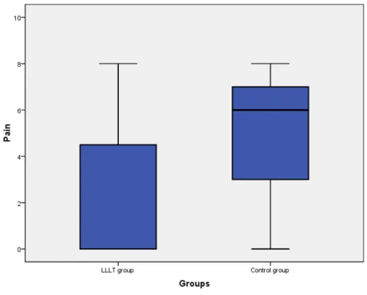
the control group, five (83.33%) patients reported pain, while in the LLLT group,
four (36.36%) reported pain. Moreover, patients from the LLLT group tended to
report a lesser intensity of pain than did those from the control group (p=0.136;
Figure 2). Patient’s lost, two females of 5 years of age and one male of 6 years
of age, presented selected mucoceles on the lower lip. No patients reported
edemas, and no infections could be observed in any patient. All patients
presented a clinical healing of the lesions within 30 days. Nonhealing cases
were not present in this study. Recurrence was also not observed within a mean
10.88-month follow-up period.
Discussion
The current study is the first of its kind to verify the influence of LLLT in
alleviating pain caused by the micro-marsupialization technique, as well as in
healing oral ranulas and selected mucoceles in a double-blind randomized
clinical trial. The main observations included: 1) the micro-marsupialization
treatment proved to be clinically effective regarding oral ranulas and selected
mucoceles and 2) the LLLT shows a tendency to reduce the
post-micro-marsupialization pain.
The current results demonstrate that each of 17 cases of oral ranulas
and selected mucoceles submitted to the micro-marsupialization technique
presented a total clinical healing with no signs of recurrence or infection in
follow-up examinations. These results are in accordance with previous studies
Ranulas and mucoceles are treated by excision of the lesion, associated
or not with removal of the affected gland [12, 13]; marsupialization [12, 14];
cryosurgery [15]; or laser surgery [2]. However, these techniques may cause 1)
injuries to the lingual nerve or submandibular duct [1]; 2) unacceptable
recurrence rates [12]; or 3) may bring additional costs to the treatment.
Micro-marsupialization treatment represents a good alternative to surgical excisions or
cryosurgery, considering that the present technique is simple to execute,
presents a low degree of invasiveness, and is generally well-tolerated by
patients. Recurrence rates of oral ranulas have proven to be variable (0% to
45%) and depend on the specific type of treatment [16]. The
micro-marsupialization technique has demonstrated no recurrence rate [3-5].
Nonhealing cases with micro-marsupialization, as reported by Delbem et al. [5]
(2000) and Sandrini et al. [4] (2007), lead to the repetition of the same
technique, which proved to be effective. In this manner, although the use of the
LLLT association presented no significant difference in the improvement of the
clinical healing of the lesions, the current study did show that no nonhealing
cases occurred. In addition, though this technique is well-tolerated by patients
[5], the LLLT association appears to be quite beneficial. In addition, LLLT has
proven to be effective in managing damaged oral tissue as well as in controlling
pain [9]. The most popularly described treatment benefit of LLLT is the healing
of wounds [17]. The effects of LLLT, when compared to the untreated areas,
showed evidence of intracytoplasmatically accumulated collagen fibrils and
electrondense vesicles within laser irradiated fibroblasts [18, 19]. In addition, the
cell reproduction, increased prostaglandin levels, and increased the
microcirculation after laser stimulation, which accelerated wound healing [17].
Concerning the controlling of pain, the use of LLLT may have significant
neuropharmacologic effects on the synthesis, release, and metabolism of a
range of neurochemicals, including serotonin and acetylcholine at central levels,
and histamine and prostaglandin at peripheral levels [20]. The effects of LLLT in
alleviating pain can also be seen through the enhanced synthesis of
endorphins, decreased c-fiber activity, bradykinin, and altered pain thresholds
[21, 22]. Other LLLT action mechanisms have suggested that photons are made
up of wave lengths of a visible or nearly infrared spectrum, which are absorbed
by chromophore enzymes, such as cytochrome c oxidase. Alterations in the
activity of the cytochrome c oxidase in turn increase the production of
adenosine triphosphate (ATP), which leads to the normalization of the cellular
function [23, 24].
Clinically, LLLT has been used successfully in the treatment of pain
associated with oral lesions [25, 26]. Biostimulation by LLLT depends on
laser-irradiation parameters, such as wavelength, laser-output power, and energy
density [27]. Ribeiro et al. [28] (2011) employed InGalAsP diode laser to aid in
alleviating the pain caused by the cryosurgical treatment of oral leukoplakia. In
a similar manner, the current study demonstrated a tendency toward the
decrease of pain associated with micro-marsupialization. Differently of the
Ribeiro et al. [28] (2011), in the current study it was used an output powers of
the 100mW. This modification was in accordance to the studies that had
demonstrated significantly great amounts of collagen and angiogenesis [29-31]
However, it is important the development of the studies that correlate pain and
major output powers.
Conclusion
InGaAsP diode laser within the parameters tested represents a good alternative
to reduce the pain caused by the post-micro-marsupialization of oral ranulas
and selected mucoceles. However, it seems not affect the healing of the lesions
after the treatment.
Acknowledgments and Author Disclosure Statement
We would like to thank the Conselho Nacional de Desenvolvimento Científico e
Tecnológico (CNPq, #309209/2010-2). The authors wish to thank DMC LTDA
(São Carlos, Brazil) for their supply of the InGaAsP diode laser 660nm and
NUPEN for their scientific assistance. RA Mesquita is a research fellow of
References
1. Baurmash HD (2003) Mucoceles and ranulas. J Oral Maxillofac Surg
61:369-378
2. Lai JB, Poon CY (2009) Treatment of ranula using carbon dioxide laser –
case series report. Int J Oral Maxillofac Surg 38:1107-1011
3. Morton RP, Bartley JR (1995) Simple sublingual ranulas: pathogenesis
and management. J Otolaryngol 24:253-254
4. Sandrini FA, Sant’ana-Filho M, Rados PV (2007) Ranula management:
suggested modifications in the micro-marsupialization technique. J Oral
Maxillofac Surg 65:1436-1438
5. Delbem AC, Cunha RF, Vieira AE, Ribeiro LL (2000) Treatment of mucus
retention phenomena in children by the micro-marsupialization
technique: case reports. Pediatr Dent 22:155-158
6. Reddy GK (2004) Photobiological basis and clinical role of low-intensity
lasers in biology and medicine. J Clin Laser Med Surg 22:141–150
7. Damante AC, Greghi SL, Santana AC, Passanezi E, Taga R (2004)
Histomorphometric study of the healing of human oral mucosa after
gingivoplasty and low-level laser therapy. Lasers Surg Med 35:377–384
8. Damante AC, Greghi SL, Santana AC, Passanezi E (2004) Clinical
evaluation of the effects of low intensity laser (Gaalas) on wound healing
after gingivoplasty in humans. J Appl Oral Sci 12:133–136
9. Amorim JCF, Souza GR, Silveira LB, Prates RA, Pinotti M, Ribeiro MS
(2006) Clinical study of the gingiva healing after gingivectomy and
10. Caraceni A, Cherny N, Fainsinger R, et al (2002) Pain measurement
tools and methods in clinical research in palliative care:
recommendations of an expert working group of the European
Association of Palliative Care. J Pain Symptom Manage 23:239–255
11. Barrêto EPR, Ferreira EF, Pordeus IA (2004) Evaluation of toothache
severity in children using a visual analogue scale of faces. Pediatr Dent
26 485-491
12. Yoshimura Y, Obara S, Kondoh T, Naitoh S (1995) A comparison of
three methods used for treatment of ranula. J Oral Maxillofac Surg
53:280-283
13. Zhao YF, Jia Y, Chen XM, Zhang WF (2004) Clinical review of 580
ranulas. Oral Surg Oral Med Oral Pathol Oral Radiol Endod 98:281-287
14. Baurmash HD (1992) Marsupialization for treatment of oral ranula: a
second look at the procedure. J Oral Maxillofac Surg 50:1274-1279
15. Toida M, Ishimaru JL, Hobo N (1993) A simple cryosurgical method for
treatment of oral mucous cysts. Int J Oral Maxillofac Surg 22:353-355
16. Harrison JD (2010) Modern management and pathophysiology of ranula:
literature review. Head Neck 32:1310-1320
17. Sun G, Tunér J (2004) Low-level laser therapy in dentistry. Dent Clin N
Am 48:1061-1076
18. Mester E, Spiry T, Szende B, Tota JG (1971) Effect of laser rays on
wound healing. Am J Surg 122:532-535
19. Kreisler M, Christoffers AB, Willerstausen B, d’Hoedt B (2003) Effect of