DOI: 10.1590/0004-282X20160144
ARTICLE
Performance of adjuvant treatment correlates
with survival in reoperated glioblastomas
Uso de tratamento adjuvante é relacionado ao aumento da sobrevida em pacientes com
glioblastoma submetidos à reoperação
Willey Gonçalves Zanovello1, Suzana M. F. Malheiros1, João Norberto Stavale2, Orestes P. Lanzoni1, Miguel M. Canteras1, Adrialdo J. Santos1, Felipe Slaviero1, Bruno Fernandes1, Sergio Cavalheiro1, Manoel A. de Paiva Neto1
Glioblastoma (GBM) is the most common primary tumor affecting the central nervous system and represents more than 50% of glial tumors1. In spite of the advances in
microsurgery to accomplish optimized resections (exten-sive, without worsening of the neurologic function), fol-lowed by standard treatment with concomitant radio-therapy (RT) and chemoradio-therapy (CT) and maintenance therapy with temozolomide (TMZ)2, the patients’ survival
remains rather limited, with small survival gains obtained in recent years3,4,5.
he main factors associated with patient survival are age and functional status before surgery6,7; the extent of resection7,8,
adju-vant treatment7,9 and methylation of the O6-methylguanine-DNA
methyltransferase (MGMT) promoter10, which also seem to be
associated with increased survival. he role of surgical treatment for recurrence or regrowth of GBM has not yet been fully estab-lished. While some authors recommend reoperation11,12,13, others
did not ind increased survival with this strategy14,15.
Despite the large number of studies published in recent years demonstrating the beneits of reoperation in patients with recurrent GBM, we were not able to locate any simi-lar study conducted in Brazil. hus, we assessed a group of patients with recurrent GBM subjected to reoperation at a Brazilian public healthcare service aiming at describing the clinical characteristics of patients and identifying the ones who might beneit from this treatment modality.
1Universidade Federal de São Paulo, Departamento de Neurocirurgia, São Paulo SP, Brasil; 2Universidade Federal de São Paulo, Departamento de Patologia, São Paulo SP, Brasil.
Correspondence: Manoel A. Paiva Neto; Universidade Federal de São Paulo, Departamento de Neurocirurgia; Rua Leandro Dupret, 847; 04025-013 São Paulo SP, Brasil; E-mail: [email protected]
Conflict of interest: There is no conlict of interest to declare.
Received 04 February 2016; Received in inal form 29 May 2016; Accepted 09 August 2016.
ABSTRACT
Objective: To analyze cases of recurrent glioblastoma subjected to reoperation at a Brazilian public healthcare service. Methods: A total of 39 patients subjected to reoperation for recurrent glioblastoma at the Department of Neurosurgery, São Paulo Hospital, Federal University of São Paulo, from January 2000 to December 2013 were retrospectively analyzed. Results: The median overall survival was 20 months (95% conidence interval – CI = 14.9–25.2), and the median survival after reoperation was 9.1 months (95%CI: 2.8–15.4). The performance of adjuvant treatment after the irst operation was the single factor associated with overall survival on multivariate analysis (relative risk – RR = 0.3; 95%CI = 0.2–0.7); p = 0.005). Conclusion: The length of survival of patients subjected to reoperation for glioblastoma at a Brazilian public healthcare service was similar to the length reported in the literature. Reoperation should be considered as a therapeutic option for selected patients.
Keywords: glioblastoma; reoperation; general surgery; survival.
RESUMO
Objetivo: Analisar o papel da reoperação em pacientes com glioblastoma recidivado em um serviço público no Brasil. Métodos: Foram analisados retrospectivamente 39 pacientes submetidos à reoperação por recorrência de glioblastoma no Departamento de Neurocirurgia da Universidade Federal de São Paulo, no período de janeiro de 2000 até dezembro de 2013. Resultados: A sobrevida global mediana foi de 20 meses (IC 95% = 14.9–25.2), e a sobrevida mediana após a reoperação foi de 9.1 meses (IC 95% = 2.8–15.4). A realização de tratamento adjuvante após a primeira cirurgia foi o único fator associado com a sobrevida global numa análise multivariada (RR = 0.3; IC 95% = 0.2–0.7; p = 0.005). Conclusão: A sobrevida dos pacientes submetidos à reoperação em um serviço público no Brasil é semelhante à reportada pela literatura. A reoperação deve ser considerada como uma opção terapêutica em pacientes selecionados.
METHODS
We performed a retrospective and descriptive analysis of patients followed up at the Neurosurgery Section, who were diagnosed with GBM (World Health Organization grade IV) from January 2000 to December 2013 and subjected to reoperation due to tumor recurrence or regrowth. he patients were followed up until February 2015.
he data were collected from the patients’ medi-cal records and the database of the Neuro-oncology unit, Discipline of Neurosurgery, UNIFESP, having been treated by the same neuro-oncologist in a linear manner (SMFM).
he study population included patients older than 18 years of age, subjected to reoperation due to recurrence or progression of tumors diagnosed as GBM upon histopatho-logical examination at the irst operation. Patients subjected to only one biopsy in one of the surgeries, those who under-went reoperation due to complications, and cases with miss-ing data were excluded from the present study.
he indication for reoperation was established in periodic multidisciplinary meetings that included neurosurgeons, neuro-oncologists, neurologists and neuroradiologists. Patients with Karnofsky Performance Status (KPS) scores equal to or higher than 70 and tumors likely to be entirely resected and located in non-eloquent brain areas were con-sidered as candidates for reoperation.
he variables analyzed were age, gender, initial symp-tom, initial KPS score, duration of symptoms, tumor local-ization, tumor laterality, extent of resection in both surgical procedures, localization of the recurrent tumor, time elapsed between the operations, adjuvant treatment after primary surgery and reoperation, delay in the onset RT, time of relapse relative to reoperation, overall survival (OS), survival after reoperation (SARp) and survival after relapse (SAR).
he extent of resection was established based on com-puted tomography and/or magnetic resonance imaging (MRI) of the brain, both with contrast and performed within the irst 72 hours after surgery. Resection was considered to be total (TR) when non-linear contrast enhancement was not found in the control test and was considered partial (PR) when enhancement was found (presence of residual lesion).
Recurrence was deined as the appearance of a new lesion in the control test of any patient subjected to TR, and regrowth as more than a 25% increase in residual lesions after the irst operation. Spectroscopy and perfusion MRI were used in the diferential diagnosis of pseudo-progression as per our service routine. Recurrence was considered to be local when it appeared in the same brain lobe as the primary tumor and as distant when it appeared in any other lobe or the contralateral brain hemisphere. Adjuvant treatment was deined as performance of RT and/or CT. Radiotherapy delay was deined as any interval longer than six weeks between the irst operation and the onset of RT.
Overall survival was deined as the time elapsed from the date of the irst operation to death and SARp as the time elapsed from the date of reoperation to death.
he study was approved by the research ethics committee of UNIFESP, ruling no. 1,093,864.
Statistical analysis
he categorical variables are expressed as percentages and the continuous ones as medians (ranges), as beitting. he event cumu-lative curves were estimated using the Kaplan-Meier method and were compared via the log-rank test. he patients lost to follow-up were censored at their last follow-up visit, and patients who com-pleted the study were censored in February 2015.
Univariate and multivariate Cox regression was performed as needed. All variables with p < 0.1 on multivariate analysis or con-sidered as having conirmed clinical relevance were included in the multivariate model. Multicollinearity problems were solved before including the variables in the model. he possible inter-actions between the variables that were kept in the model were tested. he assumption of risk proportionality was tested using the Schoenield residuals. he signiicance level was set as p < 0.05.
Characteristics of the patients
A total of 167 patients were subjected to the same sur-gical procedure. he data corresponding to 48 patients sub-jected to reoperation for tumor recurrence or regrowth were initially included. However, nine patients had to be excluded because biopsy had only been performed in the irst opera-tion in six cases and the second biopsy in the other three. hus, the sample included a total of 39 patients.
Table 1 describes the demographic and clinical character-istics of the included patients. heir median age was 49 years old (20 to 79 years old); 26 patients were male (66%). Headache was the most common initial symptom of disease, occurring in 23 patients (59%); followed by seizures, 11 cases (28%); and motor deicit, behavioral changes, dizziness and vomiting, one case each (3%). he median preoperative KPS score was 80, and the postoperative KPS score of the irst surgery was 90 (assessed at the irst follow-up visit). he median duration of symptoms from the date of the appearance of the irst symptom to the irst operation was eight weeks (ranging from one to 103 weeks).
RESULTS
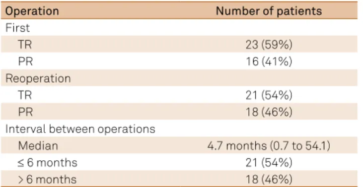
Table 2 describes the extent of resection in each operation and the interval of time between them. he irst operation was classiied as TR in 23 cases (59%) and as PR in 16 (41%). he second operation was classiied as TR in 21 cases (54%) and as PR in 18 (46%).
Five patients were subjected to a third operation, with three cases of TR and two of PR.
he morbidity rate corresponding to the irst operation was 2.6%, with no cases of perioperative death. he morbidity rate corresponding to reoperation was 20.5% (eight patients): three cases of surgical wound infection that required surgical cleansing, one of which progressed into osteomyelitis; one patient exhibited transient hemihypesthesia; and four exhibited permanent neu-rological aggravation, hemiplegia and aphasia in two cases, sleep-iness and ataxia in one, and behavioral disorders in one.
Table 3 describes the data relative to adjuvant treatment. A total of 33 patients (83%) recieved CT, 18 (55%) using
carmustine (bis-chloroethylnitrosourea, BCNU), 14 (42%) using TMZ and one (3%) using BCNU/procarbazine, lomus-tine, and vincristine (PCV). It should be noted that TMZ became available in our service starting May 2009; from that moment onwards, only one out of 15 patients used BCNU due to social reasons, while all of the others were treated with TMZ. Among the 33 patients who used TMZ, 11 (33%) received CT after reoperation only. he median time to onset of CT was 16 weeks.
A total of 35 patients (88%) were subjected to RT, with a median dose of 60 Gy. he median time to onset of RT was eight weeks. Onset of RT was delayed beyond six weeks in 29 cases (74%).
Recurrence occurred before the onset of or in the course of RT in 18 cases (46%) and after the end of RT in 21 cases (54%). It should be noted that adjuvant treatment after reopera-tion was not uniform. he patients who had received stan-dard adjuvant treatment after the irst operation (RT followed by CT with BCNU until 2009 and with TMZ afterwards) were indicated for additional CT and/or RT (maintenance of TMZ for a protracted regimen, bevacizumab or re-irradiation). In turn, the patients who had not been given adjuvant ther-apy after the irst operation were indicated for the standard regimen after reoperation.
he median survival after reoperation was 9.1 months (95%CI = 2.8–15.4), and the median OS was 20 months (95%CI = 14.9–25.2).
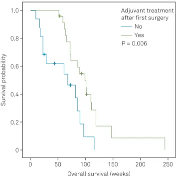
On univariate analysis, the variables associated with OS were performance of adjuvant treatment before the irst operation (Figure 1) and an interval between operations lon-ger than six months. On multivariate analysis, adjuvant treat-ment after the irst surgery was the only independent variable associated with OS (relative risk – RR = 0.3; 95%CI = 0.2–0.7; p = 0.005) after adjustment for KPS score, age and interval of time between operations (Table 4).
On univariate analysis, the variables associated with SARp were TR at reoperation (RR = 0.3; 95%CI = 0.1–0.7;
Table 1. Sociodemographic and clinical characteristics of the 39 analyzed patients.
KPS: Karnofsky performance status.
Characteristics Number of patients
Gender
Male 26 (66%)
Female 13 (34%)
Median age 49 (20–79)
Initial KPS score
Median 80
< 70 9 (23%)
≥ 70 30 (77%)
Initial tumor localization
Right hemisphere 18 (46%)
Left hemisphere 21 (54%)
Temporal 11 (28%)
Frontal 10 (26%)
Parietal 2 (5%)
Occipital 1 (3%)
Multilobar 15 (38%)
Initial symptom
Headache 23 (59%)
Seizure 11 (28%)
Other 5 (13%)
Recurrence
Local 34 (87%)
Distant 3 (8%)
Local + distant 2 (5%)
Table 2. Extent of resection in each operation and interval of time between operations.
Operation Number of patients
First
TR 23 (59%)
PR 16 (41%)
Reoperation
TR 21 (54%)
PR 18 (46%)
Interval between operations
Median 4.7 months (0.7 to 54.1)
≤ 6 months 21 (54%)
> 6 months 18 (46%)
TR: total resection; PR: partial resection.
Table 3. Characteristics of adjuvant treatment.
Adjuvant treatment Number of patients
Chemotherapy
No 9 (15%)
Yes 33 (85%)
BCNU 18 (55%)
TMZ 14 (42%)
BCNU/PCV 1 (3%)
Radiotherapy
No 4 (12%)
Yes 35 (88%)
Delay (> 6 weeks) 29 (74%)
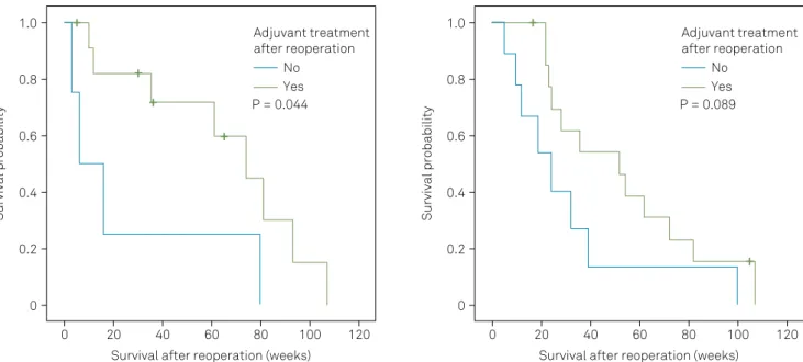
p = 0.005) and adjuvant treatment after reoperation (RR = 0.4; 95%CI = 0.2–0.8; p = 0.016). Multivariate analysis detected an interaction between those two variables. In the analysis stratiied per extent of resection (Figure 2), relative to the group that received adjuvant treatment after reoperation, the median SARp was longer in the patients with subtotal resec-tion at reoperaresec-tion (p: 0.044). Relative to the patients with TR, adjuvant treatment after reoperation was not associated with longer SARp (p = 0.089).
Delayed onset of RT was not associated with OS in the analyzed sample of patients. Separate additional analysis of the patients in which onset of RT was delayed did not show any diference in OS between the ones who had tumor relapse before/during RT (15 cases) and the ones in whom the tumor had relapsed after the end of RT (14 cases), RR = 2.0 (95%CI = 0.8-4.8; p = 0.11).
DISCUSSION
In the present study, we performed a descriptive analy-sis of patients subjected to reoperation for recurrent GBM. Survival may be longer in our sample (20 months) compared to the median survival of patients with GBM reported in the literature, which varied from six to 18 months4,16.
Role of reoperation in overall survival
In our study, the OS was 20 months, consistent with those found in the literature.McNamara et al.17 compared
104 patients who underwent repeated surgery with 397 who underwent primary surgery only and found that the OS
was 21 months in the former versus 10 months in the latter (p < 0.001). Similar results were reported by Helseth et al.5,
who analyzed a sample of 516 patients, with 56 subjected to reoperation; OS was longer in the latter compared to the ones who underwent primary surgery only, 18.4 months ver-sus 8.6 months (p < 0.001).
Role of reoperation in survival after recurrence (SAR)
Relative to the full study population, the median SARp in this study was 9.1 months. In the review Ryken et al.18
per-formed in 2014, the average SARp was nine months, which is much longer than the one reported in the review by Barbagallo et al.11 from 2008, which ranged from three to ive months.
Ringel et al.7 analyzed 503 patients who underwent reoperation
for active disease and found that the SARp was 11.9 months, while in the study by Quick et al.19, in which patients with KPS
score > 60 and tumors likely to be entirely resected were sub-jected to repeated surgery, the SARp was 13 months.
Relevance of adjuvant treatment
In our study, performance of adjuvant treatment (RT and/or CT) before reoperation behaved as a prognostic fac-tor for OS. Since 2009, all patients diagnosed with GBM at our institution have been indicated for adjuvant treatment based on the Stupp protocol2. However, as a function of problems
inherent to the Brazilian health system, the tumors recurred in 41% of the patients before the adjuvant treatment was initi-ated, a fact that was associated with reduced OS in that group compared to the cases in which the tumors recurred after adjuvant treatment (15.9 vs. 23.1 months; p: 0.008).
he impact of adjuvant treatment resulting in longer sur-vival of patients with GBM has been previously reported in the literature5,15,20,21. Stark et al.21 assessed 492 patients with
GBM and found that RT and CT alone or in combination had an impact on OS. he same conclusion was reached by Filippini et al.15 in their study of 676 patients with GBM.
Extent of resection at reoperation
Relative to survival after reoperation, the only indepen-dent factors on both univariate and multivariate analy-sis were the extent of resection at reoperation (RR = 0.3; 95%CI = 0.1–0.7; p = 0.005) and performance of adjuvant treatment after reoperation (RR = 0.4; 95%CI = 0.2 –0.8; p = 0.016). Relative to the patients with TR at reoperation, the subsequent performance of adjuvant treatment did not exert any impact on SARp on multivariate analysis (p = 0.089). However, adjuvant treatment exerted an impact on SARp in the cases with PR at reoperation (p = 0.044).
Yong et al.22 conirmed the relevance of the extent of
resection at reoperation after inding that a residual tumor volume of less than 3 cm3 after reoperation exerted an
impact on the patients’ survival: OS values were 20 months in patients with TR, 11 months in patients with residual tumor volume less than 3 cm3 and ive months when the residual Figure 1. Kaplan-Meier curve representing the overall survival
(OS) of patients who did or did not receive adjuvant treatment after the irst operation.
Overall survival (weeks)
Survival probability
1.0
0.8
0.6
0.4
0.2
0
0 50 100 150 200 250
Adjuvant treatment after first surgery
No Yes P = 0.006 +
+
+ +
+
Table 4. Univariate and multivariate analyses relative to overall survival.
Variable (n) Univariate analysis Median OS Multivariate analysis
RR 95%CI p value (Months) RR 95%CI p value
Gender
Male (26) 0.6 0.3–1.2 0.159 17 NS
Female (13) 1 -- 26
Age
≥ 50 years old (18) 1.5 0.7–3.1 0.282 20.8 NS
< 50 years old (21) 1 19.1
KPS score
≥ 70 (30) 1.3 0.5–3.3 0.528 20.1 NS
< 70 (9) 1 19.8
Laterality
Right (18) 1.1 0.5–2.2 0.862 20.1 NS
Lobe (21) 1 19.8
Frontal lobe localization
Yes (10) 1 0.5–2.0 0.929 20.8 NS
No (29) 1 20.1
Temporal lobe localization
Yes (11) 1.3 0.6–2.6 0.531 17 NS
No (28) 1 21
> 1 affected lobes
Yes (15) 0.9 0.4–1.8 0.68 19.1 NS
No (24) 1 20.8
Interval between operations
≥ 6 months (18) 0.4 0.2–0.9 0.023 23.6 NS
< 6 months (21) 1 16.8
RT delay
Yes (29) 1.3 0.6–3.0 0.467 20.1 NS
No (10) 1 17
First operation resection
TR (23) 1.4 0.7–3.1 0.369 19.8 NS
PR (16) 1 20.8
Reoperation resection
TR (21) 1.7 0.8–3.6 0.137 23.1 NS
PR (18) 1 16.8
Adjuvant therapy after irst operation
Yes (23) 0.4 0.2–0.8 0.008 23.1 0.3 0.2-0.7 0.005
No (16) 1 15.9 1
Adjuvant therapy after reoperation
Yes (26) 1 0.5–2.2 0.903 23.1 NS
No (13) 1 19.8
Recurrence
Distant (5) 2.4 0.7–8.1 0.148 25.9 NS
Local (34) 1 19.1
tumor volume was larger than 3 cm3. Following a study with
170 patients, Oppenlander et al.23 concluded that an extent
of resection larger than 80% in repeated surgery suices to increase patients’ survival.
Influence of radiotherapy delay on survival
To assess the inluence of RT delay on survival, we com-pared the patients who had started RT within the irst six weeks after surgery (no delay) to those who had started it more than six weeks afterwards (RT delay) and found no diference in their survival (RR = 1.3; 95%CI = 0.6; p = 0.467). We chose that time interval because it was the one adopted in previous studies24,25. Our indings in this regard agree with
data reported in the literature24,26. Although some authors
found that RT delay has negative efects on OS25,27 this topic
remains controversial. In turn, Loureiro et al.28 assessed the
time elapsed from surgery to onset of RT at one public and one private healthcare service in Brazil and also failed to ind any diference in OS in association with delayed RT (RR = 1.2; 95%CI = 0.8 –1.8; p = 0.470).
Delay in the onset of RT is mainly due to the increase in the demand for this treatment modality as a function of pop-ulation aging, with consequent elevations in the incidence rates of various types of cancer29. While diiculty in
meet-ing the demand is encountered in developed countries, it is much more frequent in developing countries, especially in the public healthcare setting28. In Brazil, the situation is
espe-cially worrisome, as the increasing demand for RT created an ineicient system; thus, only 65.9% of the patients need-ing RT actually receive it, havneed-ing to wait a median interval of almost four months, independent of the type of cancer28. Our
service represents a classic example of these problems: onset
of RT was delayed in 74% of the patients analyzed in the pres-ent study: the median interval from surgery to onset of RT was eight months. One of the patients in our sample had to wait 23 weeks (5.3 months) to start RT.
Nevertheless, upon analyzing the subgroup of patients with delayed RT, no diference was found in OS between the patients with tumor recurrence before/during RT and the ones in whom the tumor recurred after the end of RT (RR = 2.0; 95%CI = 0.8 –4.8; p = 0.11). hus, we are also led to believe that the patients with tumor relapse before or dur-ing RT are candidates for reoperation. hat is, reoperation of patients who relapsed before RT due to treatment delay aforded the opportunity to conduct adjuvant treatment.
Study limitations
Our study exhibits some limitations, including small sam-ple size; retrospective data collection; lack of uniformity in the adjuvant treatment instituted after the irst and second operations; selection bias, as only patients with KPS scores over 70 and lesions considered to be surgically accessible were subjected to reoperation; and the fact that we did not compare the patients subjected to re-resection to others who did not undergo a second surgery.
Future perspectives
Among the most promising treatments for GBM, tar-geted molecular therapies have been widely investigated in recent years. However, no efective results have yet been achieved. Intratumor heterogeneity and the heterogene-ity between the primary and recurrent tumors represent signiicant limitations to the success of this type of thera-peutic approach. Collecting samples of the relapsed tumor,
Figure 2. (A) Kaplan-Meier curve representing the Survival after reoperation (SARp) of patients subjected to parcial resection (PR) at reoperation and who did or did not receive adjuvant treatment after reoperation. (B) Kaplan-Meier curve representing the SARp of patients subjected to total Resection (TR) at reoperation and who did or did not receive adjuvant treatment after reoperation.
Survival after reoperation (weeks)
Survival probability
1.0
0.8
0.6
0.4
0.2
0
0 20 40 60 100 120
Adjuvant treatment after reoperation
No Yes P = 0.044
80
Survival after reoperation (weeks)
Survival probability
1.0
0.8
0.6
0.4
0.2
0
0 20 40 60 100 120
Adjuvant treatment after reoperation
No Yes P = 0.089
80 +
+ +
+
+
whenever possible, is crucial to advance not only in the assessment of the response to targeted therapies but also in the understanding of the mechanisms of resistance to treat-ment30. In this regard, reoperation, provided it is safe, will
likely become an essential component of the management of these patients in the future.
In conclusion, despite the limitations of our study, we sought to describe the actual conditions of the treatment of recurrent glioblastoma at a Brazilian public healthcare ser-vice. In many developing countries, therapeutic alternatives and targeted therapies remain obscure but keeping in mind factors such as age, KPS prior to and after surgery, degree of resection TR vs. PR and adjuvant RT and CT can still pro-vide results comparable to modern day series. In spite of the
delays and diiculties that conspired against the achieve-ment of the ideally complete treatachieve-ment, the length of survival of patients who underwent reoperation was similar to those reported in the literature. Our results indicate that reopera-tion is feasible and may be considered a treatment opreopera-tion for a select group of patients with recurrent GBM in Brazil.
Acknowledgements
Fernando A. P. Ferraz (in memoriam), M.D., PhD, founder of the Neuro-oncology Group in Federal University Of São Paulo, and Luiz Daniel Cetl, M.D., for participation in the management care of some of these patients.
References
1. Egan KM, Thompson RC, Nabors LB, Olson JJ, Brat DJ, Larocca RV et al. Cancer susceptibility variants and the risk of adult glioma in a US case-control study. J Neuroncol. 2011;104(2):535-42. doi:10.1007/s11060-010-0506-0
2. Stupp R, Mason WP, Bent MJ, Weller M, Fisher B, Taphoorn MJ et al. Radiotherapy plus concomitant and adjuvant temozolomide for glioblastoma. New Engl J Med. 2005;352(10):987-96. doi:10.1056/NEJMoa043330
3. Darefsky AS, King JT, Jr., Dubrow R. Adult glioblastoma multiforme survival in the temozolomide era: a population-based analysis of surveillance, epidemiology, and end results registries. Cancer. 2012;118(8):2163-72. doi:10.1002/cncr.26494
4. Stupp R, Hegi ME, Mason WP, Bent MJ, Taphoorn MJ, Janzer RC et al. Effects of radiotherapy with concomitant and adjuvant temozolomide versus radiotherapy alone on survival in glioblastoma in a randomised phase III study: 5-year analysis of the EORTC-NCIC trial. Lancet. Oncology. 2009;10(5):459-66. doi:10.1016/S1470-2045(09)70025-7
5. Helseth R, Helseth E, Johannesen TB, Langberg CW, Lote K, Rønning P et al. Overall survival, prognostic factors, and repeated surgery in a consecutive series of 516 patients with glioblastoma multiforme. Acta Neurol Scand. 2010;122(3):159-67. doi:10.1111/j.1600-0404.2010.01350.x
6. Krex D, Klink B, Hartmann C, Deimling A, Pietsch T, Simon M et al. Long-term survival with glioblastoma multiforme. Brain. 2007;130(10):2596-606. doi:10.1093/brain/awm204
7. Ringel F, Pape H, M, Krex D, Bock HC, Misch M et al. Clinical beneit from resection of recurrent glioblastomas: results of a multicenter study including 503 patients with recurrent glioblastomas undergoing surgical resection. Neuro-oncol. 2016;18(1):96-104. doi:10.1093/neuonc/nov145
8. Lacroix M, Abi-Said D, Fourney DR, Gokaslan ZL, Shi W, DeMonte F et al. A multivariate analysis of 416 patients with glioblastoma multiforme: prognosis, extent of resection, and survival. J Neurosurg. 2001;95(2):190-8. doi:10.3171/jns.2001.95.2.0190
9. Samis Zella MA, Wallocha M, Slotty PJ, Isik G, Hänggi D, Schroeteler J et al. Evaluation of post-operative complications associated with repeat resection and BCNU wafer implantation in recurrent glioblastoma. Acta Neurochir (Wien). 2014;156(2):313-23. doi:10.1007/s00701-013-1931-6
10. Hong B, Wiese B, Bremer M, Heissler HE, Heidenreich F, Krauss JK et al. Multiple microsurgical resections for repeated recurrence of glioblastoma multiforme. Am J Clin Oncol. 2013;36(3):261-8. doi:10.1097/COC.0b013e3182467bb1
11. Barbagallo GM, Jenkinson MD, Brodbelt AR. “Recurrent”
glioblastoma multiforme, when should we reoperate? Br J Neurosurg. 2008;22(3):452-55. doi:10.1080/02688690802182256
12. Chaichana KL, Zadnik P, Weingart JD, Olivi A, Gallia GL, Blakeley J et al. Multiple resections for patients with glioblastoma: prolonging survival. J Neurosurg. 2013;118(4):812-20. doi:10.3171/2012.9.JNS1277
13. Sughrue ME, Sheean T, Bonney PA, Maurer AJ, Teo C. Aggressive repeat surgery for focally recurrent primary glioblastoma: outcomes and theoretical framework. Neurosurg Focus. 2015;38(3):E11. doi:10.3171/2014.12.FOCUS14726
14. Clarke JL, Ennis MM, Yung WK, Chang SM, Wen PY, Cloughesy TF et al. Is surgery at progression a prognostic marker for improved 6-month progression-free survival or overall survival for patients with recurrent glioblastoma? Neuro-oncol. 2011;13(10):1118-24. doi:10.1093/neuonc/nor110
15. Filippini G, Falcone C, Boiardi A, Broggi G, Bruzzone MG, Caldiroli D et al. Prognostic factors for survival in 676 consecutive patients with newly diagnosed primary glioblastoma. Neuro-oncol. 2008;10(1):79-87. doi:10.1215/15228517-2007-038
16. Gorlia T, Stupp R, Brandes AA, Rampling RR, Fumoleau P, Dittrich C et al. New prognostic factors and calculators for outcome prediction in patients with recurrent glioblastoma: a pooled analysis of EORTC Brain Tumour Group phase I and II clinical trials. Eur J Cancer. 2012;48(8):1176-84. doi:10.1016/j.ejca.2012.02.004
17. McNamara MG, Lwin Z, Jiang H, Templeton AJ, Zadeh G, Bernstein M et al. Factors impacting survival following second surgery in patients with glioblastoma in the temozolomide treatment era, incorporating neutrophil/lymphocyte ratio and time to irst progression. J Neurooncol. 2014;117(1):147-52. doi:10.1007/s11060-014-1366-9
18. Ryken TC, Kalkanis SN, Buatti JM, Olson JJ. The role of cytoreductive surgery in the management of progressive glioblastoma :
a systematic review and evidence-based clinical practice guideline. J Neurooncol. 2014;118(3):479-88. doi:10.1007/s11060-013-1336-7
19. Quick J, Gessler F, Dützmann S, Hattingen E, Harter PN, Weise LM et al. Beneit of tumor resection for recurrent glioblastoma. J Neurooncol. 2014;117(2):365-72. doi:10.1007/s11060-014-1397-2
20. Stark AM, Nabavi A, Mehdorn HM, Blömer U. Glioblastoma multiforme-report of 267 cases treated at a single institution. Surg Neurol. 2005;63(2):162-69. doi:10.1016/j.surneu.2004.01.028
22. Yong RL, Wu T, Mihatov N, Shen MJ, Brown MA, Zaghloul KA et al. Residual tumor volume and patient survival following reoperation for recurrent glioblastoma. J Neurosurg. 2014;121(4):802-9. doi:10.3171/2014.6.JNS132038
23. Oppenlander ME, Wolf AB, Snyder LA, Bina R, Wilson JR, Coons SW et al. An extent of resection threshold for recurrent glioblastoma and its risk for neurological morbidity. J Neurosurg. 2014;120(4):846-53. doi:10.3171/2013.12.JNS13184
24. Blumenthal DT, Won M, Mehta MP, Curran WJ, Souhami L, Michalski JM et al. Short delay in initiation of radiotherapy may not affect outcome of patients with glioblastoma: a secondary analysis from the radiation therapy oncology group database. J Clin Oncol. 2009;27(5):733-9. doi:10.1200/JCO.2008.18.9035
25. Valduvieco I, Verger E, Bruna J, Caral L, Pujol T, Ribalta T et al. Impact of radiotherapy delay on survival in glioblastoma. Clin Transl Oncol. 2013;15(4):278-82. doi:10.1007/s12094-012-0916-x
26. Graus F, Bruna J, Pardo J, Escudero D, Vilas D, Barceló I et al. Patterns of care and outcome for patients with glioblastoma diagnosed during 2008-2010 in Spain. Neuro-oncol. 2013;15(6):797-805. doi:10.1093/neuonc/not013
27. Spratt DE, Folkert M, Zumsteg ZS, Chan TA, Beal K, Gutin PH et al. Temporal relationship of post-operative radiotherapy with temozolomide and oncologic outcome for glioblastoma. J Neurooncol. 2014;116(2):357-63. doi:10.1007/s11060-013-1302-4
28. Loureiro LV, Pontes LB, Callegaro-Filho D, Koch LO, Weltman E, Victor ES et al. Waiting time to radiotherapy as a prognostic factor for glioblastoma patients in a scenario of medical disparities. Arq Neuro-psiquiatr. 2015;73(2):104-10. doi:10.1590/0004-282X20140202
29. Chen Z, King W, Pearcey R, Kerba M, Mackillop WJ. The relationship between waiting time for radiotherapy and clinical outcomes: a systematic review of the literature. Radiother Oncol. 2008;87(1):3-16. doi:10.1016/j.radonc.2007.11.016