w w w . r b o . o r g . b r
Original
Article
Evaluation
of
the
acromiohumeral
distance
by
means
of
magnetic
resonance
imaging
umerus
夽
Flávio
de
Oliveira
Franc¸a
∗,
André
Couto
Godinho,
Elísio
José
Salgado
Ribeiro,
Lorenzo
Falster,
Lucas
Emanuel
Gava
Búrigo,
Rafael
Berenstein
Nunes
HospitalOrtopédicoBH,BeloHorizonte,MG,Brazil
a
r
t
i
c
l
e
i
n
f
o
Articlehistory:
Received6May2015 Accepted11June2015
Availableonline4February2016
Keywords:
Rotatorcuff
Magneticresonanceimaging Acromion
Humerus
a
b
s
t
r
a
c
t
Objective:Todemonstratetherelationshipbetweenthesize,degreeofretractionand
topog-raphyofrotatorcuffinjuriesandthedegreeofriseofthehumeralhead,andtoevaluatethe influenceofgravity,usingmagneticresonanceimaging(MRI).
Methods:Weevaluated181shoulderMRIsfrom160patientsagedover45years,between
November2013andJuly2014.Thepatientsweredividedintotwogroups:onecontrol(no lesionorpartialdamagetotherotatorcuff);andtheotherwithcompletetearsoftherotator cuff.Wemeasuredtheacromiohumeraldistanceinthesagittalplane,andestablishedthe shortestdistancebetweentheapexoftheheadandtheacromion.
Results:Inthisstudy,96examinationsonfemalepatients(53.04%)and58onmalepatients
(46.96%)wereevaluated.Themeanagewas63.27years:inthecontrolgroup,61.46;and inthegroupwithinjuries,65.19.Fromanalysisonthemeasurementsofthesubacromial space,weobservedsignificantlyhighervaluesinthecontrolgroup(7.71mm)thaninthe groupwithinjuries(6.99).Incomparingthecontrolgroupwithsomespecificsubgroup, i.e.posterosuperior(6.77),anteroposterior-superior(4.16)andretractionPatteIII(5.01),we confirmedtheimportanceoftopographyanddegreeofretractioninrelationtotheriseof thehumeralhead.
Conclusion: Theriseofthehumeralheadwasdirectlyrelatedtothesize,degreeofretraction
andtopographyoftherotatorcuffinjuries,withgreaterdegreesofriseincasesofsuperior andposteriorlesionsandanteroposterior-superior(massive)lesions.Theassessmentusing MRIwasnotinfluencedbytheforceofgravity.
©2016SociedadeBrasileiradeOrtopediaeTraumatologia.PublishedbyElsevierEditora Ltda.Allrightsreserved.
夽
WorkperformedintheShoulderGroup,HospitalOrtopédicoBH,BeloHorizonte,MG,Brazil.
∗ Correspondingauthor.
E-mails:[email protected],[email protected](F.deOliveiraFranc¸a). http://dx.doi.org/10.1016/j.rboe.2016.01.008
Palavras-chave:
Bainharotadora Imagemderessonância magnética
Acrômio Úmero
r
e
s
u
m
o
Objetivo:Demonstrararelac¸ãoentreotamanho,grauderetrac¸ãoetopografiadaslesõesdo
manguitorotadorcomograudeascensãodacabec¸aumeraleavaliarainfluênciadaforc¸a dagravidadenaressonânciamagnética.
Métodos:Avaliamos181ressonânciasmagnéticasdeombrode160pacientescommaisde45
anos,entrenovembrode2013ejulhode2014.Ospacienteseramdivididosemdoisgrupos, umdecontrole(semlesãooucomlesãoparcialdoMR)eoutrocomlesãocompletadoMR. Fizemosamensurac¸ãodadistânciaacrômio-umeralnoscortessagitaisefoiestabelecidaa menordistânciaentreoápicedacabec¸aeoacrômio.
Resultados: Foramavaliadosnesteestudo96(53,04%)examesdepacientesdosexo
fem-ininoe58 depacientesdosexomasculino(46,96%).Aidademédia foi63,27anos,ado grupocontrole61,46eadogrupocomlesão65,19.Aoanalisarasmedidasdoespac¸o sub-acromial,observamosvaloressignificativamentemaioresnogrupocontrole(7,71mm)do quenogrupocomlesão(6,99).Quandocomparamosogrupocontrolecomalguns subgru-posespecíficos,posterossuperior(6,77),anteroposterossuperior(4,16)eretrac¸ãoPatteIII (5,01),confirmamosaimportânciadatopografiaegrauderetrac¸ãoparaascensãodacabec¸a umeral.
Conclusão:Aascensãodacabec¸aumeraltemrelac¸ãodiretacomotamanho,grauderetrac¸ão
eatopografiadaslesõesdomanguitorotador,comgrausmaioresdeascensãonaslesões posterossuperioreseanteroposterossuperiores(extensas).Aavaliac¸ãofeitapela ressonân-ciamagnéticanãosofreinfluênciadaforc¸adagravidade.
©2016SociedadeBrasileiradeOrtopediaeTraumatologia.PublicadoporElsevier EditoraLtda.Todososdireitosreservados.
Introduction
The rotator cuff acts as a compressor of the head of the humerusagainsttheglenoid,thusenablingshoulder move-mentsindifferentspatialplanes.Diseaseoftherotatorcuff maycausethisequilibriumtobebrokenandmayculminatein moreadvancedstagesofinjurywithconsequentascensionof thehumeralhead.1–4Thisphenomenoncanbequantifiedby
measuringthedistancefromthehumerustotheacromion.5
Themechanismforascensionofthehumeralheadisstill notcompletelyclear.Itisbelievedthatthetractionexertedby thedeltoidmusclewithoutthestabilizingactionoftherotator cuffmightexplainthesefindings.5Inthisregard,failureofthe
infraspinatusandthedepressorfunctionofthehumeralhead wouldallowascensionbymeansofaninjuredsupraspinatus thatwouldnotoccupythesubacromialspace.6–9
Clinically,thismeasurementcanbeusedtoevaluatethe functionoftherotatorcuffandaidinchoosingthetypeof therapytobeused.10,11Adistancefromthehumerustothe
acromionoflessthanorequalto7mmmeasuredon radio-graphsinanteroposteriorviewsuggeststhattherotatorcuff injuryislarge,whichdiminishesthelikelihoodofsuccessful surgicaltreatment.10,12Ithasalsobeenshownthatthe
ascen-sionofthehumeralheadisrelatedtofattydegenerationof therotatorcuff.11–13
Useofmagneticresonanceimaging(MRI)forestimating theascensionofthehumeralheadiscurrentlyundergreat discussion.Oneofthemainissuesinvolvedisthefactthat MRI is performed in dorsal decubitus. It is believed that
thereductionoftheforceofgravityonthelimbduringthe examinationmightoverestimatethisradiologicalfinding.
Theobjectivesofthisstudywere:(1)todemonstratethat thedegreeofascensionofthehumeralheadisrelatedtothe sizeoftherotatorcuffinjuryanditsdegreeofretraction;(2) toevaluatewhetherthe ascensionofthehumeralheadon MRI hasany relationship withgravity; and (3) toascertain whetherthelocationoftheinjuryinfluencesthisascension ofthehumeralhead.
Materials
and
methods
ThisstudywasapprovedbytheResearchEthicsCommittee of our institution. Prospective evaluations were conducted on181MRIsoftheshoulder, from160patientswho under-wentthisexaminationbetweenNovember2013andJuly2014. Theseevaluationswereperformedinaradiologyclinic.
Forinclusioninthisstudy,thepatientsneededtobemore than45yearsofageandtohaveundergoneMRIfor investiga-tionofnon-traumaticpathologicalconditionsoftheshoulder. Patients withhistories of fractures or previous surgery on the shoulder thatwas now undergoingexamination,cases involvingmagneticresonancearthrographyandpatientswith glenohumeralarthrosisthathadalreadybecomeestablished wereexcluded.
Fig.1–Apexofthehumeralhead,seenonacoronalslice(imageonleft).Measurementoftheshortestdistancebetween theapexofthehumeralheadandtheacromion,seenonasagittalslice(imageonright).
subdividedaccordingtothelocationoftheinjury (anterosupe-rior,superior,posterosuperiororanteroposterosuperior)and thedegreeoftendonretractionaccordingtotheclassification ofPatte.14
All the examinations were performed inMRI machines of1.5T.Theexaminationswereevaluatedandthe measure-ments were made bytwophysicians undergoingspecialist trainingintheshouldersurgeryservice,withassistancefrom a radiologist who was a specialist in osteomuscular MRI. Themeasurementsonthedistancefromthehumerustothe acromionweremadeonsagittalslicesandtheshortest dis-tancebetweentheapexoftheheadandthe acromionwas established(Fig.1:howthemeasurementwasmade).Through thismeasurement,theaimwastomaketheexaminationas reproducibleaspossibleinconsultationoffices.
Thestatisticalanalysisrelatingtothesizeofthelesionwas doneusingtheANOVAparametrictest.
Toevaluatethedifferencesbetweenthegroupsoflesion sizeandthevariableofsubacromialspace,whichcomprised morethanthreegroups,eithertheANOVAparametrictestor Student’st testforindependentsampleswasusedto com-parethemeansbetweenthegroups.Thisanalysiswasalso performedforthesubacromialspaceinrelationtothedegree oftendonretractionaccordingtotheclassificationofPatte(I, IIorIII).
ThedataofthisstudywereprocessedusingthePredictive AnalyticsSoftware(PASW18).Inallthestatisticaltests,the significancelevelwastakentobe5%.
Results
Amongthe181shouldersevaluated(160patients),88(48.6%) presentedcompleterotatorcufftearsandformedtheinjury group,while93(51.3%)formedthecontrolgroup.
Injurygroup
Thepatients’meanagewas65years,andtheagerangewas from 45 to 89 years. There were 43 males (48.8%) and 45 females(51.1%);55rightshoulderswereaffected(62.5%)and sevenpatientswereaffectedbilaterally(7.95%).
Regarding the locations of the injuries, 40 shoulders (45.45%) were injured in a superior location, eight (9.09%) anterosuperior,27 (30.68%) posterosuperiorand 13 (14.77%) anteroposterosuperior. The mean ages in these subgroups were63,66,65and70years,respectively(Table1).
Inanalyzingtheinjuriesregardingtheirdegreeof retrac-tion, based on the classification of Patte,14 there were 29
shoulders(32.95%)withgradeI,32(36.36%)withgradeIIand 27 (30.68%)withgradeIII.Themeanagesinthesubgroups were,respectively,62,64and69years(Table2).
Controlgroup
Thepatients’meanagewas61yearsandtheagerangewas from 45to79.Therewere 42males(45.1%)and51 females
Table1–Demographicsoftheinjurygroup.
n(%) Meanage(range) Meansubacromialspace(range)
Injurygroup 88(100) 65.19(36–89) 6.97(1.81–13.5)
Superior 40(45.45) 63.47(36–89) 7.78(4.15–11.58)
Anterosuperior 8(9.09) 66.25(51–85) 8.28(5.73–13.5)
Posterosuperior 27(30.68) 65.03(51–88) 6.77(5.04–9.91)
n(%) Meanage(range) Meansubacromialspace(range)
Injurygroup 88(100) 65.19(36–89) 6.97(1.81–13.5)
PatteI 29(32.95) 62.31(36–78) 8.56(4.22–13.5)
PatteII 32(36.36) 64.28(51–89) 7.23(4.19–11.73)
PatteIII 27(30.68) 69.37(51–88) 5.01(1.81–10.11)
Table3–Correlationofsubacromialspacebetweenthecontrolandinjurygroups.
Group N Meansubacromialspace SD ttest p-Value
Control 93 7.71 1.58 2.418 0.017a
Injury 88 6.99 2.37
a Theprobabilitiesofsignificance(p-values)refertoStudent’sttestforindependentsamples.
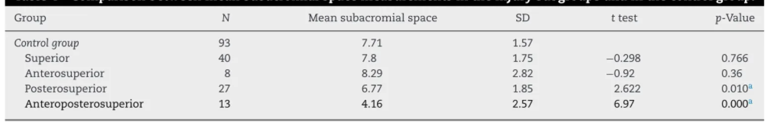
Table4–Comparisonbetweenmeansubacromialspacemeasurementsintheinjurysubgroupsandinthecontrolgroup.
Group N Meansubacromialspace SD ttest p-Value
Controlgroup 93 7.71 1.57
Superior 40 7.8 1.75 −0.298 0.766
Anterosuperior 8 8.29 2.82 −0.92 0.36
Posterosuperior 27 6.77 1.85 2.622 0.010a
Anteroposterosuperior 13 4.16 2.57 6.97 0.000a
a Theprobabilitiesofsignificance(p-values)refertoStudent’sttestforindependentsamples.
(54.8%).Therightshoulderwasevaluatedin49cases(52.6%) andbothshoulderswereusedintheevaluationin13cases (13.9%).
Incomparing the meansubacromial space betweenthe injury and control groups, it was foundto be significantly largerinthecontrolgroup(7.71)thanintheinjurygroup(6.99), withp<0.05(Table3).
Amongthesubgroupsofpatientswithinjuries(superior, anterosuperior,posterosuperiorandanteroposterosuperior), itwasseenthatthemeansubacromialspace wasdifferent betweenthefoursubgroups.Itwaspossibletomake compar-isonsbetweenpairsofsubgroupsinordertoidentifythereal differencebetweenthem.
In analyzing the difference inmean subacromial space measurements between these subgroups and the control group,no statistical differences betweenthe controlgroup andthepatientswitheithersuperiororanterosuperiorlesions wereobserved(p>0.05).Ontheotherhand,inrelationtothe individualswithposterosuperiorandanteroposterosuperior, it was noted that the subacromial space was statistically smaller than in the individuals inthe control group or in the subgroups with superior and anterosuperior injuries. In comparing the differences in mean measurements of
the subacromial space between the subgroups of postero-superior andanteroposterosuperior injuries,which showed lower means than the other subgroups, we observed that the subgroupofanteroposterosuperiorinjuriespresenteda significantly lower mean (4.16) (p<0.05), in relation to the subgroupofposterosuperiorinjuries(6.77)(Table4).
Inrelationtotheanalysisonthemeanmeasurementson thesubacromialspacebetweentheinjurysubgroups classi-fied inaccordance with Patte and the controlgroup, there wasastatisticaldifference(p<0.05)inrelationtoPatte sub-group I, which presenteda largersubacromial space (8.57) thanthecontrolgroup(7.71);andinrelationtoPattesubgroup III,inwhichthemeansubacromialspacemeasurementwas
smaller(5.01)than inthe controlgroup(7.71).Thepatients withinjuriesclassifiedasPatteIIpresentedamean subacro-mialspacemeasurementof7.23,whichwasnotstatistically differentfromthecontrolgroup(Table5).
In evaluating thesesubgroupsinrelation toeach other, it was observedthat the individuals withPatte III injuries presentedmeansubacromialspacemeasurementsthatwere smaller than those ofindividuals with Patte I and Patte II injuries. Likewise,the subacromial space in individuals in PattesubgroupIIwassmallerthanthespaceinindividuals
Table5–ComparisonbetweenmeansubacromialspacemeasurementsinthePattesubgroupsandinthecontrolgroup.
Group N Meansubacromialspace SD ttest p-Value
Controlgroup 93 7.71 1.58
PatteI 29 8.57 1.91 −2.425 0.017a
PatteII 32 7.23 1.55 1.484 0.14
PatteIII 27 5.01 2.25 7.063 0.000a
Table6–Pair-by-paircomparisonofmeansubacromialspacemeasurementsinrelationtothePatteclassification.
Group N Meansubacromialspace SD ttest p-Value
I 29 8.57 1.91 3.009 0.004a
II 32 7.23 1.55
I 29 8.57 1.91 6.386 0.000a
III 27 5.01 2.25
II 32 7.23 1.55 −4.472 0.000a
III 27 5.01 2.25
a Theprobabilitiesofsignificance(p-values)refertoStudent’sttestforindependentsamples.
withPatte I injuries. Both ofthese comparisonspresented statisticallysignificantdifferences(p<0.05)(Table6).
Inevaluating the subacromial space ofindividuals with posterosuperiorinjuriesand comparingtheminrelationto the Patte classification, it was observed that those with posterosuperior injuries classified as Patte III presented a mean subacromial space measurement of5.74,which was significantlysmallerthanthespaceinindividualswith pos-terosuperiorinjuriesclassifiedasPatteIandII(7.48).
Discussion
Evaluationofthe subacromialspace bymeans ofMRImay provideinformationonthebiomechanicsoftheshoulderand the prognosisforrotator cuffinjuries. In astudy inwhich subacromialspacemeasurementsfromconventionalX-rays andmagneticresonancearthrographywerecompared,Saupe etal.5concludedthatthereductionindistancewas
associ-atedwithrotatorcuffinjuriesandfattydegenerationofthe tendons.Thesameauthorsconcludedthatinvolvementofthe infraspinatustendoncausedgreatervariationinthe subacro-mialspace. Inourstudy,patientswithrotator cuffinjuries presented decreased distances from the humerus to the acromion,incomparisonwiththecontrolgrouppatients,with astatisticallysignificantdifference.Incomparingthe subacro-mialspaceinpatientswithrotatorcuffinjuriesindifferent locationswithpatientsinthecontrolgroup,wefoundthat thesubacromialspacewassmallerinpatientswithinjuriesin theposterosuperiorandanteroposterosuperiorregions,witha statisticallysignificantdifference.Thesefindingsshowedthe importanceofthetendonoftheinfraspinatusinmaintaining thesubacromialspace.
Inthestudies byPatte14 andGerber etal.,15the
biome-chanicalfunctionoftorn,retractedanddegeneratedtendons maybeimpaired,therebyenablingascensionofthehumeral head.15,16Throughcomparingthevariationofthesubacromial
spacewiththeextentoftherotatorcuffinjuryaccordingto Patte’sclassification,itwasconcludedfromthepresentstudy thatPatte IIIinjuriesevolvedwithgreater ascensionofthe humeralhead.TheassociationbetweenPatte’sclassification andfattydegenerationiswellknown.
Theprognosticvalueofthedistancebetweenthehumerus andtheacromioniswellknown andcanbeusedtoaid in therapeuticdecision-making.WeinerandMacnab17werethe
firsttodescribeanassociationbetweenreductionofthe sub-acromialspaceandrotatorcuffinjuries.Theyobservedthat if the distancewas ≤7mm on conventional radiographs, a
completetendontearwouldbepresent.Subsequently,itwas observed that this measurement ofthe subacromial space might alsobeassociated witha lower successrate in sur-gical treatment.18,19 Werner etal.20 believedthatthecutoff
point forevaluating theascension ofthe humeralheadon MRIshouldbe≤6mm,i.e.lowerthanonconventional radio-graphs.Theyattributedthistogeometricalattributesandto the patient’s positionduringtheexamination,which could causealterationstomuscletonus.21Thepresentstudyfounda
meansubacromialspacemeasurementof7.71mminthe con-trolgroup,whichdoesnotcorroboratethefindingsofWerner etal.,20and6.97mminpatientswithrotatorcuffinjuries.IN
evaluatingthetendongroupsaffected,weobservedthatonly the patients who presented anteroposterosuperior injuries hadsubacromialspaces≤6mm(meanof4.15mm).
Conclusions
Ascension ofthe humeral head (reduction ofthe subacro-mial space)isdirectly relatedtothelocationand extentof theinjuries.Thus,extensiveinjuriesorthoseina posterosu-periorlocationpresentascension ofthehumeralhead, and thisworsenswhenitisassociatedwithadvanceddegreesof tendonretraction.
TheascensionofthehumeralheadassessedthroughMRI doesnotinfluencetheseveritybut,rather,thelocationand extentofrotatorcuffinjuries.
Posterosuperiorandanteroposterosuperiorinjuries (exten-siveinjuries)presentascensionofthehumeralheadonMRI. Thegreaterthedegreeoftendonretractionassociatedwith theseinjuriesis,thegreatertheascensionwillbe.
Conflicts
of
interest
Theauthorsdeclarenoconflictsofinterest.
r
e
f
e
r
e
n
c
e
s
1.HurovJ.Anatomyandmechanicsoftheshoulder:reviewof currentconcepts.JHandTher.2009;22(4):328–42.
2.ZemanCA,ArcandMA,CantrellJS,SkedrosJG,BurkheadWZ Jr.Therotatorcuff-deficientarthriticshoulder:diagnosisand surgicalmanagement.JAmAcadOrthopSurg.
1998;6(6):337–48.
OrthopRelatRes.1992;(284):144–52.
5. SaupeN,PfirrmannCW,SchmidMR,JostB,WernerCM, ZanettiM.Associationbetweenrotatorcuffabnormalities andreducedacromiohumeraldistance.AJRAmJRoentgenol. 2006;187(2):376–82.
6. Nové-JosserandL,EdwardsTB,O’ConnorDP,WalchG.The acromiohumeralandcoracohumeralintervalsareabnormal inrotatorcufftearswithmuscularfattydegeneration.Clin OrthopRelatRes.2005;(433):90–6.
7. Nové-JosserandL,LévigneC,NoëlE,WalchG.L’espace sousacromial.Étudedesfacteursinfluenc¸antsahauteur.Rev ChirOrthopReparatriceApparMot.1996;82(5):379–85. 8. BlaimontP,TaheriA.Analysefonctionnelledesprincipaux
musclesdel’épaule.In:BlaimontP,TaheriA,editors. Biomécaniquedel’épaule,delathéorieàlapratique.Paris: Springer-Verlag;2006.p.57–70.
9. ComtetJJ,AuffrayY.Physiologyoftheelevatormusclesofthe shoulder.RevChirOrthopReparatriceApparMot.
1970;56(2):105–17.
10.JostB,PfirrmannCW,GerberC,SwitzerlandZ.Clinical outcomeafterstructuralfailureofrotatorcuffrepairs.JBone JointSurgAm.2000;82(3):304–14.
11.NorwoodLaBarrackR,JacobsonKE.Clinicalpresentationof completetearsoftherotatorcuff.JBoneJointSurgAm. 1989;71(4):499–505.
12.PeterssonCJ,Redlund-JohnellI.Thesubacromialspacein normalshoulderradiographs.ActaOrthopScand. 1984;55(1):57–8.
itsheight.RevChirOrthopReparatriceApparMot. 1996;82(5):379–85.
14.PatteD.Classificationofrotatorcufflesions.ClinOrthop RelatRes.1990;(254):81–6.
15.NeerCS2nd,CraigEV,FukudaH.Cuff-teararthropathy.J BoneJointSurgAm.1983;65(9):1232–44.
16.GerberC,PenningtonSD,NyffelerRW.Reversetotalshoulder arthroplasty.JAmAcadOrthopSurg.2009;17(5):284–95. 17.WeinerDS,MacnabI.Superiormigrationofthehumeral
head.Aradiologicalaidinthediagnosisoftearsoftherotator cuff.JBoneJointSurgBr.1970;52(3):524–7.
18.EllmanH,HankerG,BayerM.Repairoftherotatorcuff: end-resultstudyoffactorsinfluencingreconstruction.JBone JointSurgAm.1986;68(8):1136–44.
19.MarechalE.Rupturesdegenerativesdelacoiffedesrotateurs del’epaule:evaluationfonctionnelle;resultatsdutraitement chirurgical[these].Lyon,France:UniversireClaudeBernard; 1990.
20.WernerCM,ConradSJ,MeyerDC,KellerA,HodlerJ,GerberC. Intermethodagreementandinterobservercorrelationof radiologicacromiohumeraldistancemeasurements.J ShoulderElbowSurg.2008;17(2):237–40.
21.GraichenH,BonelH,StammbergerT,EnglmeierKH,ReiserM, EcksteinF.Subacromialspacewidthchangesduring