INTRODUCTION
Laryngopharyngeal reflux of gastric content may cause laryn-geal inflammation, with consequent symptoms including hoarse-ness, throat pain, sensation of a lump in the throat, cough, repeti-tive throat cleaning, excessive phlegm, dysphagia, odynophagia, heartburn and voice fatigue(1). There is a well-established associa-tion between gastroesophageal reflux disease (GERD) and reflux laryngitis syndrome(2,3); however, laryngitis may also be caused by tobacco, alcoholic beverages, allergies, rhinopharyngeal infections and traumatic lesions(4).The diagnosis of laryngopharyngeal reflux of gastric content is not easy(3,5) and treatment includes proton pump inhibitors, which may or may not improve the symptoms(6,7).
Dysphagia is present in patients with GERD and in patients with laryngitis(1,6,8). In patients with GERD, the symptom may be related to ineffective esophageal motility and also individual sensitivity(2,6,9). Pharyngeal phase of swallowing may be impaired, with slower bolus transit through the mouth, pharynx and upper esophageal sphincter(10,11). In laryngitis, a possible cause of phar-yngeal dysphagia is larphar-yngeal inflammation.
Our objective in this investigation was to evaluate oral and pharyngeal bolus transit time in patients with laryngitis, regardless of its cause. Our hypothesis was that bolus transit time through mouth and pharynx of patients with laryngitis had a different duration than in normal volunteers.
METHODS
We evaluated by videofluoroscopy the oral and pharyngeal phases of swallowing in 21 patients with laryngitis and 21 healthy
Swallowing in patients with laryngitis
Isabela
MODA
, Hilton Marcos Alves
RICZ
, Lilian Neto
AGUIAR-RICZ
and Roberto Oliveira
DANTAS
Received 21/8/2017 Accepted 5/10/2017
ABSTRACT – Background – Dysphagia is described as a complaint in 32% of patients with laryngitis. Objective – The objective of this investigation
was to evaluate oral and pharyngeal transit of patients with laryngitis, with the hypothesis that alteration in oral-pharyngeal bolus transit may be involved with dysphagia. Methods – Videofluoroscopic evaluation of the swallowing of liquid, paste and solid boluses was performed in 21 patients with laryngitis, 10 of them with dysphagia, and 21 normal volunteers of the same age and sex. Two swallows of 5 mL liquid bolus, two swallows of 5 mL paste bolus and two swallows of a solid bolus were evaluated in a random sequence. The liquid bolus was 100% liquid barium sulfate and the paste bolus was prepared with 50 mL of liquid barium and 4 g of food thickener (starch and maltodextrin). The solid bolus was a soft 2.2 g cookie coated with liquid barium. Durations of oral preparation, oral transit, pharyngeal transit, pharyngeal clearance, upper esophageal sphincter opening, hyoid movement and oral-pharyngeal transit were measured. All patients performed 24-hour distal esophageal pH evaluation previous to videofluoroscopy.
Results – The evaluation of 24-hour distal esophageal pH showed abnormal gastroesophageal acid reflux in 10 patients. Patients showed longer oral
preparation for paste bolus and a faster oral transit time for solid bolus than normal volunteers. Patients with laryngitis and dysphagia had longer preparation for paste and solid boluses, and a faster oral transit time with liquid, paste and solid boluses. Conclusion – A longer oral preparation for paste and solid boluses and a faster transit through the mouth are associated with dysphagia in patients with laryngitis.
HEADINGS – Laryngitis. Deglutition disorders. Pharynx. Esophageal pH monitoring. Fluoroscopy.
Declared conflict of interest of all authors: none Disclosure of funding: no funding received
Departamento de Oftalmologia, Otorrinolaringologia, Cirurgia de Cabeça e Pescoço e Departamento de Clínica Médica, Faculdade de Medicina de Ribeirão Preto – Universidade de São Paulo, Ribeirão Preto, SP, Brasil.
Correspondence: Roberto Oliveira Dantas. Faculdade de Medicina de Ribeirão Preto – Universidade de São Paulo. Av. Bandeirantes, 3900 – Campus da USP – CEP: 14049-900 – Ribeirão Preto, SP, Brasil. E-mail: [email protected]
controls. Both groups were similar with respect to sex distribution (14 women and 7 men), and age (mean 50 years, 35-65 years). The control volunteers did not report symptoms, had no chronic or acute diseases, movement limitations, eating restrictions, no previous use of tobacco, alcoholic beverages, no history of aller-gies, rhinopharyngeal infections and traumatic lesions. Patients with laryngitis came to the hospital with voice and/or throat complains. They also had no history of use of tobacco, alcoholic beverages, allergies, rhinopharyngeal infections and traumatic le-sions. They were submitted to laryngoscopic examination, which found laryngeal lesions; the most frequent were hyperemia and posterior laryngeal edema, hyperemia and edema of vocal folds, and pachydermia laryngitis (TABLE 1). The most frequent complaints were respiratory and upper digestive symptoms, hoarseness, and constant throat clearing (TABLE 2). Dysphagia was a complaint in ten patients (47.6%), five of them with odinophagia. They were untreated on test days.
TABLE 1. Laryngoscopic findings in the patients with laryngitis (n=21)
N %
Hyperemia and posterior laryngeal edema 15 71.4 Hyperemia and vocal folds edema 6 28.6 Posterior laryngeal pachydermia 4 19.0
Cordite 1 4.8
Nodule 1 4.8
Cyst 1 4.8
Laryngeal polyps 1 4.8
Reinke’s edema 1 4.8
Before videofluoroscopy all patients were submitted to a 24-hour esophageal pH test for detection of acid gastroesophageal reflux. The pH sensor was positioned 5 cm above the manometri-cally determined lower esophageal sphincter. Acid gastroesophageal reflux was considered excessive when the percentage of time of distal esophageal exposure to pH below 4 was greater than 4.5% of the measurement period, in addition to a DeMeester score greater than 14.7(12).
Videofluoroscopic evaluation of swallowing was performed with Arcomax angiograph system (Phillips, model BV 300, Veen-pluis, The Netherlands). The examination was performed in the lat-eral position with patients and volunteers seated on a chair. Images were recorded at 30 frames/second and stored for posterior analysis.
Two swallows of 5 mL liquid bolus, two swallows of 5 mL paste bolus and two swallows of a solid bolus were evaluated in random sequence. The liquid bolus was 100% liquid barium sulfate and the paste bolus was prepared with 50 mL of liquid barium and 4 g of food thickener (starch and maltodextrin). The liquid bolus consistency was classified as level 3 (moderately thick) and the paste bolus as level 4 (extremely thick) in the IDDSI flow test proposed by the International Dysphagia Diet Standardization Initiative (IDDSI) (13). The liquid was given to the subjects by gradu-ated syringe and the paste bolus by spoon. The solid bolus was a soft 2.2 g cookie coated with liquid barium, which needed to be chewed before swallowing, and classified as level 6 in the category of transitional foods of the IDDSI classification(13).
The following time parameters were assessed: (1) time the complete bolus volume was inside the mouth; (2) onset of the propulsive movement of the tongue tip at the maxillary incisors; (3) passage of the bolus head through the tongue base; (4) pas-sage of the bolus tail through the tongue base; (5) onset and end of hyoid movement; (6) onset and offset of upper esophageal sphincter (UES) opening. In addition, the duration of the follow-ing movements were measured: (a) oral preparation time (OPT): time during which complete bolus volume was maintained inside
the mouth, before the propulsion of the bolus through posterior oral cavity; (b) oral propulsive transit time (OTT): time interval between the onset of the propulsive movement of the tongue tip at incisors to complete passage of the bolus tail over the tongue base; (c) pharyngeal transit time (PTT): time interval between ar-rival of the bolus tail at the tongue base to complete passage of the bolus tail through the UES; (d) pharyngeal clearance time (PCT): time interval between arrival of the bolus head at the tongue base to complete passage of the bolus tail through the UES; (e) UES opening (UESO): time interval between arrival of the bolus head at UES to complete passage of the bolus tail through the UES; (f) duration of hyoid movement (HM): time interval between the beginning and end of hyoid movement; (g) oral-pharyngeal transit time (OPTT): time elapsed from the onset of tongue tip movement at incisors to complete passage of the bolus tail through the UES.
The investigation was approved by the Human Research Com-mittee of the General Hospital of Ribeirão Preto Medical School, University of São Paulo, protocol number 3498/2011. Written informed consent was obtained from each participant and the anonymity of each volunteer and patient was preserved.
Statistical analysis was performed by a linear model with mixed effects (random and fixed effects), in which the responses of the same subject were grouped and the assumption of independence between observations within the same group was not appropri-ate(14). The results are shown, in milliseconds (ms), as mean and 95% confidence interval (95% CI). The differences were considered significant when P≤0.05.
RESULTS
The 24-hour esophageal pH test revealed that 10 patients had abnormal exposure of distal esophagus to acid reflux. The mean duration of distal esophageal exposure to pH below 4 was 11.3% (7.6%-28.1%) of measurement time, and mean DeMeester score was 41.5% (19.3-102.1). In 11 patients, time duration of acid exposure was below 4.5% of the measurement time.
Premature posterior spillage was more frequent in patients than in controls, for liquid (52.4% of patients and 9.5% of controls,
P=0.006), and paste (71.4% of patients and 9.5% of controls,
P=0.001) bolus. No statistical significance was found between the groups for solid (61.9% of patients and 33.3% of controls, P=0.122). There was no difference in oral (OTT) or pharyngeal transit time (PTT) of liquid bolus between patients and controls ( TA-BLE 3). Oral preparation for paste bolus was longer in patients
TABLE 2. Symptoms of patients with laryngitis (n=21)
N %
Hoarseness 18 85.7
Throat clearing 18 85.7
Heartburn 16 76.2
Acidic regurgitation 16 76.2
Globus sensation 15 71.4
Multiple swallows to clear a swallowed bolus 13 61.9
Throat pain 12 57.1
Coughing after swallows 12 57.1
Choking 11 52.4
Dysphagia 10 47.6
Non-cardiac chest pain 9 42.9
Respiratory distress during swallows 7 33.3
Chronic cough 7 33.3
Odynophagia 5 23.8
Loss of taste sensation 5 23.8
Difficult in starting swallowing 5 23.8 Vocal alterations after swallowing 5 23.8
Alteration in diet 2 9.5
Nasal reflux 1 4.8
Vomit 1 4.8
TABLE 3. Duration of oral and pharyngeal events, in milliseconds (ms), in patients with laryngitis (n=21) and healthy controls (n=21) after swallowing of the liquid bolus
Controls Laryngitis
P value
Mean 95% CI Mean 95% CI
OPT 1184 1008-1360 1675 1300-2050 0.161
OTT 730 500-960 540 438-641 0.397
PTT 355 328-382 376 350-400 0.332
PCT 513 483-544 556 507-604 0.369
UESO 342 325-360 343 321-364 0.982
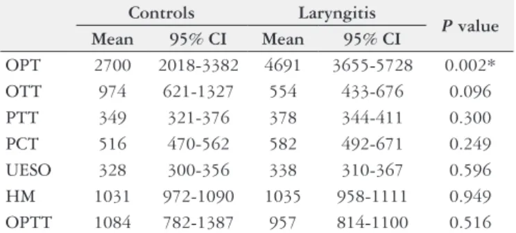
with laryngitis (4691 ms) than in controls (2700 ms) (P=0.002,
TABLE 4), with no difference for liquid or solid bolus (FIGURE 1). OTT for solid bolus was faster in patients (548 ms) than in con-trols (933 ms) (P=0.012, TABLE 5), with no difference for liquid
or paste bolus (FIGURE 2). PTT, PCT, UESO, HM and OPTT
were not different between patients and controls for none of the three bolus swallowed.
In patients with dysphagia (n=10) and without dysphagia (n=11) oral preparation for the paste bolus was longer than in con-trols. The difference was also observed in patients with dysphagia for solid bolus (P<0.050, FIGURE 3A). Duration of oral transit in patients with dysphagia was shorter for liquid (controls: 730 ms; patients: 472 ms), paste (controls: 974 ms; patients: 464 ms) and solid (controls: 933 ms; patients: 397 ms) boluses than in controls (P<0.050, FIGURE 3B). There was no difference in oral or phar-yngeal transit time between individuals with increased esophageal acid exposure and individuals without this alteration (P>0.350).
TABLE 4. Duration of oral and pharyngeal events, in milliseconds (ms), in patients with laryngitis (n=21) and healthy controls (n=21) after swallowing of the paste bolus
Controls Laryngitis
P value Mean 95% CI Mean 95% CI
OPT 2700 2018-3382 4691 3655-5728 0.002*
OTT 974 621-1327 554 433-676 0.096
PTT 349 321-376 378 344-411 0.300
PCT 516 470-562 582 492-671 0.249
UESO 328 300-356 338 310-367 0.596
HM 1031 972-1090 1035 958-1111 0.949 OPTT 1084 782-1387 957 814-1100 0.516
OPT: oral preparation time; OTT: oral transit time; PTT: pharyngeal transit time; PCT: pharyngeal clearance time; UESO: upper esophageal sphincter opening; HM: hyoid movement; OPTT: oropharyngeal transit time. *P<0.050.
TABLE 5. Duration of oral and pharyngeal events, in milliseconds (ms) in patients with laryngitis (n=21) and healthy controls (n=21) after swallowing of solid bolus
Controls Laryngitis
P value Mean 95% CI Mean 95% CI
OPT 18381 15928-20834 20471 18180-22761 0.242
OTT 933 518-1500 548 419-677 0.012*
PTT 325 295-354 358 324-393 0.199
PCT 509 464-554 659 437-880 0.092
UESO 258 242-274 287 257-316 0.167
HM 1066 991-1141 1047 973-1121 0.753 OPTT 1043 875-1211 1081 795-1367 0.942
OPT: oral preparation time; OTT: oral transit time; PTT: pharyngeal transit time; PCT: pharyngeal clearance time; UESO: upper esophageal sphincter opening; HM: hyoid movement; OPTT: oral-pharyngeal transit time. *P<0.050.
FIGURE 1. Duration of oral preparation time, in milliseconds (ms), in patients with laryngitis and healthy controls during the swallowing of liquid, paste and solid boluses. Horizontal bars represent the means.
FIGURE 2. Duration of oral transit time, in milliseconds (ms), in patients with laryngitis and healthy controls during the swallowing of liquid, paste and solid boluses. Horizontal bars represent the means.
DISCUSSION
It was observed that patients with laryngitis have longer OPT for the paste bolus and a faster OTT for the solid bolus than con-trols. There was no difference between the groups for swallowing parameters of liquid bolus. In addition, patients with dysphagia have a faster OTT with liquid, paste and solid bolus than healthy volunteers.
Dysphagia, commonly associated with GERD(8), is a frequent complaint in patients with laryngitis, described in 32% of patients in a previous study(15) and in 47.6% of patients in this investigation. The longer oral preparation and faster oral bolus transit may be caused by altered coordination of pharyngeal-esophageal phase rather than alteration of the oral swallowing phase, considering these patients have higher sensitivity to reflux episodes, mainly in the proximal esophagus, than people without GERD(16-18). Also, the oral phase of swallowing is a voluntary stage, which allows the individuals to control it as desired. There is an interaction between the pharynx and the esophagus, and abnormalities in such coordination may result in esophageal symptoms or disease that affect both oral and pharyngeal phases of swallowing(19,20). In this context, the longer oral preparation may be the cause of the more frequent premature spillage seen in the patients. Important to consider the possibility that pain during swallowing (odynophagia) causes longer oral bolus preparation and faster oral transit. The number of patients with odynophagia (five) was not enough to reach any conclusion about the influence of pain on swallow dynamics. However, make sense the possibility that pain during swallows as the cause of alterations of oral dynamics of patients with dysphagia.
In our study it is impossible to say that the patients had supra-esophageal manifestations of GERD. The diagnosis of GERD as the cause of pharyngeal manifestations of reflux is not easy(3,5,21), and there is no strong evidence that a positive response to treat-ment with proton bomb inhibitors means that the laryngitis was caused by GERD(22).
A previous study described that patients with GERD had a longer transit through the mouth, pharynx and upper esophageal sphincter(10) which could be an adaptive mechanism to prevent GERD-related esophageal symptoms and protect the esophageal body from the ongoing bolus. In our study, although the patients with laryngitis showed a faster OTT, no changes in PTT was ob-served, which may be an attempt to minimize symptoms during the pharyngeal passage of the bolus.
The longer oral preparation may cause the more frequent pre-mature bolus spillage seen in patients, which may be the cause of the
faster oral transit, an interaction between the oral and pharyngeal phases of swallowing(19,20,23).
Our results, together with those reported by studies investigat-ing swallowinvestigat-ing dynamics in patients without neurologic diseases or anatomic alterations of mouth or pharynx(24,25), indicate that in cases when only changes in pharyngeal or in esophageal mucosa are seen, there is no important clinical alterations in the oral or pharyngeal phases of swallowing. Although dysphagia may be as-sociated with neurologic impairment and anatomic diseases that can affect these swallowing stages, the symptom may also result from esophageal hypersensitivity and/or hypervigilance, without any alteration in the oral, pharyngeal or esophageal transit(9,26).
In patients with non-erosive GERD, proximal and distal es-ophageal mucosa have more superficial afferent nerves compared with no reflux disease or patients with erosive disease and patients with Barrett esophagus(27). Patients with extra esophageal mani-festations of gastroesophageal reflux frequently have non-erosive disease in esophagus(15), with the possibility to have more superficial afferent nerves and hypersensitivity in proximal esophageal body, explaining the possibility of dysphagia in these patients. This hypothesis needs more advanced investigation with methodology that permits a clear characterization of supra esophageal reflux of gastric content and association with symptoms. Chronic laryngitis is a disease with heterogeneous causes, with GERD as one cause or an aggravating factor(7).
The limitation of this investigation was that it was not possible to distinguish laryngitis caused by GERD from other causes, since the influence of different causes on swallowing may not be the same. However, patients did not have clinical history which suggested other cause for laryngitis. In addition, the controls did not have a nasofibroscopic evaluation, and it is known that even in supposedly normal individuals changes in the hypopharynx associated with GERD may be found(28). None of the control volunteers reported any complaints suggestive of GERD.
In conclusion, patients with laryngitis have a longer oral prepa-ration before swallows of the paste bolus and a faster transit of a solid bolus through the mouth compared to healthy subjects. A longer oral preparation for paste and solid boluses, a faster transit through the mouth, and perhaps pain during swallows, are associ-ated with dysphagia in patients with laryngitis.
Authors’ contributions
Moda I, Ricz HMA, Aguiar-Ricz LN, Dantas RO. Deglutição em pacientes com laringite. Arq Gastroenterol. 2018;55(1):50-4.
RESUMO – Contexto – Disfagia é uma queixa presente em 32% dos pacientes com laringite. Objetivo – O objetivo desta investigação foi avaliar o
trân-sito oral e faríngeo de pacientes com laringite, com a hipótese de que a alteração no trântrân-sito do bolo pela boca e faringe pode estar envolvida com a queixa de disfagia. Métodos – A avaliação videofluoroscópica da deglutição de bolos líquido, pastoso e sólido foi realizada em 21 pacientes com laringite, 10 deles com disfagia e 21 voluntários normais da mesma idade e sexo. Duas deglutições de 5 mL de bolo líquido, duas deglutições de bolo pastoso e duas deglutições de bolo sólido foram avaliadas em sequência casual definida por sorteio. Bolo líquido foi sulfato de bário 100%, e o bolo pastoso foi preparado com 50 mL de bário líquido e 4 g de espessante alimentar (amido e maltodextrina). O bolo sólido foi 2,2 g de uma bolacha macia embebida em bário líquido. A duração da preparação oral, trânsito oral, trânsito faríngeo, depuração da faringe, abertura do esfíncter superior do esôfago, movimento do hióide e do trânsito oral-faríngeo foram medidas. Precedendo a videofluoroscopia todos pacientes realizaram exame de pHmetria de 24 horas. Resultados – O registro do pH intraesofágico distal revelou resultado anormal em 10 pacientes. Pacientes com laringite apre-sentaram maior duração da preparação oral para bolo pastoso e um tempo de trânsito oral mais rápido para bolo sólido. Os pacientes com laringite e disfagia tiveram uma preparação oral mais longa para bolo pastoso e sólido e tempo de trânsito oral menor com bolos líquido, pastoso e sólido.
Conclusão – Preparação oral mais longa para bolos pastoso e sólido e trânsito mais rápido através da boca são situações associadas com a presença
de disfagia em pacientes com laringite.
DESCRITORES – Laringite. Transtornos de deglutição. Faringe. Monitoramento do pH esofágico. Fluoroscopia.
REFERENCES
1. Saritas Yuksel E, Vaezi MF. Extraesophageal manifestations of gastroesoph-ageal reflux disease: cough, asthma, laryngitis, chest pain. Swiss Med Wkly. 2012;142:w13544.15.
2. Richter JE. The many manifestations of gastroesophageal reflux disease: presen-tation, evaluation, and treatment. GastroenterolClin N Am. 2007;36:577-99. 3. Tsoukali E, Sifrim D. Investigation of extraesophageal gastroesophageal reflux
disease. Ann Gastroenterol. 2013;26:290-5.
4. Diamond L. Laryngopharyngeal reflux – it is not GERD. JAAPA 2005;18:50-3. 5. Herbella FAM, Andolfi C, Vigneswaran Y, Patti MG, Pinna BR. Importance of esophageal manometry and pH monitoring for the evaluation of otorhinolat-ingologic (ENT) manifestations of GERD. A multicenter study. J Gastrointest Surg. 2016;20:1673-8.
6. Asaoka D, Nagahara A, Matsumoto K, Hojo M, Watanabe M. Current perspec-tive on reflux laryngitis. Clin J Gastroenterol. 2014;7:471-5.
7. Wang AJ, Liang MJ, Jiang AY, Lin JK, Xiao YL, Peng S, et al. Comparison of patients of chronic laryngitis with and without troublesome reflux symptoms. J Gastroenterol Hepatol. 2012;27:579-85.
8. Kidambi T, Toto E, Ho N, Taft T, Hirano I. Temporal trends in the relative prevalence of dysphagia etiologies from 1999-2009. World J Gastroenterol. 2012; 18:4335-41.
9. Lazarescu A, Karamanolis G, Aprile L, Oliveira RB, Dantas R, Sifrim D. Percep-tion of dysphagia: lack of correlaPercep-tion with objective measurements of esophageal function. Neurogastroenterol Motil. 2010;22:1292-e337.
10. Cassiani RA, Mota GA, Dantas RO. Oral and pharyngeal bolus transit in gas-troesophageal reflux disease. Esophagus. 2015;12:345-51.
11. Mendell DA, Logemann JA. A retrospective analysis of the pharyngeal swallow in patients with clinical diagnosis of GERD compared with normal controls: a pilot study. Dysphagia. 2002;17:220-6.
12. Jamieson JR, Stein HJ, DeMeester TR, Bonavina L, Schwiger W, Hinder RA, et al. Ambulatory 24-hour esophageal pH monitoring: normal values, optimal thresholds, specificity, sensitivity and reproducibility. Am J Gastroenterol. 1992; 87:1102-11.
13. Cichero JAY, Lam P, Steele CM, Hanson B, Chen J, Dantas RO, et al. Develop-ment of international terminology and definitions for texture – modified foods used in dysphagia management: the IDDSI framework. Dysphagia. 2017;32: 293-314.
14. Schall R. Estimation in generalized linear models with random effects. Biometrika. 1991;78:719-27.
15. Stein DJ, Noordizi JP. Incidence of chronic laryngitis. Ann Otol Rhinol Laryngol. 2013;122:771-4.
16. Patel A, Sayuk GS, Gyawali CP. Prevalence, characteristics, and treatment outcomes of reflux hypersensitivity detected on pH impedance monitoring. Neurogastroenterol Motil. 2016;28:1382-90.
17. van Hoeij FB, Weijenborg PW, Weerman MAB, van den Wijngaard RMJGJ, Verheij J, Smouth AJPM, et al. Mucosal integrity and sensitivity to acid in prox-imal esophageus in patients with gastroesophageal reflux disease. Am J Physiol. 2016;311:G117-22.
18. Weijenberg PW, Smouth AJPM, Bredenoord AJ. Esophageal acid sensitivity and mucosal integrity in patients with functional heartburn. Neurogastroenterol Motil. 2016;28:1649-54.
19. Massey BT. Pathological pharyngo-esophageal interactions. Dysphagia. 1995;10:232-4.
20. Triadafilopoulos G, Hallstone A. Nelson-Abbot H, Bedinger K. Oropharyngeal and esophageal interrelationships in patients with non obstructive dysphagia. Dig Dis Sci. 1992;37:551-7.
21. Madanick RD. Extraesophageal presentations of GERD: where is the science? Gastroenterol Clin N Am. 2014;43:105-20.
22. Vaezi MF. Reflux and laryngeal symptoms: a sea of confusion. Am J Gastroen-terol. 2016; 111:1525-7.
23. Cassiani RA, Santos CM, Parreira LC, Dantas RO. The relationship between the oral and pharyngeal phases of swallowing. Clinics. 2011;66:1385-8. 24. Duca AP, Dantas RO, Rodrigues APC, Sawamura R. Evaluation of swallowing
in children with vomiting after feeding. Dysphagia. 2008;23:177-82.
25. Santos CM, Cassiani RA, Dantas RO. Videofluoroscopic evaluation of swallowing in Chagas’ disease. Dysphagia. 2011;26:361-5.
26. Aziz Q, Fass R, Gyawali CP, Miwa H, Pandolfino JE, Zerbib F. Esophageal disorders. Gastroenterology. 2016;150:1368-79.
27. Woodland P, Ooi JL, Grassi F, Nikaki K, Lee C, Evans JA, et al. Superficial esophageal mucosal afferent nerves may contribute to reflux hypersensitivity in nonerosive reflux disease. Gastroenterology. 2017;153:1230-9.