www.rbo.org.br/
issn/$–see front matter © 2013 Sociedade Brasileira de Ortopedia e Traumatologia. Published by Elsevier Editora Ltda. All rights reserved. doi: 10.1016/j.rboe.2012.10.003
A RT I C L E I N Fo
Article history:
Received on May 21, 2012 Approved on October 3, 2012
Keywords: Simvastatin Tibial fractures Rats
Fracture healing
a b s t r a c t
Objective: Evaluate the effects of simvastatin in the process of fracture healing in rat tibia. Methods: Thirty-six rats were subjected to diaphyseal fracture of the leg bones and divided in the statin group (GE) and control group (GC), being subdivided into three subgroups according to days post-fracture (7th, 14th and 28th day) to assess bone healing. In GE was
administered by gavage a solution of simvastatin to the sacrifice. In the control group was administered saline by the same route of SG. Immobilization was not used. After the sacrifice was made to limb amputation in the distal femur and conducted the clinical, radiological and histological analysis. Clinical evaluation was made as to the mobility of the fracture. Then the samples were radiographed and evaluated for callus diameter. Histological examination was performed with cuts of 5 micrometers and stained with hematoxylin-eosin, Masson’s trichrome and Alcian blue pH 2.5. The level of significance to exclude the null hypothesis was 5%. Results: All GE animals showed greater stability of the fracture and higher callus area. There were no significant changes in the histological study. Conclusion: Simvastatin accelerates the consolidation process by increasing the callus, but does not alter the histology of the newly formed bone.
© 2013 Sociedade Brasileira de Ortopedia e Traumatologia. Published by Elsevier Editora Ltda. All rights reserved.
Original Article
Evaluation of simvastatin in the process of fracture healing in
tibiae of rats
José Wanderley Vasconcelos,
1,*Leopoldina Milanez da Silva Leite,
2Gerusa de Arruda Vasconcelos,
3Igor Marcelo Souto Maior Araujo,
4Letácio Santos Garcia Ferro
51PhD in Surgery from Universidade Federal do Rio de Janeiro; Associate Professor at Universidade Federal do Maranhão (UFMA), São Luís,
MA, Brazil.
2PhD in Clinical and Experimental Physiopathology from Universidade do Estado do Rio de Janeiro; Professor at UFMA, São Luís, MA,
Brazil
3Nurse; MSc in Health Sciences from São Luís, MA, Brazil. 4MD from UFMA, São Luís, MA, Brazil.
5MD from UFMA, São Luís, MA, Brazil.
Work performed in the Laboratory of the Maranhão Academic League for Experimental Surgery (LabLacema), UFMA, São Luís, MA, Brazil.
*Corresponding author at: Av. dos Holandeses, quadra 19, Residencial Monet, apt. 1202, Olho D’Água, CEP 65 065-180, São Luís, MA, Brazil. Tel.: (98) 3248-6319 and 8119-0794.
Introduction
Many studies have indicated that statins act towards remodeling and formation of bone tissue and are thus osteoinductive substances.
Mundy et al.1 reported from studies on rodents that statins
have anabolic effects on bone, with stimulation of bone tissue formation. Statins are believed to act by strengthening the expression of bone morphogenic protein-2 (BMP-2) in osteoblasts. However, in attempting to reproduce the experiments of Mundy et al.,1 Maritz et al.2 did not find the
expected effect from statins. On the contrary, they reported that simvastatin produced an inhibitory effect with regard to bone formation, with increased bone reabsorption.
Several clinical investigations have reexamined the study series by means of observational and cohort experiments, in an attempt to evaluate the effect of statins on bone metabolism, fracture risk and bone consolidation.
Staal et al.3 reported that statins act by inhibiting the
mevalonate pathway. Mevalonate is the precursor for formation of steroids and the isoprenoids of steroids. These are involved in cell membrane biogenesis, DNA reproduction and protein glycosylation in a variety of cells. Statins inhibit the activity of 3-hydroxy-3-methylglutaryl-coenzyme A reductase (HMG-CoA), thereby reducing cholesterol synthesis, which is required for conversion of HMG-CoA to mevalonate, and this is the same pathway as in bisphosphonate action. In acting to diminish mevalonate synthesis, statins interfere with cell proliferation and activity.
Baek et al.4 studied the effect of simvastatin on the
proliferation and differentiation of cells of the bone marrow stroma in culture medium. They isolated mononuclear cells and cultured an osteoblastic lineage from the bone marrow of healthy volunteers. In the first culture medium, simvastatin diminished the average size of the colonies that give shape to fibroblastic units (colony-forming units, CFU-Fs) and emphasized calcification of the matrix. These cells were subcultured and the alkaline phosphatase activity of each group was measured in relation to time. Simvastatin increased the alkaline phosphates activity in a dose-dependent manner, and the effect was most evident over the initial periods of culturing. It was concluded from that study that simvastatin has a stimulatory effect on bone formation by means of osteoblastic differentiation.
Researchers from the Heart Protection study did not reported any difference in hospital treatment for the same type of fractures caused by car accidents, between the group that used simvastatin (40 mg/day) and the placebo group.5
Through these studies, contradictory results regarding the effect of statins on bone metabolism have been generated.6
Moreover, the action of this drug on fractures, mineral density and bone remodeling in humans is uncertain. Thus, a path towards new studies has been opened up, through the need for further observational, clinical, randomized and experimental studies on statin use at appropriate doses.
The aim of this paper was to evaluate the effect of statins on fracture healing in rats.
Methodology
Thirty-six adult male Wistar rats (Rattus norvegicus albinus; Rodentia: Mammalia), aged 45 to 65 days and weighing 200 to 300 g, were used. They were housed in the Laboratory of the Maranhão Academic League of Experimental Surgery (LabLacema), in polypropylene boxes (0.15 m2), with a
maximum of five animals in each box. This study followed the standards of the Nomina Anatomica Veterinaria7 and the
ethical principles for animal experimentation (Cobea), and it had been approved by the Ethics Committee of the Veterinary Department of Universidade Estadual do Maranhão.
Each animal was anesthetized with ketamine at a dose of 75 mg/kg and xylazine at a dose of 8 mg/kg intramuscularly in the left gluteal region. Antisepsis was performed on the right hind leg using a degerming solution and iodine-based antisepsis.
To produce the tibial fracture, pliers with three pressure points were used, in accordance with the technique established by Otto et al.8 The rats then remained housed in their respective boxes, without any type of immobilization or restriction of weight-bearing on the fractured limb.
The animals were distributed randomly by means of a draw into two groups of 18, which were named the statin group (SG) and control group (CG). Each group was divided into three subgroups (SSG and SCG) that were defined with numerical suffixes corresponding to the number of days between the surgical procedure and sacrifice, thus:
SSG-07 and SCG-07: (sacrifice on the 7th day) – five rats in each subgroup;
SSG-14 and SCG-14: (sacrifice on the 14th day) – five rats in each subgroup;
SSG-28 and SCG-28: (sacrifice on the 28th day) – eight rats in each subgroup.
These intervals between fracture production and sacrifice of the animals followed the experimental model proposed by Padula et al.9
Each rat was weighed and identified in its box with stripes made using a CD pen on the animal’s tail. The rat identified as R1 was marked with one transverse stripe, R2 with two transverse stripes, R3 with three transverse stripes, R4 with one longitudinal stripe and R5 was not marked but was only subjected to the stress using a pen without ink. For SSG-28 and SCG-28, there were four animals in four boxes (two in each subgroup), thus making a total of 16 animals, which were identified as R1 to R4.
The SG rats were subjected to oral gavage with a simvastatin solution at a dose of 20 mg/kg, 12 hours after fracture production, and this was repeated every 24 hours. In the CG, gavage was performed using placebo. Gavage was performed every day until the date of sacrifice.
Grade 4 Complete bone union
Grade 3 Bone union incomplete due to presence of small quantity of cartilage in the callus
Grade 2 Well-formed hyaline cartilage bridge can be seen, uniting the main fragments (complete cartilaginous union
Grade 1 Incomplete cartilaginous union, with retention of fibrous elements in the chondral plate
Grade 0 Absent or delayed fracture repair, characterized by presence of cartilage between the fragments and remains of hematoma or other fluid (pseudarthrosis)
Chart 1 - Histomorphological classification proposed by Allen et al. for staging bone consolidation.
third of the femur in order to study the bone callus. The skin around the musculature of the tibia was then removed without damaging the muscle group.
The mobility of the fracture focus was clinically evaluated. Grade 0 was attributed to consolidated fractures, grade 1 to intermediate patterns of movement and grade 2 to cases of clear movement.10 All the examiners were defined by means
of a draw and they were unaware of which group they were going to examine. After the clinical evaluation of mobility of the fracture focus, the surgical specimen was fixed in 10% formol. The area of the bone callus was assessed by means of analysis on digital radiographs, using the Auto CAD® 2007 software (Fig. 1). The entire callus area was delimited and measured in mm2.
For the histological analysis, the fractured tibiae and their surrounding soft tissues were decalcified for 12 to 15 hours and then dehydrated, clarified and embedded in paraffin. Sections of thickness 5 micrometers were cit sagittally to the fracture plane, using a microtome. These sections were stained with hematoxylin-eosin (HE), Masson’s trichrome (MT) and Alcian blue pH 2.5 (AB). HE and MT were used to study the cellularity of the extracellular matrix, while AB showed the sulfated and non-sulfated acid proteoglycans, which reflect the chondrogenic activity. All the laminas were analyzed by means of optical microscopy. The stage of bone consolidation was determined in accordance with the classification system proposed by Allen et al.11 (Chart 1).
Fig. 1 - Use of Autocad® for calculating the area of the callus.
Descriptive statistical techniques were applied, thus enabling exploratory analysis through parameter estimates (mean standard deviation and median) for all the variables investigated in this study. Following this, bone consolidation was assessed by means of the dependent variable (bone area) in relation to the independent variables of days and groups.
Considering that the sample size was small and that the data did not present normal distribution, the nonparametric Mann-Whitney test was performed. To analyze the histological data, quantitative assessment was performed using Fisher’s exact test. For all the tests, the significance level for rejecting the nullity hypothesis was 5%.
Results
All the animals accepted the diet well and could move around after receiving the fracture. They presented claudication of the fractured limb after an average of six to eight days, and there was no difference between the statin group (SG) and the control group (CG). None of the animals presented an operative wound at the fracture site.
Clinical analysis
Regarding the mobility of the fracture focus after seven days, in SCG-07 there were four animals with intermediate mobility (grade 1) and one with clear mobility (grade 2), while in SSG-07 all five animals had intermediate mobility (grade 1).
In the subgroup evaluated 14 days after fracturing, there were four animals in SCG-14 without mobility (grade 0) and one with intermediate mobility (grade 1), while all five animals in SSG-14 had mobility grade 0.
All the animals that were evaluated 28 days after receiving the fracture presented mobility grade 0 on examination.
Radiological analysis
All the animals in the subgroups that were sacrificed on the seventh day presented fractures of the tibia and fibula, thus demonstrating that the fracture closure took place in a standardized manner. No radiological bone callus was found in any of the rates at this time.
Among the subgroups that were sacrificed on the 14th and
28th days, bone calluses and bone bridges were seen in all the
rats. In SSG-14, the mean area of the callus was 66.65 mm², while in SCG it was 55.29 mm². SSG-28 also had a greater mean area of bone callus than SCG-28, with values of respectively 67.46 mm2 and 56.09 mm2 (Fig. 2). Therefore, the simvastatin
group presented a mean bone callus size that was greater than in the control group, both in the 14 and 28-day evaluations and in the overall assessment.
There was a significant difference in relation to callus area (p = 0.043), such that the average area in the statin group (67.1 mm2) was greater than the area in the control group (55.4 mm2).
four grade 3, two grade 2 and one grade 1. In SSG-28, there was bone callus grade 3 in six animals (Fig. 5) and grade 1 in two. In all the animals of this subgroup, bone tissue with presence of osteocytes and osteoblasts was found.
In the quantitative evaluation, Fisher’s exact test was used and no statistically significant difference was found between the groups.
Discussion
The bone consolidation process is influenced by a variety of factors that have been studies in depth from the times of Hippocrates to the present day.
In the present study, simvastatin was chosen from among various inhibitors of HMG-CoA reductase because like atorvastatin and cerivastatin, it presents greater in vitro effects on bone than seen with lovastatin and pravastatin.3
Oxlund and Andreassen12 found that treatment with
simvastatin at a dose of 20 mg/kg orally partially impeded bone loss in female Wistar rats that were subjected to ovariectomy, and also increased cortical bone formation. Based on these authors’ results, we used this dose taking into account simvastatin metabolism in the liver, in order to investigate its effect on bone tissue.
Some authors, like Skoglund et al.,13 used much larger
doses of the drug, of 120 mg/kg, in order to demonstrate the effects of statins on bone consolidation in animals. They concluded that animals only presented increased bone callus area and noted that simvastatin acts on the mechanism for mobilization of cells involved in the fracture repair process, thereby leading to increased bone callus size.
Fig. 3 - Photomicrographs of sections from animals in the 14-day subgroups, grade 1, SSG-14. (a) Presence of many neoformed vessels in fibrous connective tissue, with presence of formation of immature bone tissue. H&E x4. (b) With MT.
Histological analysis
All the rats were histologically evaluated by the pathologist responsible, in a random manner without access to or knowledge of the control group, the group divisions or the respective days of sacrifice of each subgroup.
In SCG-07, there were elements of fibrous connective tissue, absence of or only small focal areas of bone tissue, with bone calluses of grade 1 observed in three animals and grade 0 in two. In SSG-07, there was fibrous connective tissue and cartilaginous tissue, with the periphery of the callus composed of elements of mature and immature bone tissue. In four animals, grade 1 bone callus was seen and in one, grade 0.
In SCG-14, four rats presented fibrous connective tissue and all of them had elements of chondrogenesis. There was grade 1 bone callus in four of these animals and grade 2 in only one. In SSG-14, three rats were classified as grade 2 and two as grade 1, and bone tissue was present on the periphery of the callus (Figs. 3 and 4).
In SCG-28, there was greater variation regarding the Allen classification. In this subgroup, one had bone callus of grade 4,
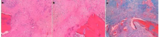
Fig. 4 - Photomicrographs of sections from animals in the 14-day subgroups, grade 2. (A) SCG-14. Bone extremity with mature cartilaginous tissue, with complete union. HE x4. (B) SSG-14. Extremity with mature cartilaginous, forming complete hyaline bridge. HE x4. (C) SSG-14. Opposite extremity with vascular neoformation. MT x4.
Fig. 5 - Photomicrographs of sections from animals in SSG-28, grade 3. Formation of incomplete bone tissue due to presence of cartilaginous tissue in focal area
In our study, we also observed that the bone callus area increased, thus corroborating these authors’ affirmation.13
The acceleration of the consolidation process in the statin group may have occurred in SSG-07, but this could not be seen in our study because the callus is radiotransparent at this stage of radiographic evaluation. On the other hand, both SSG-14 and SSG-28 presented large areas of bone callus, thus demonstrating acceleration of the consolidation process.
However, in the study by Skoglund et al.,13 the results
relating to the callus in the rats treated with simvastatin for 14 days were better than ours, with a cross-sectional area 53% larger than that of the control. However, after 21 days of treatment, the fracture consolidation did not present any differences between the control group and simvastatin group. In our study, SSG-28 presented bone callus size greater than that of the control, thus disagreeing with the above authors.
The findings of Maritz et al.,2 in which statins increased
the bone metabolism rate, thus increasing both bone formation and bone reabsorption, seem to favor bone consolidation when statins are used.
We also agree with the findings of Tsubone et al.,14
regarding the capacity of statins to promote bone formation and improve the consolidation process. These authors studied cerivastatin used in bone transplantations in rat tibiae and found through histological analyses that it increased the formation of new bone tissue and increase bone union.
As already described, the mechanism for inducing bone consolidation triggered by simvastatin is mediated by increased production of BMP-2. Therefore, it is possible to compare the results from studies that used BMP-2 rh in the consolidation process and those that used statins.15
Stein et al.15 studied the effects of rhBMP-2 on consolidation
of unstable and stable fractures of the tibia in rabbits, administered in biodegradable particles, in collagen gel or by injection. When rhBMP-2 was injected, the callus of mechanically unstable fractures developed more rapidly. In the same way as in our study on statins, these authors concluded that although BMP-2 accelerates the callus development rate and cortical bone union, it does not affect the quantity of bone and cartilage produced. In our study, there was no statistically significant difference regarding histomorphological classification when 7, 14 and 28 were compared. However, there were certain variations in histological grade between the different subgroups.
Conclusion
Simvastatin promoted greater clinical stability for the fracture focus and increased the size of the bone callus, 14 and 28 days after the fracturing event, such that this difference was statistically significant. It also produced a difference in the histological analysis on the callus, but without statistical significance.
Conflicts of interest
The authors declare that there was no conflict of interests in conducting this study.
R E F E R E N C E S
1. Mundy G, Garret R, Harris S, Chan J, Chen D, Rossini G et al. Stimulation of bone formation in vitro and in rodents by statins. Science. 1999;286(5446):1946-9.
2. Maritz FJ, Conradie MM, Hulley PA, Gopal R, Hough S. Effect of statins on bone mineral density and bone histomorphometry in rodents. Arterioscler Thromb Vasc Biol. 2001;21(10):1636-41. 3. Staal A, Frith JC, French MH, Swartz J, Güngör T, Harrity TW et al. The ability of statins to inhibit bone resorption is directly related to their inhibitory effect on HMG-CoA reductase activity. J Bone Miner Res. 2003;18(1):88-96.
4. Baek KH, Lee WY, Oh KW, Tae HJ, Lee JM, Lee EJ et al. The effect of simvastatin on the proliferation and differentiation of human bone marrow stromal cells. J Korean Med Sci. 2005;20(3):438-44.
5. Rosenson RS, Tangney CC, Langman CB, Parker TS, Levine DM, Gordon BR. Short-term reduction in bone markers with highdose sinvastatin. Osteoporos Int. 2005;16(10):1272-6. 6. Rizzo M, Rini GB. Statins, fracture risk, and bone remodeling:
what is true? Am J Med Sci. 2006;332(2):55-60.
7. International Committee on Veterinary Gross Anatomical Nomenclature. Nomina Anatomica Veterinária. Columbia: Editorial Committee Hannover, 1983.
8. Otto TE, Patka P, Haarman HJ. Closed fracture healing: a rat model. Eur Surg Res. 1995;27(4):277-84.
9. Padula EOC, Andrade ML, Giordano V, Ramalho MV. Aspectos morfológicos do processo de consolidação de fratura em ratos diabéticos. Rev Bras Ortop. 2003;38(3):127-36. 10. Pozenato LC, Santana PJ, Guarniero J, Oliveira LAA,
Domingues BL. Efeitos da ipriflavona sobre a consolidação de fraturas em ratas com desnutrição protéica: trabalho experimental. Rev Bras Ortop. 2004;39(7):390-7.
11. Allen HL, Wase A, Bear WT. Indomethacin and aspirin: effect of nonsteroidal anti-inflammatory agents on the rate of fracture repair in the rat. Acta. Orthop. Scand. 1980;51(4):595-600.
12. Oxlund H, Andreassen TT. Simvastatin treatment partially prevents ovariectomy-induced bone loss while increasing cortical bone formation. Bone. 2004;34(4):609-18.
13. Skoglund B, Forslund C, Aspenberg P. Simvastatin improves fracture healing in mice. J Bone Miner Res. 2002;17:2004-2008. 14. Tsubone T, Shigetomi M, Ihara K, Ikeda K, Merida L, Ohno T et
al. Hypertrophy of vascularized bone isograft in rats treated with cyclosporine A. Calcif Tissue Int. 2003;73(4):393-9. 15. Stein D, Lee Y, Schmid MJ, Killpack B, Genrich Ma, Narayana