www.rbo.org.br/
issn/$–see front matter © 2013 Sociedade Brasileira de Ortopedia e Traumatologia. Published by Elsevier Editora Ltda. All rights reserved. doi: 10.1016/10.1016/j.rboe.2012.10.002
*Corresponding author at: Av. São José 300, Cristo Rei, CEP: 80050-350, Curitiba, PR, Brazil. E-mail: [email protected]
A RT I C L E I N Fo
Article history:
Received on July 1, 2012 Approved on October 3, 2012
Keywords:
Knee Dislocation/diagnosis Knee Dislocation/surgery Knee Dislocation/epidemiology Knee Injuries/diagnosis Knee Injuries/surgery
a b s t r a c t
Objective: Describe the ligamentous and associated injuries that occur in the traumatic knee dislocation, relating them to the mechanisms of trauma and to identify patterns of injuries. Methods: Twenty three knee dislocations were described in the period between March 2010 and March 2011. After the diagnosis of the lesions, the reduction and transarticular external fixation of the dislocated knees were done. At the second moment, the patients were evaluated with physical examination under anesthesia and the surgical exploration of peripheral lesions was perfomed by a surgeon of the knee surgery group of this institution. The patients data with the description of the injuries were found and registered. Results: 65% of patients were male, the average age was 35 years and the most common mechanism of trauma was the motorcycle accident (60%). The lesion of the anterior cruciate ligament (ACL) occurred in 75% of the cases, and the lesion of posterior cruciate ligament (PCL) in 95%. The medial peripheral injuries happened in 65% of the dislocations, and the lateral lesions in 40%. The most common dislocations were classified as KDI (25%) and as KDIIIm (25%). The arterial injury was present in 15% of the cases, and the nervous injury where registered in one patient (5%). At the initial radiographic evaluation, 45% of the dislocations presented reduced. Conclusion: The characteristics of the knee dislocations described showed a great range of variability demonstrating that an individualized evaluation of each case is mandatory. The surgeon should be able to recognize and choose the correct treatment to these lesions.
© 2013 Sociedade Brasileira de Ortopedia e Traumatologia. Published by Elsevier Editora Ltda. All rights reserved.
Original Article
Knee dislocation: descriptive study of injuries
Fabiano Kupczik,
1Marlus Eduardo Gunia Schiavon,
2Lucas de Almeida Vieira,
3,*Daniel Pundek Tenius,
4Rodrigo Caldonazzo Fávaro
51MSc in Surgery, Pontifícia Universidade Católica do Paraná (PUC-PR); Head of the Knee Surgery Group, Hospital Universitário Cajuru, and
Preceptor of Medical Residence in Orthopedics and Traumatology, Hospital Universitário Cajuru, Curitiba, PR, Brazil.
2Orthopedist and Traumatologist; Member of the Knee Surgery Group, Hospital Universitário Cajuru, Curitiba, PR, Brazil. 3Orthopedist and Traumatologist; Fellow of Knee Surgery, Hospital Universitário Cajuru, Curitiba, PR, Brazil.
4Orthopedist and Traumatologist; Member of the Knee Surgery Group, Hospital Universitário Cajuru, Curitiba, PR, Brazil. 5Orthopedist and Traumatologist; Fellow of Knee Surgery, Hospital Universitário Cajuru (2012), Curitiba, PR, Brazil.
Introduction
Traumatic dislocation of the knee is one of the most severe injuries to this joint. In the literature, some authors have stated that injury to two ligament structures is needed in order to define this dislocation.1 Other papers have suggested
that damage to the joint capsule with loss of femorotibial congruence characterize this dislocation.2,3
Historically, it was described as a rare and unusual injury4,5
totaling 0.2% of orthopedic injuries and 0.5% of dislocations.2,6
However, its incidence has increased over recent years because of high-energy trauma and improvements in care for multiple trauma patients.6,7 This incidence could even be higher
because of cases of spontaneous reduction, which are rarely registered.8,9
From the statistics in the literature, these dislocations are most common among young male patients. They are related to motor vehicle accidents in up to half of the cases, followed by sports injuries and low-energy trauma.3
In addition to the joint instability caused by ligament injury, associations with artery and nerve injury are another factor that might add severity to knee dislocations.10 Injury
to the common fibular nerve gives rise to loss of function of the musculature of the anterior and lateral compartments of the lower leg, and also sensory changes to the corresponding dermatomes.11,12 Injury to the popliteal artery requires early
attendance, to avoid the risk of ischemia of the lower limb and the possibility of amputation.13
The classifications most used are those of Kennedy14 and
Schenck.7 The first of these describes the anatomical position
of the distal segment in relation to the proximal segment. The second is based on involvement of the injured ligamentous structures in the dislocation.
There continues to be controversy regarding the treatment for these injuries, and several protocols have been described. However, surgical treatment followed by physiotherapeutic rehabilitation has shown better results than conservative treatment.3,10,11,15,16
Joint stiffness secondary to arthrofibrosis and failure of ligament reconstruction are the commonest early complications. Over the long term, more than 50% of the patients will develop post-traumatic osteoarthrosis.3 This
implies great limitations for patients who are mostly young and socioeconomically active, and treatment and rehabilitation for these dislocations requires large investments from the public and/or supplementary healthcare systems.6
The objectives of this study were to describe the ligament injuries and associated injuries that occur in knee dislocations and to correlate them with the trauma mechanisms and identify the injury patterns.
Materials and methods
The medical files of 23 patients with traumatic knee dislocations who were attended at the emergency service of an institution within the Brazilian public healthcare system (Sistema Único de Saúde, SUS), over a 12-month period
from March 2010 to March 2011, were analyzed. This study had previously been approved by the institution’s ethics committee under the registration number 0005182/11, through a report dated August 3, 2011. Patients with a diagnosis of acute dislocation of the knee joint who were attended at the emergency service due to loss of joint congruence, and whose data available from the medical files answered the questions raised in this investigation, were included. Medical files with incomplete data were excluded from the study.
All the patients were assessed at the emergency service through history-taking, physical examination and initial radiographs. The patients were initially attended by an emergency physician, who then activated the on-call orthopedics and traumatology team. The team was composed of one orthopedist who was a member of staff of this institution and three resident physicians of orthopedics and traumatology who were accredited by the Ministry of Education (MEC) and by the Brazilian Society of Orthopedics and Traumatology (SBOT). A vascular surgery assessment was requested for all the patients as soon as the diagnosis of knee dislocation was suspected. At the institution, on-call orthopedics and traumatology care is provided on a precautionary basis, and thus the assessment took place at the surgical center, to which the patients were taken after the initial assessment. After the initial orthopedic procedure, the vascular surgery team performed arteriography on all these patients.
The diagnosis of knee dislocation was made through history-taking with regard to complaints and the history of the trauma, in association with the physical examination on admission, presence of local deformity and functional incapacity, and radiographic evaluations. In the physical examination, special attention was given to joint instability, neurological function and the pulse and perfusion of the extremity affected.
After the initial attendance, the patients were taken to the surgical center where, under anesthesia, they were again examined. Joint reduction was performed, with stabilization using a transarticular external fixator in the lower limb affected, in order to control the initial orthopedic damage.
Peripheral ligamentous injuries and knee joint fractures, when present, were dealt with in a second procedure up to 15 days later. In this, the peripheral ligament was repaired or reconstructed and fracture fixation was implemented. This new surgical intervention was scheduled in accordance with the description from the physical examination under anesthesia. The initial and control radiographs after reduction and external fixation were also evaluated in this preoperative planning, along with the computed tomography scans that were produced in cases of fracture-dislocation after the initial surgical treatment. Magnetic resonance imaging (MRI) was not available for evaluating these patients through SUS at the institution.
collateral ligaments were tested using valgus and varus stress at 0º and 30º. The posterolateral corner was examined using the recurvatum test, dial test and posterolateral drawer test. The ligaments of the central pivot were not dealt with at the acute moment. The peripheral ligaments, when injured, were explored and their injuries were described.
After release from hospital, the patients were sent to the knee surgery outpatient clinic for appropriate follow-up and scheduling of arthroscopic reconstruction of the central pivot injuries, on an elective basis.
To analyze the patients’ medical files, the following items were gathered: 1) name, 2) sex, 3) age, 4) trauma mechanisms, 5) associated injuries, 6) intra-articular ligamentous injuries, 7) medial extra-articular ligamentous injuries, 8) lateral extra-articular ligamentous injuries, 9) arterial injuries, 10) neurological injuries, 11) other injuries, 12) associated injuries and 13) characteristics of the initial radiographic service (classified as reduced, dislocated or fracture-dislocation) (Fig. 1).
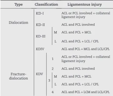
With the ligamentous injuries described by means of the physical examination and surgical exploration of the peripheral injuries, and with the presence or absence of intra-articular fractures, the dislocations were classified in accordance with Schenck7 (Table 1). In the presence of vascular injuries, the
letter “C” was added and in the presence of neurological injury, the letter “N”.9,7
Out of the 23 patients with a diagnosis of knee dislocation that were attended, 21 underwent initial surgical treatment at the orthopedics emergency sector, with a physical examination under anesthesia, closed reduction of the dislocation and transarticular external fixation.
The other two patients were cases of fracture-dislocation, in which the physical examination under anesthesia, the approach towards the peripheral ligamentous injuries and the fixation of joint fractures were done in the same initial surgical procedure, by one of the members of the knee surgery group who was part of the on-call orthopedics team at the time that these two patients were attended.
Three patients who were attended in the emergency service with the initial surgical procedure described above were excluded from the evaluation of this study: two patients because they were transferred to other hospitals and one patient because of associated cranioencephalic and abdominal trauma that necessitated a long recovery period in the intensive care unit.
Thus, out of the 23 cases of knee dislocation attended during this period at our institution, the injuries of 20 patients were described and analyzed.
Results
The dislocations occurred predominantly in male patients: 13 cases (65%). The right side was more affected: 11 patients (55%). The patients’ mean age was 35 years (range: 17-58).
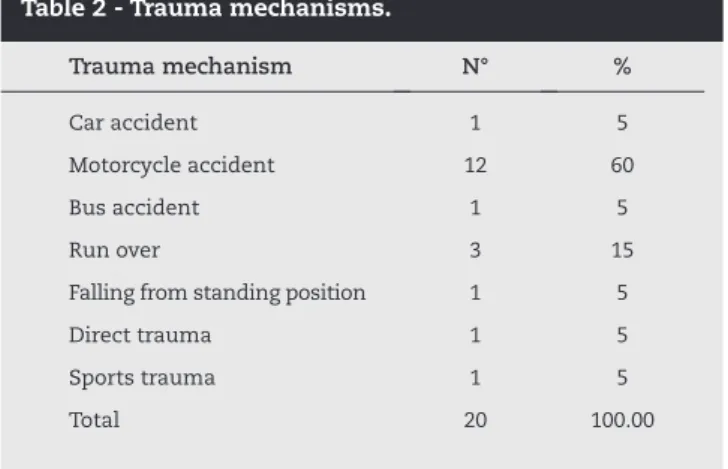
The commonest trauma mechanism was motorcycle accidents (60%). The trauma mechanisms are described in Table 2.
Table 3 shows the percentages of dislocations in relation to the Schenck classification.
ACL injuries occurred in 15 patients (75%) and were related to PCL lesions in 93.33%. PCL injuries occurred in 19 patients (95%) of the cases and 73.68% were correlated with ACL injuries. Injuries to the medial collateral ligament (MCL) occurred in 13 cases (65%), and these were related to PCL injuries in 92.30% and ACL injuries in 84.51%. Lateral peripheral injuries occurred in eight cases (40%) and 100% were related to PCL injuries. Among the lateral structures, injuries to the lateral collateral ligament (LCL) and the popliteal tendon occurred in all the patients and injuries to the biceps tendon in half of them.
Table 4 shows the frequencies of associations between the ligamentous injuries among the 20 cases for which the descriptions were complete.
Six cases of dislocation with involvement of only one of the cruciate ligaments were described (Table 5), and one of them was a fracture-dislocation (Schatzker IV).
Arterial injuries occurred in three patients (15%), classified as KDI, KDIIIL and V3M, which on radiography presented as reduced, dislocated and fracture-dislocation, respectively. The frequencies of their ligamentous injuries are described in Table 6.
Nerve injury was recorded in only one case (5%), classified as KDIIIL, which on the initial radiographic examination presented as reduced.
Fig. 1 - (A) Knee dislocation – Schenck KDIIIm. (B) Knee dislocation – Schenck KDV3m.
Type Classification Ligamentous injury
Dislocation
KD-I ACL or PCL involved + collateral ligament injury
KD-II ACL and PCL involved
KD-III M
ACL and PCL + MCL L ACL and PCL + LCL / CPL KDIV ACL and PCL + MCL and LCL/CPL
Fracture-dislocation
KDV
1 ACL or PCL involved + collateral ligament injury
2 ACL and PCL involved
Trauma mechanism N° %
Car accident 1 5
Motorcycle accident 12 60
Bus accident 1 5
Run over 3 15
Falling from standing position 1 5
Direct trauma 1 5
Sports trauma 1 5
Total 20 100.00
Table 2 - Trauma mechanisms.
Type Classification No %
Dislocation
KD-I 5 25
KD-II 0
-KD-III M 5 25
L 4 20
KDIV 2 10
Fracture-dislocation KDV
1 1 5
2 0
-3 M 3 15
L 0
-4 0
-Total 20 100
Table 3 - Schenck classification: real and percentage values of dislocations.
Associated ligamentous injuries
Ligament injury ACL PCL MEDIAL LATERAL
ACL (15 knees) - 14 (93.33%) (73.33%)11 6 (40%)
PCL (19 knees) 14 (73.68%) - (63.15%)12 8 (42.10%)
Medial (13 knees) 11 (84.51%) 12 (92.30%) - 2 (15.38%)
Lateral (8 knees) 6 (75%) 8 (100%) 2 (25%) -Table 4 - Associated ligamentous injuries.
Dislocation with involvement of only one cruciate ligament
Central pivot Medial Lateral Other injuries
Schenck I
ACL (one case) 1 - Patellar tendon PCL (four
cases) 2 2
-Schenck V1 PCL (one case) - - Iliotibial tract/Schatzker IV Table 5 - Injuries with involvement of one of the cruciate ligaments.
Frequency of ligamentous injury in
the three cases of arterial injury N° %
ACL 2 66.66%
PCL 3 100%
Medial 2 66.66%
Lateral 1 33.33%
Table 6 - Frequencies of ligamentous injuries in the three cases of arterial injury.
Knee joint fractures were described in five cases (25%). There were three cases of fracturing of the tibial plateau, classifies as Schatzker II (one case) and Schatzker IV (two cases). There was also one case of fracturing of the medial femoral condyle and of avulsion fracture of the tibial spine. In addition to these joint fractures, there was one case of fracturing of the fibular head. No fractures of the patella were encountered.
The following injuries to other structures relating to the knee were reported: medial meniscus, four cases (20%); lateral meniscus, two cases (10%); patellar ligament, two cases (10%); and iliotibial tract, two cases (10%). Injuries originating in the medial head of the gastrocnemius, medial patellofemoral ligament and pes anserinus were described in only one case (5%).
Other orthopedic injuries were found in five cases. In the lower limbs, there were two fractures of the acetabulum, one fracture of the pelvis, one floating knee, one patellar dislocation and one fracture of the contralateral tibial plateau. In the upper limbs, there was one glenohumeral dislocation and one fracture of the distal third of the radius. The initial radiographic evaluation showed that nine dislocations (45%) were presented reduced, seven (35%) were dislocated and four (20%) were cases of fracture-dislocation.
Out of the nine reduced dislocations, four of them (44.44%) were classified as KDIIIL, three (33.33%) as KDI and two (22.22%) as KDIIIM.
Among the seven cases in which the initial radiography showed dislocated knees, three cases (42.85%) were classified as KDIIIM, two (28.57%) as KDI and another two (28.57%) as KDIV (10% of all the dislocations).
Discussion
Acute knee dislocation is a severe injury with a potential risk of complications because of the neurovascular injuries that may be associated, thus requiring a high degree of suspicion at the time of the clinical evaluation.3 The dislocation is
associated with other musculoskeletal injuries and injuries to other systems, as presented in our results. However, these should not deflect attention from dealing with the knee, which requires careful physical examination, since almost half of the dislocations are presented reduced in the initial clinical and radiographic evaluation.
Comparing the classical studies on knee dislocation with the current studies, it is noticeable that there has been an increase in incidence but also an improvement in functional results over the last few years.6,14 Nonetheless, this injury
is still considered to be rare, and it presents in a variety of forms, which indicates that there is a need for individualized assessment and treatment for each case.
Clark and Engebretsen17 presented a table with 21 studies
published between 1987 and 2010. In this, they showed the number of patients and the length of the study. The largest sample comprised 21 cases of multi-ligament injury per year and the smallest comprised 1.1 dislocations per year. The mean was 4.9 cases per year. This analysis was difficult to do because of the diversity of the information cited: for example, the number of centers in which these studies were conducted, or whether multi-ligament injuries should be included along with cases of dislocation.
The sample of the present study, with 20 cases described and 23 diagnosed over a one-year period, was larger than the average in studies in the literature, probably because our hospital is a reference center for trauma, and also because of the high degree of suspicion at the first attendance of these injuries. These injuries were more common in motorcycle accidents, which demonstrates the increase in severity of motor vehicle trauma and the vulnerability of the lower limbs.1 They occurred more frequently in males, which was in
accordance with the data in the literature.1,17
In 1963, Kennedy published a classification of knee dislocations based on the position of the tibia in relation to the femur.14 Occurrences of spontaneous joint reduction, i.e.
before radiographic documentation could be produced (as seen in 45% of the cases of the present study), have made this classification unviable. The classification proposed by Schenck7
is more descriptive, grouping the injuries according to the ligamentous structures injured, presence or absence of joint fractures and presence or absence of vascular or nerve injuries, which this aids in planning the treatment. KDI and KDIIIM were the most frequent injury types in this sample. The injury types KDI or KDV1 indicate any dislocation with injury to only one of the cruciate ligaments, which includes injuries with lateral or medial peripheral involvement, or both of these. This may represent a limiting factor for the classification. Subtypes at these levels would provide a better description of dislocations with involvement of one or more cruciate ligament.
The susceptibility to arterial injury is explained by the local anatomy. The fibrous tunnel of the hiatus of the adductors and
the fibrous arch of the soleus muscle form two fixation points, thus creating an area that may be subject to stress caused by the dislocation or by hyperextension of the popliteal artery.10,13
There is controversy regarding the rate of these injuries and also regarding the main trauma mechanism for this. The incidence ranges from 7% to 64%, thus indicating that there is a need for vascular evaluation using protocols for investigating arterial injuries.10,16,18 The incidence in the present study was
15% (three cases), and in all of these the PCL was injured, but there was no correlation with the classification of the dislocations.
The common fibular nerve is the one most frequently injured in knee dislocations, particularly in posterolateral dislocations, when the nerve structures are strained.19 In the
present study, only one case was found (5%), in a knee that was presented reduced in the initial radiographic assessment. However, the incidence in the literature has ranged from 14% to 40%, and the final clinical result is worse when this injury is present. Presence of paresthesia should also signal the differential diagnosis with compartment syndrome. Both of these injuries may be present, and this necessitates physical examination in order to assess pain, mobilization of the extremity and edema. Perfusion, reflexes and neurological function are of prime importance. If necessary, the pressure of the compartment in question should be measured.6,10,11,20
Levy et al.21 recommended a sequenced approach,
using external fixation at the emergency attendance and ligament reconstruction three to six weeks after the injury, with reconstruction of all the ligaments by means of grafts from tissue banks. Clark and Engebretsen17 indicated
arthroscopic reconstruction of the central lesions and repair or reconstruction of the peripheral lesions as early as possible. Howells et al.3 indicated initial use of an external fixator in
cases of vascular injury, joint fracture and gross instability. Definitive surgery is recommended within two weeks.
In the present case series, the initial treatment consisted of joint reduction on an emergency basis and stable fixation using a transarticular external fixator in all the patients. This had the objectives of promoting better soft-tissue control, maintaining the joint reduction and facilitating patient management with regard to angiographic studies and nursing care.
The forms of definitive surgical treatment have varied in relation to the time of the surgery, open or arthroscopic reconstruction of the central pivot, and reconstruction or repair of the peripheral ligaments.22,23 There is controversy regarding
the management strategies, and recent studies have indicated acute reconstruction of the cruciate ligaments.6,17 There is a
consensus that surgical treatment with repair or reconstruction of the injured ligaments, followed by early gain of range of motion, provides better results for patients with traumatic knee dislocation than does conservative treatment.3,15,16,24,25
In the present study, extra-articular reconstructions were performed at the initial stage, as recommended in protocols for stepwise approaches.3,16,21 However, arthroscopic
reconstruction of the central pivot was scheduled as a second procedure, after rehabilitation, based on gain of range of motion and muscle strengthening.2,4,21,26 The objective was to diminish
patient who, because of social issues, have difficulty in the physiotherapeutic rehabilitation process.
Despite the diversity of treatment protocols and the better results from surgical treatment,27 there is still no reference
point regarding which injuries are the ones most encountered in cases of dislocation, along with their incidence and characteristics. In our sample of 20 cases over a 12-month period, we observed that PCL injuries occurred in 95% of the cases and ACL injuries in 75%, and that the frequency of medial injuries was greater than that of lateral injuries. The commonest type of fracture-dislocation was Schatzker IV.
Twaddle et al.1 described 63 dislocations over a five-year period, i.e. an average of 12.6 cases per year, in two large centers in the United States and New Zealand, and they showed that motor vehicle accidents were the main cause of dislocations, followed by sports trauma, thus demonstrating that sports practice may also have sufficient trauma energy for complex knee injuries. They also highlighted the great variety of injuries involved in traumatic knee dislocation and found that 67% of the dislocations reduced spontaneously at the time of hospital attendance. These authors stated that for dislocation to occur, injury to at least two ligaments would be needed, without depending on injuries to the two cruciate ligaments.
In the present study, a case of fracture-dislocation with an isolated injury to the PCL and peripheral ligament integrity was described. However, there was injury to the iliotibial tract and, in the medial part, instability due to fracturing of the tibial plateau. The rate of reduced dislocations was smaller (45%). However, if the fracture-dislocations are excluded and only the cases of dislocated and reduced knees are evaluated, the percentage of injuries that were shown to be reduced on the initial radiograph would be 56%, i.e. closer to the value of that series, in which fracture-dislocations were not mentioned.
The dislocations were evaluated without using MRI because this examination was available to SUS patients at our institution. This is one of the limiting factors of the present study, with regard to better description of the injuries.
MRI is not a replacement for a good physical examination under anesthesia, and should therefore be used as an adjuvant method for detection and description of injuries.24 Lonner et
al.28 stated that the physical examination under anesthesia is
more accurate than MRI for describing ligamentous injuries in cases of knee dislocation. However, in some treatment protocols, this imaging examination is said to be fundamental and is done on all patients.21,23
Thus, with a physical examination under anesthesia, initial radiographs and surgical exploration, it was possible to describe the ligamentous and associated injuries and demonstrate their great variability. This variability means that each patient requires individualized treatment.
Conclusion
Knee dislocation is a severe injury that presents challenges to orthopedists, who need to be able to recognize them and provide emergency and specific treatment for the various structures that have been damaged.
The diversity of injury patterns and high percentage of associated injuries indicated that multidisciplinary individualized assessment is needed for each case. There needs to be a high degree of suspicion in order to recognize reduced dislocations and detect complications such as neurovascular injuries.
Conflicts of interest
The authors declare that there was no conflict of interests in conducting this study.
R E F E R E N C E S
1. Twaddle BC, Bidwell TA, Chapman JR. Knee dislocation: Where are the lesions? J Orthop Trauma. 2003;17(3):198-202. 2. Seroyer ST, Musahl V, Harner CD. Management of the
acute knee dislocation: The Pittsburgh experience. Injury. 2008;39(7):710-8.
3. Howells NR, Bruntona LR, Robinsona J, Porteusa AJ, Eldridgea JD, Murraya JR. Acute knee dislocation: An evidence based approach to the management of the multiligament injured knee. Injury. 2010;42(11):1198-204.
4. Roberston A, Nutton RW. The dislocated knee. Curr Orthop. 2006;20:95-120.
5. Meyers M, Harvey JP Jr. Traumatic dislocation of the knee joint. J Bone Joint Surg Am. 1971;53(1):16-29.
6. Engebretsen L, Risberg MA, Robertson B, Ludvigsen TC, Johansen S. Outcome after knee dislocations: a 2-9 years follow-up of 85 consecutive patients. Knee Surg Sports Traumatol Arthrosc. 2009;17(9):1013-26.
7. Schenck RC Jr. The dislocated knee. Instr Course Lect. 1994;43:127-36.
8. Siliski JM. Evaluation and treatment of dislocation of the knee. Mediguide Orthop. 1991;10:1-7.
9. Schenck RC Jr. Classification of knee dislocations. Oper Tech Sports Med. 2003;11:193-8.
10. Schenck RC Jr, Stannardl JP, Wascher DC. Dislocations and fracture: dislocations of the Knee. In: Court-Brown C, Heckman JD, Bucholz RW. Rockwood and Greens fractures in adults. 6th. Lippincott: Williams Wilkins; 2006.
11. Bonneviallea P, Dubranab F, Galaudc B, Lustigd S, Barbiere O, Neyretd P, et al. Common peroneal nerve palsy complicating knee dislocation and bicruciate ligaments tears. Orthop Traumatol Surg Res. 2010;96(1):64-9.
12. Cush G, Irgit K. Drop foot after knee dislocation: evaluation and treatment. Sports Med Arthrosc. 2011;19(2):139-46. 13. Green NE, Allen BL. Vascular injuries associated
with dislocation of the knee. J Bone Joint Surg Am. 1977;59(2):236-9.
14. Kennedy JC. Complete dislocation of the knee joint. J Bone Joint Surg Am. 1963;45:889-904.
15. Werier J, Keating JF, Meek RN. Complete dislocation of the knee. The long-term results of ligamentous reconstruction. Knee. 1998;5:255-60.
16. Levy BA, Fanelli GC, Whelan DD, Stannard JP, Macdonald PA, Boyd JL, et al. Controversies in the treatment of knee dislocations and multiligament reconstruction. J Am Acad Orthop Surg. 2009;17(4):197-206.
18. Hollis JD, Daley BJ. 10-year review of knee dislocations: is arteriography always necessary? J Trauma. 2005;59(3):672-5. 19. Sisto DJ, Warren RF. Complete knee dislocation. Clin Orthop
Relat Res. 1985;(198):94-101.
20. Eranki V, Begg C, Wallace B. Outcomes of operatively treated acute knee dislocation. Open Orthop. 2010;4:22-30.
21. Levy BA, Krych AJ, Shah JP, Morgan JA, Stuart MJ. Staged protocol for initial management of the dislocated knee. Knee Surg Sports Traumatol Arthrosc. 2010;18(12):1630-7. 22. Dedmond BT, Almekinders LC. Operative versus
nonoperative treatment of knee dislocations: a meta-analysis. Am J Knee Surg. 2001;14(1):33-8.
23. Harner CD, Waltrip RL, Bennett CH, Francis KA, Cole B, Irrgang JJ. Surgical management of knee dislocations. J Bone Joint Surg Am. 2004;86(2):262-73.
24. Stannardl JP, Schenck RC Jr , Fanelli GC. Dislocation and fracture-dislocation of the knee. In: Bucholz RW, Heckman jD, Court-Brown CM, Tornetta P. Rockwood and Green’s fractures in adults. 7th ed. Philadelphia: Lippincott Williams & Wilkins; 2010. p.1833-64.
25. Peskun CJ, Whelan DB. Outcomes of operative and nonoperative treatment of multiligament knee injuries: an evidence-based review. Sports Med Arthrosc. 2011;19(2):167-73. 26. Ibrahim SA, Ahmad FH, Salah M, Al Misfer AR, Ghaffer SA,
Khirat S. Surgical management of traumatic knee dislocation. Arthroscopy. 2008;24(2):178-87.
27. Hirschmann MT, Zimmermann N, Rychen T, Candrian C, Hudetz D, Lorez LG, et al. Clinical and radiological outcomes after management of traumatic knee dislocation by open single stage complete reconstruction/repair. BMC Musculoskelet Disord. 2010;27;11:102.