RBCCV 44205-1421 DOI: 10.5935/1678-9741.20120096
Effect of exercise associated with stem cell
transplantation on ventricular function in rats
after acute myocardial infarction
Efeito do exercício associado ao transplante de células-tronco sobre a função ventricular de ratos
pós-infarto agudo do miocárdio
Simone Cosmo
1, Julio César Francisco
2, Ricardo Correa da Cunha
3, Rafael Michel de Macedo
4,
José Rocha Faria-Neto
5, Rossana Simeoni
6, Katherine Athayde Teixeira de Carvalho
7, Marcia
Olandoski
8, Nelson Itiro Miyague
9, Vivian Ferreira do Amaral
10, Luiz César Guarita-Souza
111. Biologist, Master's Degree student of Pontiical Catholic University of Paraná (PUCPR), Curitiba, PR, Brazil. Project design.
2. Biochemist at PUCPR. Project design, bone-marrow blood collection. 3. PhD, Positivo University, Curitiba, PR, Brazil. Guidance on
methodology of swimming physical activity.
4. PhD, PUCPR, Curitiba, PR, Brazil. Development of methodology and discussion.
5. PhD, PUCPR, Curitiba, PR, Brazil. Review of the article, methodology and discussion.
6. MD, PhD; PUCPR, Curitiba, PR, Brazil. Isolation of mononuclear cells.
7. MD, PhD, Pequeno Príncipe Faculty Institute and Paraná Federal University, Curitiba, PR, Brasil. Aid in the methodology of cell isolation.
8. PhD, PUCPR, Curitiba, PR, Brazil. Statistical analysis.
9 MD, PhD, PUCPR, Curitiba, PR, Brazil. Performance of ecocardiographic exams.
10. PhD, PUCPR, Curitiba, PR, Brazil. Paper discussion.
11. PhD, Adjunct Professor, PUCPR, Curitiba, PR, Brazil. Guidance on the project.
This study was carried out at Pontiical Catholic University of Paraná, Curitiba, PR, Brazil.
Correspondence address: Luiz César Guarita-Souza
Rua Rosa Kaint Nadolny, 190 – 5º andar – Curitiba, PR Brazil – Zip code: 81200-525
E-mail: [email protected]
Support: CNPq
Article received on May 8th, 2012 Article accepted on September 27th, 2012
Abstract
Objective: To assess the functional and anatomical-pathological effect of transplantation of bone marrow mononuclear cells associated to aquatic physical activity after myocardial infarction in rats.
Methods: Twenty-one rats were induced by myocardial infarction, through left coronary artery ligation. After a week, the animals were subjected to echocardiography for evaluation of left ventricle ejection fraction (LVEF, %) and dyastolic and end systolic volume of the left ventricle
Results: Two months after the transplant, were observed decrease in FE in the control group (35.20 to 23.54 P=0.022) and addition of LVEF and stabilization of ventricular remodeling in the group trained with cells (29.85 to 33.43% P=0.062 and 0.71 to 0.73 ml, P=0.776, respectively). Identiied
the reduction of collagen ibers, myocardial ibrosis regions
in the group trained with and without cells.
Conclusion: The group trained with cells improves ventricular function compared to the control group,
suggesting the beneit of associated cell therapy will physical
activity.
Descriptors: Myocardial infarction. Stem cell transplantation. Exercise.
Resumo
Objetivo: Avaliar o efeito da associação terapêutica entre o transplante autólogo de células-tronco e o exercício físico aquático, sobre a fração de ejeção do ventrículo esquerdo (FEVE) de ratos com disfunção ventricular pós-infarto agudo do miocárdio (IAM).
Métodos: Foram induzidos ao IAM, por ligadura da artéria coronária esquerda, 21 ratos Wistar. Os animais
foram submetidos à ecocardiograia para avaliação da FEVE (%) e dos volumes diastólico e sistólico inais do ventrículo
esquerdo (VDF, VSF, ml), randomizados e ao transplante das células-tronco mononucleares. Os animais foram divididos em quatro grupos: grupo sedentário sem células (n=5), sedentário com células (n=5), treinado sem células (n=5) e treinado com células (n=6). O treinamento físico foi iniciado 30 dias após o IAM e realizado em piscina adaptada durante
30 dias. No início e no inal do protocolo de treinamento físico,
foram realizadas dosagens de lactato. Os animais foram
submetidos a nova ecocardiograia após 60 dias do IAM.
Resultados: Comparação dos valores de FEVE 30 dias e 60 dias pós-IAM, respectivamente: sedentário sem (35,20 ± 7,64% vs. 22,39 ± 4,56% P=0,026), com células (25,18 ± 7,73% vs. 23,85 ± 9,51% P=0,860) e no treinado sem (21,49 ± 2,70% vs. 20,71 ± 7,14% P=0,792), treinado com células (28,86 ± 6,68 vs. 38,43 ±7,56% P=0,062). Identiicou-se a diminuição de
ibras colágenas, nas regiões de ibrose miocárdica no grupo
treinado com e sem células.
Conclusão: A associação terapêutica entre exercício físico
e o transplante autólogo de células-tronco foi benéica contra as ações do remodelamento ventricular.
Descritores: Infarto do miocárdio. Transplante de células-tronco. Exercício.
Abbreviations, acronyms and symbols
ANOVA Analysis of variance
LVEF Left ventricular ejection fraction H&E Hematoxylin-eosin
AMI Acute myocardial infarction IMDM Iscove’s Modiied Dulbecco’s Media PUCPR Pontiical Catholic University of Paraná rpm Rotations per minute
FBS Fetal bovine serum
SD with Infarcted group without exercise, sedentary, with cells SD without Infarcted group without exercise, sedentary
TR with Infarcted group, trained, with cells TR without Infarcted group, trained, without cells EDV End-diastolic volume
LV Left ventricle ESV End-systolic volume
INTRODUCTION
Cardiovascular disease, the leading cause of death worldwide, constitute the major causes of morbidity and mortality, and acute myocardial infarction (AMI) is among the most frequent ischemic heart disease. Technological advances in the diagnosis and treatment have greatly increased the survival of patients, but the available options for the treatment of AMI are still palliative and limited, highlighting the need to develop new therapeutic modalities [1,2].
Although some authors suggesting that there mitotic division of the heart, the vast majority of cardiomyocytes has no capacity for regeneration after AMI and, when this occurs, there is deterioration of contractile activity and,
with the extensive area of AMI, ventricular remodeling can occur and heart failure [3].
Experimental studies indicate the possibility of myocardial regeneration through stem cell transplantation as an alternative for the treatment of this disease. In experimental models of acute and chronic myocardial ischaemia, implantation of bone marrow mononuclear cells was capable of improving myocardial perfusion and contraction. These results have been replicated in recent clinical studies in humans [4,5].
the functional capacity of the organs cellular structures and their systems. Pronounced changes have been observed in relation to the mechanisms of control of bodily functions and metabolic processes, including cellular self-regulation, neural and hormonal levels [6].
Therefore, we sought to assess whether aquatic physical activity associated with the transplantation of bone marrow mononuclear cells in AMI also offers this same beneit. The aim of this study is to assess the functional and pathological effect of transplantation of stem cells from the bone marrow associated with aquatic physical activity after MI with left ventricular dysfunction in rats.
METHODS
The research was performed at the Center for Surgery and Diagnosis of the Laboratory of Experimental Surgical Technique of the Pontiical Catholic University of Paraná (PUCPR), after having been approved by the Research Ethics Committee (registration at CEUA/PUCPR n°434), with animals from PUCPR vivarium, according to the principles of the Brazilian College of Animal Experimentation [7].
We used 30 male Wistar rats weighing between 260 and 300g. Rats were grouped and kept stored in polypropylene cages at ambient with controlled temperature (22ºC - 24ºC) and light, under light/dark (12/12h) cycles, and water ad libitum. All animals underwent induction of MI. After seven days, they underwent echocardiography and animals that had ejection fraction (LVEF) below 35% were included in the study (n=21). The remaining nine animals died from extensive AMI. At this moment, the animals were randomly divided into four groups, listed below:
• Group I: infarcted without exercise, sedentary (SD without). The animals were monitored for 60 days (n = 5);
• Group II: infarcted without exercise, sedentary, cell (SD with). The animals were transplanted with bone marrow mononuclear cells and monitored for 60 days (n = 5);
• Group III: infarcted, trained without cells (RT without). The animals underwent physical activity for 15 minutes three times a week and monitored for 60 days (n = 5);
• Group IV: infarcted, trained with cell (TR with). The animals were transplanted with bone marrow mononuclear cells and underwent physical activity for 15 minutes three times a week and monitored for 60 days (n = 6).
Physical activity was initiated 30 days after induction of MI, after a period of aquatic adaptation.
Description of the procedure
All rats in the experiment underwent general anesthesia with a combination of ketamine and xylazine (50 mg/kg) intramuscularly. After induction of anesthesia,
we observed absence of ocular relex eyelid. With the relaxation of the animal, we obtained a deinitive airway by tracheal intubation with peripheral venous line number 14. After each surgery, the airway patency was checked by connecting the catheter to the mechanical ventilation system with volume of 2.5 ml (O2/min.) And frequency of 50 cycles per minute, observing the lungs expansion.
Then, there were chest antisepsis using topical povidone-iodine and left thoracotomy in the third intercostal space. After opening the left pleura, the animal was connected to the mechanical ventilation system. We used the volume respirators (Harvard®, Inc., model 683 respirator, Massachusetts, USA) for small animals, with 21% oxygen (room air). The pericardium was opened for dislocation and better visualization of the area to be approached. After exteriorization of the heart, the left atrium was removed and the left coronary artery ligated using polypropylene monoilament nonabsorbable blue 7.0 suture, between the outlow of the pulmonary artery and left atrium. The infarcted region was immediately visualized by differential staining of the affected area. Then the heart was repositioned to the chest, the hyperinlated lungs and the chest wall sutured in layers using monoilament nylon nonabsorbable monoilament 4.0 suture. After recovery from anesthesia, the animals were kept in cages and fed with standard commercial diet and had free access to water.
Echocardiography
the same observer in a blinded method, with the inal result the average of the three.
Obtaining blood from bone marrow
After 7 days of AMI, bone marrow blood was obtained. For this purpose, we used the aspiration puncture method in the bone marrow of mice in autologous way, always preceded by anesthesia: ketamine (50 mg/kg) and xylazine (10 mg/kg). The animals were placed in the lateral position, with the lower leg bent and the upper straight. The puncture-aspiration was performed in the posterior iliac crest of the femur with disposable syringe (BD Plastipak®-) 5 mL, with 0.2 mL of heparin (5000 IU/mL), using 21 mm needle 25x8 G1 (BD-Precision Glide®); approximately 1 mL of blood from bone marrow was collected from each rat, followed by identiication of syringes[8].
Isolation of mononuclear bone marrow stem cells For the isolation of the mononuclear fraction we used density gradient (d=1.077 g/m3) (Ficoll-Hypaque Sigma, St. Louis, MO) according to Böyum, on modiied-Dulbecco's Iscove's media (IMDM GIBCO-BRL) supplemented with 1% antibiotics (penicillin and streptomycin) and 20% buffer solution. The material collected from each rat was placed in plastic sterile centrifuge tube of 15 ml. Immediately after completed this tube up to 12 ml with IMDM culture media (Iscove's Modiied Dulbecco's Media), it was supplemented with 4% buffer and 1% antibiotics (penicillin and streptomycin) and homogenated. In a plastic tube of 15 ml, 3 mL of density gradient separation (d= 1.077) (Ficoll-Hypaque) were placed and hence then added to the homogenate containing animal bone marrow and IMDM culture medium carefully so it does not mix [8,9].
This tube was taken and subjected to centrifuge at 1400 revolutions per minute (rpm) for 40 minutes at 22°C. Soon after, it was led to low again, and removed the ring formed between the middle and gradient. This homogenate was withdrawn, which mononuclear stem cells were, and were placed into another plastic tube of 15 mL. It was completed with IMDM medium up to 15mL and centrifuged again by 1500 rpm for 10 minutes at 22°C. After removal of the centrifuge tube, it was performed quickly removal of discard from the supernatant. A precipitate was observed at the bottom of the bottle, which were mononuclear stem cells. After repeating the previous step and placing 13 mL of medium in the tube, the precipitate was resuspended and centrifuged again at 1500 rpm for 10 minutes at 22°C. After this phase, the supernatant was discarded, placed 3 mL of medium in the tube and resuspended the cells to count them. This count was performed in a Neubauer chamber and examined under an optical microscope Olympus® CX31 in a 40X objective [8].
Cell transplantation
Cells derived from bone marrow mononuclear fraction were suspended in IMDM containing 20% fetal bovine serum (FBS, Gibco BRL, Grand Island, NY) and 1% antibiotic (100 μ/ml penicillin and 100/μL streptomycin). The cell transplantation was performed on the same day of bone marrow puncture, in the transition area between the AMI and the intact myocardium, in the anterior wall of the left ventricle. The infusion was 15 μl of cells at a concentration of 5 x 10⁶, using Hamilton syringe (LT 1701, Hamilton Bonaduz AG).
Exercise
Adaptation Protocol
Physical activity with swimming was performed in temperature-controlled environment at 30°C, using a pool of 85 cm long by 30 cm wide by 50 cm high. The animals were adapted before training for 10 minutes at three different levels of water column: 20 cm with the irst day, second day with 30 cm and last day with 40 cm [10].
Training protocol
Physical activity occurred with swimming for 30 days in trained groups. The animals were exercised for 15 minutes a day, three days a week. The onset of activity occurred after 30 days of AMI. The same time was observed for the group of control animals, or that is, without exercising [10].
Lactate blood assessment
Blood samples (25 µl) were collected from the tail of the animal, to quantify the exercise. Samples were collected during two periods: the irst day of physical activity, being considered baseline data and 30 days after physical activity. The rats which exercised were tested immediately before and after physical activity. Trained groups were assessed with and without cells. Lactate concentration was determined using the portable lactimeter (Accutrend). The sedentary groups with and without cells underwent lactate concentration assessment, aiming to expand the control sample.
Euthanasia
All animals euthanized received the lethal dose (LD50) (148 mg/kg) of the anesthetic ketamine [11]. The samples were sent for histopathological analysis.
Pathological study
Hearts were preserved in vials containing 10% formalin for 24 hours. After this period, the hearts were cleaved into four crossed equal parts in the microtome (Leica RM2145 model) with a thickness of 5 mm.
underwent successive baths in 70%, 80% and 90% alcohol, three baths in 100% alcohol (Leica TP1020 model) for one hour. Thereupon, liquid parafin was impregnated in the sections through three baths at 65°C in the same apparatus. Then the sections were mounted on slides and stained with hematoxylin-eosin (H&E) and Picrosirius Red. Two blades from each fragment were performed with four cuts and the mentioned colorings.
Morphometric analysis
The morphometric analysis was performed on the 60th day after AMI, once the markers were directed at the chronic phase of healing. We performed analysis of collagen, coloring the histological cuts using H&E technique and examining under an optical microscope. The slides were examined with knowledge of known identity in a light microscope (Olympus BX40), increase of 200 X, coupled to a Sony® camera and a computer. We used the Image-Pro Plus® for Windows software for digital image analysis.
Slide images were captured for later analysis on a computer and, using the Image-Pro Plus® software, the selected areas of interest were measured.
With the dropper tool, we selected the objects of interest and the program automatically generated the measure. Since the total area of the exam was constant, we selected statistics of the program that provided the percentage of the area occupied by the object of study, or that is, the collagen.
Ten ields were measured in histologic sections of each blade in the area of AMI, obtaining then an average reading of those cuts.
Statistical Analysis
To compare the pre- and post moments within each group, we used the Student's t test for paired samples. To compare the groups with respect to the results of pre-moments, we used the analysis of variance model (ANOVA) with one factor. To compare the groups regarding the post-assessment results and regarding the differences between pre and post was used analysis of covariance, considering the pre-measure and the result of lactate as covariates. P values <0.05 were considered statistically signiicant. Data were organized into an Excel spreadsheet and analyzed using the Statistica software v.8.0.
RESULTS
Intragroup echocardiographic analysis
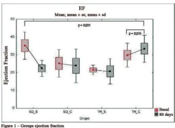
Regarding LVEF, we identiied a decrease in this parameter in the sedentary groups with and without cells and in the trained groups without cells 60 days after AMI (35.20 ± 7.64% vs. 22.39 ± 4.56% P= 0.026 and 25.18 ±
7.73% vs. 23.85% P= 9.51 ± 0.860 and 21.49 ± 2.70% vs. 20.71 ± 7.14% P= 0.792, respectively). Regarding the group
trained with cells, we identiied increasing of this parameter
vs. 29.85 ± 6.68%. 33.43 ± 7.56%, P=0.246 (Figure 1).
With respect to SV, we identiied an increase in this parameter in the sedentary groups with and in the trained group without cells 60 days after AMI (0.39 ± 0.15 ml vs. 0.65 ± 0.12 ml P=0.020, 0.50 ± 0.07 ml vs. 0.98 ± 0.12 ml P=0.018, 0.50 ± 0.09 ml vs. 0.64 ± 0.05 ml P=0.014, respectively). Regarding the trained group with cells, we identiied that this parameter decreased from 0.61 ± 0.14 ml vs. 0.59 ± 0.22 ml (P=0.872).
Regarding the EDV, we identiied increase in this parameter in the four groups 60 days after acute myocardial infarction: sedentary with and without cells and trained with and without cells (0.59 ± 0.19 ml vs. 0.83 ± 0.13
P=0.117 ml, 0.89 ± 0.13 vs. ml. 1.25 ml ± 0.20 P=0.033
vs. 0.70 ± 0.14 ml. 0.82 ml ± 0.09 P=0.058; 0.71 ± 0.13 ml
vs. 0.73 ml ± 0.06 P=0.776, respectively).
Intergroup echocardiographic analysis
Comparing the four groups together, statistically signiicant difference in echocardiographic values 30 days after AMI was found in the LVEF and EDV parameters of the LV.
Fig. 1 - Left ventricular ejection fraction
Regarding LVEF 30 days after AMI, we identiied that the groups were not homogeneous (P=0.022), so we
used an analysis of covariance. When assessing the results in 60 days after AMI, we found statistically signiicant difference only when comparing sedentary groups without cells with trained with cells (P=0.031) and trained groups
Regarding the EDV parameter 30 days after AMI, we identiied that the groups were also not homogeneous (P=0.040), thus we used analysis of covariance. When
assessing the results in 60 days we observed statistically signiicant differences between the sedentary groups with and without cells (P<0.001), sedentary with cells and
trained without cells (P <0.001) and sedentary and trained
with cells (P <0.001).
Regarding the ESV parameter 30 days after AMI, we identiied that the groups were homogeneous (P=0.052).
After 60 days there were statistically signiicant differences between the sedentary groups with and without cells (P=0.008), sedentary with cells and trained without cells
(P=0.007) and sedentary and trained with cells (P=0.002).
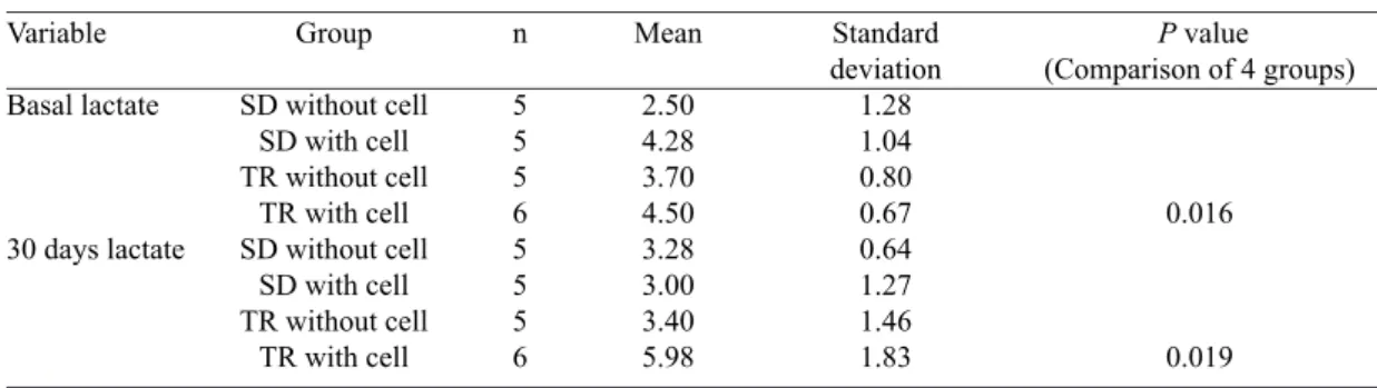
Table 2. Lactate
Comparison of groups two by two SD without x SD with
SD without x TR without SD without x TR with SD with x TR without SD with x TR with TR without x TR with
Pre 0.009 0.065 0.003 0.354 0.710 0.187
Post 0.762 0.897 0.007 0.666 0.003 0.009 Table 1. Lactate
Variable
Basal lactate
30 days lactate
Group
SD without cell SD with cell TR without cell
TR with cell SD without cell
SD with cell TR without cell
TR with cell
n
5 5 5 6 5 5 5 6
Mean
2.50 4.28 3.70 4.50 3.28 3.00 3.40 5.98
Standard deviation
1.28 1.04 0.80 0.67 0.64 1.27 1.46 1.83
P value (Comparison of 4 groups)
0.016
0.019
Table 3. Collagen Variable AMI area
Group SD without cell
SD with cell TR without cell
TR with cell
n 5 5 5 6
Mean 4.2 4.2 9.7 3.8
Standard deviation 3.8 2.5 2.7 3.5
P value
<0.001
AMI= acute myocardial infarction
Fig.2. Collagen
Lactate assessment
The values obtained after lactate are shown in Tables 1 and 2.
Collagen assessment
DISCUSSION
Physical activity can produce changes in myocardial perfusion. Coronary low is inversely proportional to vascular resistance exerted specially by the vessels situated in the microcirculation. The increase in cardiac metabolism produced by exercise promotes the reduction of vascular tone (microcirculation), consequently improving myocardial perfusion. This improvement can be considered signiicant, even when considered other components involved in the process, such as endothelial function, microcirculation, regression of coronary atherosclerotic lesions, increased collateral circulation, reduce blood viscosity and increased diastolic perfusion time [12,13].
The transplantation of bone marrow mononuclear stem cells in ischemic cardiomyopathy has been performed with results that suggest improved myocardial function, especially for the mechanism of angiogenesis at the site of transplantation [14].
Physical activity also has the potential of regional vasodilation in regions close to AMI, which enables improvement of infarcted myocardium perfusion and also overall recovery of LV function. These two treatment options have been applied in patients with associated heart failure. In this study, we used a similar experimental model because it included animals with established ibrosis and severe left ventricular dysfunction.
The association between the two therapies showed a trend of better control over the deleterious factors of ventricular remodeling, since there was no signiicant difference in LVEF values obtained before and 60 days post-training. There was also increase in absolute values.
With respect to functional analysis in the pre-transplant, the four groups had the LV LVEF and EDV parameters with statistical difference between them, hence, the LVEF and EDV were assessed as covariance, in an attempt to homogenize them.
The control group, or that is, sedentary without cells, showed signiicant deterioration of LVEF after 60 days of AMI, conirming the effect of muscle necrosis and, as a consequence, the development of heart failure, which was already expected. It was identiied small drop of LVEF, both in the sedentary group with cell as in the trained group without cells, suggesting stabilization of cardiac function. This stabilization may be justiied by the beneit of physical activity in the trained group and the action of bone marrow mononuclear stem cells in the sedentary group, which has also been identiied in other studies [15].
Regarding the trained group with cells, we identiied functional beneit, which can be explained by the action of both mononuclear stem cells as physical activity, by the mechanisms above justiied. Comparing the sedentary
groups without cell (control) and trained with cells, this result was evident. However, when comparing the trained groups with and without cells it was also identiied beneit in the group with cells, suggesting that the beneit of bone marrow mononuclear stem cells may have been more signiicant.
Assessing only the animals that underwent physical activity, or that is, those trained, it was found that the animals that received the bone marrow mononuclear stem cells showed myocardial protective effect compared to animals that received no cells. It is true that when assessed the intragroup LVEF parameter, even in the trained group without cells, there was stabilization of LVEF as a decrease of 21.49% to 20.71% it is considered irrelevant. Thus, we suggest that the protective effect of physical activity in this model, although it was identiied higher percentage of collagen, or that is, myocardial ibrosis in this group.
When we assessed the sedentary animals, it was observed that the animals that received no cells can be considered the control group, as they were infarcted, did not practice physical activity and also not received cells. In the sedentary group that received cells, we identiied a decrease in LVEF from 25.18% to 23.85%, variation also considered no signiicant, suggesting a protective mechanism of transplanted myocardial cells.
Regarding LV EDV, the four groups showed an increase over the 60 days of AMI. In the control group, as a result of deterioration of ventricular function after AMI, it already was expected that ventricular remodeling would happen. However, the sedentary group with cells showed statistically signiicant ventricular dilation, which was also identiied in a study published by the same group [14]. Regarding the trained group with cells, although ventricular dilation is identiied, it was considered that there was stabilization of ventricular remodeling.
Regarding LV ESV, the sedentary groups with and without cells and trained group without cells showed an increase over the same period, suggesting a loss of contractile capacity, however the trained group with cells identiied a decrease in this parameter. Although not signiicant, it is suggested a functional protective capability of the combined treatment proposed.
As animals with LVEF less than 35% were included in the study and as already had signiicant ventricular dysfunction with increased ventricular volumes before transplantation, it is dificult to understand that transplantation of cells can exert an antiremodeling because treatment is only regional. It is believed that the beneits associated with physical activity can justify these results.
abnormal composition of peripheral skeletal striated muscle ibers, abnormalities of blood low and the chemorelex ventilatory control. All these changes result in lower exercise tolerance and lower functional capacity.
Guimarães et al. [17] described the limiting effects to training in patients with heart failure, highlighting the behavior of central and peripheral chemoreceptors. In these patients, there is increased sensitivity in peripheral chemoreceptors, which results in greater activation of the sympathetic nervous system, increasing blood pressure, ventilation and peripheral vascular resistance. This phenomenon is described as mecanorelex. Moreover, during the exercises, patients with heart failure present early ventilatory muscle fatigue, resulting in higher demand of afferent stimuli to the central nervous system by the ibers of the phrenic nerve, activating the sympathetic nervous system and triggering peripheral vasoconstriction and lower tolerance to training. This phenomenon was described as metaborelex [17,18].
Due to mecanorelex and metaborelex, there is a change in the composition of peripheral muscle ibers of patients with heart failure. According to Schulze et al. [19], patients with heart failure suffer tonic iber atrophy, due to the reduction in the number of mitochondria and myoglobin present in these ibers as a result of low blood low allowed by increased peripheral vascular resistance. Thus, the type II muscle ibers (phasic) become more active, and the anaerobic glycolysis as the main power supply to perform movements, resulting in increased lactic acidosis and lower exercise tolerance. Thus, patients with heart failure tend to have higher lactate production at rest, compared to patients without left ventricular dysfunction. This behavior is similar in rats.
Aerobic exercise and or respiratory muscle training reduce the effects caused by metaborelex and the mecanorelex. Chiappa et al. [20] showed that individuals who have respiratory muscle training reduced peripheral vascular resistance, improving perfusion due to better conditioning and diaphragmatic inhibition of the sympathetic nervous system action. Ferraz et al. demonstrated that aerobically trained patients present similar response [16-20].
Therefore, in this study it was expected that, with aerobic training, the animals presented decrease in lactate values at rest, according to Li et al. [21]. Furthermore, with the injection of mononuclear stem cells and possible improve of ventricular function, the effects of heart failure could be minimized and composition of peripheral muscle ibers reorganized properly, allowing better utilization as oxidative energy substrate [21].
The remarkable point is that the rats that received cells and trained increased lactate at rest, which can be a sign that in this group the effects caused by the mecanorelex
and metaborelex were not controlled. The difference between lactate values at rest before and after 30 days of physical activity was signiicant with P=0.019, but it is
important to note that in both groups there was an increase in home values.
When comparing the groups two by two with respect to lactate parameter, signiicant differences were found when compared the sedentary group without cells with the trained group with cells. This result suggests a readaptation to oxidative system of energy sources from peripheral muscle ibers in the group receiving cells. Another signiicant difference was found between the sedentary group that received cells and trained group with cells. This difference was signiicant, since the amount of lactate at rest of sedentary mice that received cells decreased, whereas those who have been trained increased. This demonstrates favorable peripheral adaptation of the ibers of the rats that receive cells, while the group that received training did not improve peripheral blood perfusion.
Ferraz et al. [16] demonstrated that patients undergoing low intensity training (anaerobic threshold intensity-equivalent) improved aerobic capacity more than those who trained at high intensity (near the ventilatory compensation point). This may have happened because the trained group with cells trained during the period at an intensity above lactate threshold or anaerobic threshold.
A limiting factor of this study was the fact of not having been determinded the point of lactate threshold or training stable state. Accordingly, the rats may have been trained constantly and anaerobically, which may have resulted in little improvement in exercise tolerance or lactate levels at rest.
Through the technique of morphometric analysis, quantitative assessments of collagen in hearts after 60 days of AMI were performed. The main result of this study with regard to the collagen assessment in the area of AMI was observed as a signiicantly less quantity in the group trained with cells. It is believed that factors released due to paracrine effect of stem cells can promote reduction of the ibrosis area, suggesting recovery of left ventricular function, which can be corroborated with improved LVEF and limiting ventricular remodeling. These two mechanisms may also be explained by the angiogenic potential of bone marrow mononuclear stem cells, previously described in other studies and the mechanisms of vasodilation produced by physical activity [14,15].
A study by Bolli et al. [22] demonstrated, after direct injection of mesenchymal stem cells in ischemic hearts, decreased ibrosis, apoptosis, and increased LVEF.
that early training after MI reduces the metalloproteinases expression. In this study, this inding was not identiied because there was a greater presence of collagen in the trained group without cells. However, it was found reduced expression of collagen when associated with mononuclear cell transplantation. These results suggest that physical training with the help of bone marrow mononuclear stem cells improved physical and functional capacity of these animals.
CONCLUSION
Based on this study we can conclude that, after 60 days of AMI, we found that transplantation of bone marrow mononuclear stem cells associated with training minimized the deleterious effects of ventricular remodeling.
REFERENCES
1. Baena CP, Olandoski M, Luhm KR, Costantini CO, Guarita-Souza LC, Faria-Neto JR. Tendency of mortality in acute myocardial infarction in Curitiba (PR) in the period of 1998 to 2009. Arq Bras Cardiol. 2012;98(3):211-7.
2. Mendez GF, Cowie MR. The epidemiological features of heart failure in developing countries: a review of the literature. Int J Cardiol. 2001;80(2-3):213-9.
3. Kajstura J, Leri A, Finato N, Di Loreto C, Beltrami CA, Anversa P. Myocyte proliferation in end-stage cardiac failure in humans. Proc Natl Acad Sci U S A. 1998;95(15):8801-5.
4. Assmus B, Schachinger V, Teupe C, Britten M, Lehmann R, Dobert N, et al. Transplantation of Progenitor Cells and Regeneration Enhancement in Acute Myocardial Infarction (TOPCARE-AMI). Circulation. 2002;106(24):3009-17.
5. Stamm C, Westphal B, Kleine HD, Petzsch M, Kittner C, Klinge H, et al. Autologous bone-marrow stem-cell transplantation for myocardial regeneration. Lancet. 2003;361(9351):45-6.
6. Queen RM, Weinhold PS, Kirkendall DT, Yu B. Theoretical study of the effect of ball properties on impact force in soccer heading. Med Sci Sports Exerc. 2003;35(12):2069-76.
7. COBEA. Princípios éticos na experimentação animal. Disponível em: http:// www.cobea.org.br/. Acesso: maio de 2008
8. Carvalho KA, Cunha RC, Vialle EN, Osiecki R, Moreira GH, Simeoni RB, et al. Functional outcome of bone marrow stem cells (CD45(+)/CD34(-)) after cell therapy in acute spinal cord injury: in exercise training and in sedentary rats. Transplant Proc. 2008;40(3):847-9.
9. Boyum A. Isolation of mononuclear cells and granulocytes from human blood. Isolation of monuclear cells by one centrifugation, and of granulocytes by combining centrifugation and sedimentation at 1 g. Scand J Clin Lab Invest Suppl. 1968;97:77-89.
10. Freimann S, Scheinowitz M, Yekutieli D, Feinberg MS, Eldar M, Kessler-Icekson G. Prior exercise training improves the outcome of acute myocardial infarction in the rat. Heart structure, function, and gene expression. J Am Coll Cardiol. 2005;45(6):931-8.
11. Rebuelto M, Ambros L, Montoya L, Bonafine R. Treatment-time-dependent difference of ketamine pharmacological response and toxicity in rats. Chronobiol Int 2002;19(5):937-45.
12. McArdle WD. Fisiologia do exercício: energia, nutrição e desempenho humano. 5ª ed. Rio de Janeiro: Guanabara;2003.
13. Negrão CE, Middlekauff HR. Exercise training in heart failure: reduction in angiotensin II, sympathetic nerve activity, and barorelex control. J Appl Physiol. 2008;104(3):577-8.
14. Guarita-Souza LC, Carvalho KAT, Rebelatto C, Senegaglia A, Hansen P, Furuta M, et al. A comparação entre o transplante de células tronco mononucleares e mesenquimais no infarto do miocárdio. Rev Bras Cir Cardiovasc. 2005;20(3):270-8.
15. Guarita-Souza LC, Carvalho KA, Rebelatto C, Senegaglia A, Hansen P, Furuta M, et al. Cell transplantation: differential effects of myoblasts and mesenchymal stem cells. Int J Cardiol. 2006;111(3):423-9.
16. Ferraz AS, Yazbel-Junior P. Prescrição do exercício físico para pacientes com insuiciência cardíaca. Rev Soc Cardiol RS. 2006;XV:1-13.
17. Guimarães GV, Belli JFC, Bacal F, Bocchi EA. Behavior of central and peripheral chemorelexes in heart failure. Arq Bras Cardiol. 2011;96(2):161-7.
18. Li J, Sinoway AN, Gao Z, Maile MD, Pu M, Sinoway LI. Muscle mechanorelex and metaborelex responses after myocardial infarction in rats. Circulation. 2004;110(19):3049-54.
20. Chiappa GR, Roseguini BT, Vieira PJ, Alves CN, Tavares A, Winkelmann ER, et al. Inspiratory muscle training improves blood low to resting and exercising limbs in patients with chronic heart failure. J Am Coll Cardiol. 2008;51(17):1663-71.
21. Li M, Zheng C, Sato T, Kawada T, Sugimachi M, Sunagawa K. Vagal nerve stimulation markedly improves long-term survival after chronic heart failure in rats. Circulation. 2004;109(1):120-4.
22. Bolli R, Chugh AR, D’Amario D, Loughran JH, Stoddard MF, Ikram S, et al. Cardiac stem cells in patients with ischaemic cardiomyopathy (SCIPIO): initial results of a randomised phase 1 trial. Lancet. 2011;378(9806):1847-57.