Non-aneurysmal spontaneous subarachnoid
hemorrhage: perimesencephalic versus
non-perimesencephalic
INTRODUCTION
Subarachnoid hemorrhage (SAH) refers to bleeding into the space between the arachnoid and pia-matter. It accounts for approximately 5% of strokes and occurs at a relatively young age.(1)
Luís Guilherme Bastos Silva Aguiar Coelho1, José Manuel Dias Costa1, Elsa Irene Peixoto Azevedo Silva2
1. Department of Neuroradiology, Centro Hospitalar de São João - Porto, Portugal. 2. Department of Neurology, Centro Hospitalar
de São João - Porto, Portugal. evolution of perimesencephalic Objective: To compare the clinical
subarachnoid hemorrhage and non-perimesencephalic subarachnoid hemorrhage.
Methods: he study was conducted retrospectively in a tertiary hospital center in the north region of Portugal. Included patients had no identiiable cause for subarachnoid hemorrhage. Several epidemiologic, clinical and imaging aspects were statistically analyzed, taking into account the diferences in perimesencephalic subarachnoid hemorrhage and non-perimesencephalic subarachnoid hemorrhage.
Results: Sixty-two patients met the inclusion criteria (46.8% - perimesencephalic subarachnoid hemorrhage; 53.2% - non-perimesencephalic subarachnoid hemorrhage). Demographic and clinical background characteristics were similar in both groups. Complications were more frequent in patients with non-perimesencephalic subarachnoid hemorrhage - 84.8% of the patients had at least one complication versus 48.3% in perimesencephalic subarachnoid
Conflicts of interest: None.
Submitted on March 1, 2016 Accepted on April 29, 2016
Corresponding author:
Luís Guilherme Silva Aguiar Coelho Rua Aurélia de Sousa, 196, apto. 305 4465-283 Sao Mamede de Infesta, Portugal E-mail: [email protected]
Responsible editor: Felipe Dal Pizzol
Hemorragia subaracnóidea espontânea não aneurismática:
perimesencefálica versus não perimesencefálica
ABSTRACT
Keywords: Tomography, x-ray computed; Vasospasm, intracranial; Subarachnoid hemorrhage;
Hydrocephaly; Angiography, digital subtraction; Inpatients
hemorrhage. Vasospasm, infection and hydrocephaly were the most common complications (each was detected more frequently in the non-perimesencephalic subarachnoid hemorrhage group than in perimesencephalic subarachnoid hemorrhage group). Two patients died, both had a non-perimesencephalic subarachnoid hemorrhage. he median inpatient time was longer in the non-perimesencephalic subarachnoid hemorrhage group (21 versus 14 days). No incidents of rebleeding were reported during the follow-up period (mean time of 15 ± 10.3 months).
Conclusion: Perimesencephalic subarachnoid hemorrhage and non-perimesencephalic subarachnoid hemorrhage are two diferent entities that have diferent clinical outcomes, namely in terms of complication rate and median inpatient time. he management of these patients should respect this diference to improve treatment and optimize health care resources.
Generally, SAH occurs subsequent to the rupture of an aneurysm or of a vascular malformation, but in 15 to 20% of cases the cause remains unknown even after two or more angiographic studies(2-4) (non-aneurysmal SAH). In 1985, van Gijn et al.(4) subdivided this entity in two groups with diferent clinical outcomes. he division was made according to blood distribution observed during the initial cerebral computed tomography (CT), performed on irst 24 hours after clinical ictus. Perimesencephalic SAH (PM-SAH) is characterized by blood in the perimesencephalic cisterns anterior to the brainstem that could extend to the ambiens cisterns and basal parts of the sylvian issures; the non-perimesencephalic SAH (NPM-SAH) pattern has a more difuse blood distribution that exceeds the previously mentioned regions.(4,5)
Generally, studies evaluating non-aneurysmal SAH describe a higher prevalence of PM-SAH, but this is not a universal inding and there are some studies indicating that
NPM-SAH is more common.(3,6,7) here are no published
studies evaluating the prevalence and clinical outcome of patients with PM-SAH or NPM-SAH in Portugal.
he goal of this study was to evaluate the prevalence and clinical prognosis of patients with perimesencephalic subarachnoid hemorrhage and non-perimesencephalic subarachnoid hemorrhage.
METHODS
his was a retrospective study that was approved by
the Ethics Committee of Centro Hospitalar de São João
(#232-15) and did not require informed consent. he current study encompassed a six year period in a tertiary hospital center in the northern region of Portugal. he patients included in the study had been discharged with a diagnosis of SAH of unidentiiable cause despite undergoing an angiographic study.
Patient data included the following: gender, age, clinical background, clinical presentation symptoms/signs, admission evaluation scales (Hunt & Hess (H&H), World Federation of Neurological Surgeons (WFNS) and Fisher) imaging studies, complications, length of hospital stay, patient assessment at follow-up appointments and Modiied Rankin Scale (mRS) evaluation at three months.
PM-SAH and NPM-SAH groups were established according criteria deined by van Gijn et al.(4) using an initial CT scan (within the irst 24 hours of hospital admission).
he complications of vasospasm and hydrocephaly were considered present when clinical evidence was found and/or when diagnostic complementary exams
documented complications; transcranial Doppler ultrasound criteria(8) was used to examine vasospasm and imaging studies (usually CT) were used for hydrocephaly.
Overall outcome was evaluated using the mRS three months after hospital discharge.
Data were statistically analyzed with Statistical Package for Social Science (SPSS) version 20©: the chi square test was used for categorical variables, Student’s t test was used to evaluate variables with a normal distribution (age, granted by the Kolmogorov-Smirnov and Shapiro-Wilk tests) and Mann-Whitney test was used to evaluate variables without a normal distribution (inpatient period). We considered diferences to be statistically signiicant when the p value was lower than 0.05 and diferences to have a statistically signiicant trend when the p value was between 0.05 and 0.08. Items without requisites for statistical analysis were presented descriptively.
RESULTS
Sixty-ive patients fulilled inclusion criteria; three were excluded because the diagnosis of SAH was established without evidence of blood on the CT image (lumbar puncture) (n = 2) or patients had a convexity SAH pattern (n = 1), resulting in a inal number of 62 patients. his number represented 18.1% of all spontaneous SAH cases admitted to our institution during the study period; 29 patients (46.7%) were diagnosed with PM-SAH and 33 patients (53.3%) were diagnosed with NPM-SAH.
Demographic characteristics (Table 1) were similar in both groups; no signiicant diferences were found in terms of age (mean age PM-SAH 52.41 ± 11.76 years and NPM-SAH 56.82 ± 12.66 years) or gender (ratio male to female was 1:1.25 in both groups). Clinical background was also identical, except for the prevalence of diabetes mellitus, which was higher in the NPM-SAH group (p = 0.017).
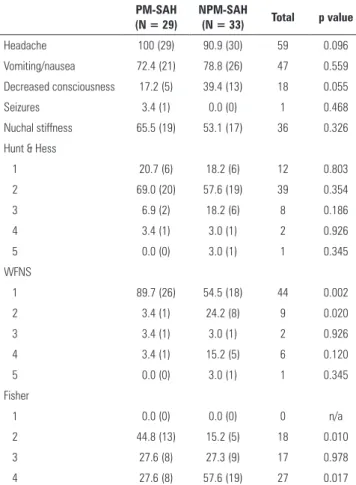
Headache was the most common symptom, reported by 100% of patients with PM-SAH and 90.9% of patients with NPM-SAH. Decreased consciousness was more common in patients with NPM-SAH, with a p value close to statistical relevance (0.055); no other symptom/sign was remarkably diferent between the two groups (Table 2).
detected on the CT scan); a score of 2 was calculated in 13 patients with PM-SAH (44.8%) versus in ive patients with NMPM-SAH (15.2%) (p = 0.010), and a score of 4 was calculated in eight patients with PM-SAH (27.6%) versus 19 patients with NPM-SAH (57.6%) (p = 0.017).
Angiographic studies included CT angiography on admission for every patient. Digital subtraction angiography was performed subsequently in 91.9% (n = 57) of patients and 42.1% of these (n = 24) patients repeated the exam. Patients who did not undergo digital subtraction angiography (n = 5) were from the PM-SAH group: two patients were evaluated with repeated CT angiography, two patients underwent magnetic resonance imaging angiography and one patient did not require any additional exam.
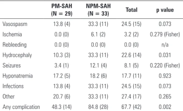
Vasospasm, infection and hydrocephaly were the most common complications (n = 15 for the former two and n = 14 for the latter) in all patients. Incidents of rebleeding were not reported.
Complications (Table 3) were more common in patients with NPM-SAH; 84.8% of patients experienced at least one complication, versus 48.3% of patients in the PM-SAH group (p = 0.02), representing a 6-fold increased complication risk in patients in the NPM-SAH group. Every complication was more common in the NPM-SAH group. his diference was statistically signiicant in regards to hydrocephaly (p = 0.031); patients with NPM-SAH exhibited a 4.3 times higher risk of developing hydrocephaly. Diferences in the occurrence of vasospasm or infections exhibited a trend without statistical signiicance (p = 0.073).
he median inpatient period was 14 days in the PM-SAH group (50% of cases between 12 and 16.5 days) and 21 days in the NPM-SAH group (50% of cases between 15 and 28 days). his diference was statistically signiicant (p < 0.001).
Two patients died during the hospital stay due to infectious complications (cerebral ventriculitis and pneumonia); both patients had been diagnosed with NPM-SAH (mortality rate of 7.1%).
Forty-nine patients (82% of the surviving patients) had follow-up appointments in our institution. he remaining 11 were lost to follow-up, as they had been transferred to other centers. he mean time of follow-up was 15.8 ± 10.3 months; no incidents of rebleeding were reported during this period. Excellent recovery (mRS 0 - 1) at three months after hospital discharge was found in 95.7% of patients diagnosed with PM-SAH and 85.7% of patients diagnosed with NPM-SAH (diference statistically irrelevant, p = 0.235).
Table 1 - Demographic data
PM-SAH (N = 29)
NPM-SAH
(N = 33) Total p value
Age 52.4 ± 11.8 56.8 ± 12.7 54.76 ± 12.2 0.163
Gender
Male 44.8 (13) 45.5 (15) 28 0.961
Female 55.2 (16) 54.5 (18) 34
Dyslipidemia 37.9 (11) 42.4 (14) 25 0.719
Diabetes mellitus 10.3 (3) 37.4 (12) 15 0.017
Smoker/ex-smoker 34.5 (10) 24.2 (8) 18 0.375
Obesity 20.7 (6) 15.2 (5) 11 0.569
Stroke 6.9 (2) 3.0 (1) 3 0.479
Other 44.8 (13) 57.6 (19) 32 0.316
PM-SAH - perimesencephalic subarachnoid hemorrhage; NPM-SAH - non-perimesencephalic subarachnoid hemorrhage. Results are expressed as number (%) or as the mean ± standard deviation.
Table 2 - Clinical presentation and admission scales
PM-SAH (N = 29)
NPM-SAH
(N = 33) Total p value
Headache 100 (29) 90.9 (30) 59 0.096
Vomiting/nausea 72.4 (21) 78.8 (26) 47 0.559
Decreased consciousness 17.2 (5) 39.4 (13) 18 0.055
Seizures 3.4 (1) 0.0 (0) 1 0.468
Nuchal stiffness 65.5 (19) 53.1 (17) 36 0.326
Hunt & Hess
1 20.7 (6) 18.2 (6) 12 0.803
2 69.0 (20) 57.6 (19) 39 0.354
3 6.9 (2) 18.2 (6) 8 0.186
4 3.4 (1) 3.0 (1) 2 0.926
5 0.0 (0) 3.0 (1) 1 0.345
WFNS
1 89.7 (26) 54.5 (18) 44 0.002
2 3.4 (1) 24.2 (8) 9 0.020
3 3.4 (1) 3.0 (1) 2 0.926
4 3.4 (1) 15.2 (5) 6 0.120
5 0.0 (0) 3.0 (1) 1 0.345
Fisher
1 0.0 (0) 0.0 (0) 0 n/a
2 44.8 (13) 15.2 (5) 18 0.010
3 27.6 (8) 27.3 (9) 17 0.978
4 27.6 (8) 57.6 (19) 27 0.017
PM-SAH - perimesencephalic subarachnoid hemorrhage; NPM-SAH - non-perimesencephalic subarachnoid hemorrhage; WFNS - World Federation of Neurological Surgeons; n/a - non-aplicable. Results are expressed as number (%).
DISCUSSION
his study conirms that spontaneous SAH is not a homogeneous disease. PM-SAH and NPM-SAH are distinct diseases with difering clinical progressions that commonly fall under the same designation, non-aneurysmal SAH. NPM-SAH exhibits a more aggressive clinical course, with a higher rate of complications and a longer hospital stay.(9) he length of hospital stay is an aspect that was often neglected in previous studies evaluating non-aneurysmal SAH, but it is of crucial importance due to inherent personal and economic costs.
Globally, our indings are in line with previous international studies, although in Portugal, information regarding this issue was lacking.
We reported a higher number of females in both groups, similar to Ildan et al.(10) and contrary to the majority of previous studies that reported a slight male predominance.(6,7)
Diabetes mellitus(8) is not recognized as a major risk factor for SAH,(11) but has been related to a higher risk
of vasospasm.(12) We did not observe that diabetes was
associated with vasospasm in the current study.
As expected, patients diagnosed with NPM-SAH had higher Fisher scale scores(13) and a higher risk of vasospasm,(14,15) which is in line with notion that patients with an aggressive clinical presentation and a greater amount of blood detected in the subarachnoid space have a poorer prognosis.(16)
In the current study, the incidence of vasospasm in the PM-SAH and NPM-SAH groups was higher than in the majority of other studies.(6,7,13,17,18) his diference may be explained by the fact that we considered the existence of vasospasm when transcranial Doppler ultrasound
Table 3 - Complications
PM-SAH (N = 29)
NPM-SAH
(N = 33) Total p value
Vasospasm 13.8 (4) 33.3 (11) 24.5 (15) 0.073
Ischemia 0.0 (0) 6.1 (2) 3.2 (2) 0.279 (Fisher)
Rebleeding 0.0 (0) 0.0 (0) 0.0 (0) n/a
Hydrocephaly 10.3 (3) 33.3 (11) 22.6 (14) 0.031
Seizures 3.4 (1) 12.1 (4) 8.1 (5) 0.220 (Fisher)
Hyponatremia 17.2 (5) 18.2 (6) 17.7 (11) 0.923
Infections 13.8 (4) 33.3 (11) 24.5 (15) 0.073
Other 20.7 (6) 33.3 (11) 27.4 (17) 0.265
Any complication 48.3 (14) 84.8 (28) 67.7 (42) 0.002
PM-SAH - perimesencephalic subarachnoid hemorrhage; NPM-SAH - non-perimesencephalic subarachnoid hemorrhage; n/a - non-applicable. Results are expressed as number (%).
blood low velocity criteria were met and not just when clinical vasospasm was observed, which is in contrast to previous studies. Transcranial Doppler ultrasound is the recommended method for monitoring SAH patients for the development of vasospasm(19) and it has been proven to be accurate.(20) Hydrocephaly was also detected more often in our study compared to previous investigations. We identiied the presence of hydrocephaly when an imaging study documented it at any time during the inpatient period,(1) whereas in other studies hydrocephaly was recorded only when permanent. We decided to report these complications as positive, even when no clinical signs were observed, because these patients have a diferent clinical approach (type of unity care, medical care supervision), which could possibly interfere with the inal clinical outcome. Nevertheless, the diference in hydrocephaly incidence between the two groups (higher in NPM-SAH) is similar to the results reported in other studies.(7,13,21)
In spite of the diferences in clinical progression between the two groups, patient outcomes at three months generally indicated a good prognosis both for NPM-SAH patients and PM-SAH patients. Our indings suggest that even though NPM-SAH exhibits a more aggressive clinical presentation, patients with NPM-SAH had a better prognosis compared to patients with aneurysmal SAH.(22)
Patients with convexity SAH were not included in the study, because this disease has a diferent pathophysiology and clinical evolution than non-aneurysmal SAH.(23-26)
Patients in whom a diagnosis of SAH was made based on a positive lumbar puncture alone were not included in the study, the SAH pattern could not be determined.
he limitations of this study include the fact that this is a retrospective analysis, the size of the cohort is small, the absence of two digital subtraction angiographies performed in every patient and the limited data from follow-up appointments.
CONCLUSION
he perimesencephalic subarachnoid hemorrhage and non-perimesencephalic subarachnoid hemorrhage are two diferent diseases with diferent clinical evolutions.
Non-perimesencephalic subarachnoid hemorrhage has a more difuse subarachnoid blood distribution, a more aggressive clinical presentation and a higher probability of complications.
Non-perimesencephalic subarachnoid hemorrhage has a longer inpatient period with higher economical costs for health care systems. In non-aneurysmal subarachnoid hemorrhage, the initial computed tomography scan might have therefore impact in patient’s management, prognosis, inpatient period, outcome and economical costs for healthcare systems.
Objetivo: Comparar a evolução clínica da hemorragia suba-racnóidea perimesencefálica com a da hemorragia subaracnói-dea não perimesencefálica.
Métodos: Estudo retrospectivo, que incluiu pacientes por-tadores de hemorragia subaracnóidea sem causa conhecida em um hospital terciário localizado na região norte de Portugal. Os dados epidemiológicos, clínicos e de imagem foram analisados estatisticamente, levando em conta a divisão dos pacientes em duas categorias: hemorragia subaracnóidea perimesencefálica e hemorragia subaracnóidea não perimesencefálica.
Resultados: Cumpriram os critérios de inclusão 62 pacien-tes, 46,8% deles com hemorragia subaracnóidea perimesencefá-lica e 53,2% com hemorragia subaracnóidea não perimesence-fálica. As caraterísticas demográicas, assim como os anteceden-tes clínicos, foram similares entre os grupos. As complicações foram observadas mais comumente no grupo com hemorragia subaracnóidea não perimesencefálica, sendo que 84,8% desses pacientes tiveram, no mínimo, uma complicação, comparados a 48,3% dos pacientes com hemorragia subaracnóidea peri-mesencefálica. Vasoespasmo, infecções e hidrocefalia foram as
complicações mais comuns - todas observadas mais frequente-mente nos pacientes com hemorragia subaracnóidea não peri-mesencefálica. Dois pacientes vieram a falecer, ambos com he-morragia subaracnóidea não perimesencefálica. A mediana do tempo de permanência no hospital foi maior nos pacientes com hemorragia subaracnóidea não perimesencefálica (21 dias, em comparação aos 14 dias observados nos pacientes com hemorra-gia subaracnóidea perimesencefálica). Não se observaram recidi-vas de sangramento durante o acompanhamento (tempo médio de 15 ± 10,3 meses).
Conclusão: As hemorragias subaracnóideas perimesencefá-lica e não perimesencefáperimesencefá-lica tiveram formas diferentes de evolu-ção clínica, principalmente no que se referiu à taxa de compli-cações e ao tempo mediano de permanência no hospital. Assim, a abordagem dessas duas formas de hemorragia subaracnóidea deve ser distinta, tanto em busca de melhorar o tratamento dos pacientes quanto para obter um melhor aproveitamento dos re-cursos de saúde.
RESUMO
Descritores: Tomograia computorizada por raios x; Vasoes-pasmo intracraniano; Hemorragia subaracnóidea; Hidrocefalia; Angiograia digital; Pacientes internados
REFERENCES
1. van Gijn J, Kerr RS, Rinkel GJ. Subarachnoid haemorrhage. Lancet. 2007;369(9558):306-18. Review.
2. Rinkel GJ, Wijdicks EF, Hasan D, Kienstra GE, Franke CL, Hageman LM, et al. Outcome in patients with subarachnoid haemorrhage and negative angiography according to pattern of haemorrhage on computed tomography. Lancet. 1991;338(8773):964-8.
3. Kim YW, Lawson MF, Hoh BL. Nonaneurysmal subarachnoid hemorrhage: an update. Curr Atheroscler Rep. 2012;14(4):328-34.
4. van Gijn J, van Dongen KJ, Vermeulen M, Hijdra A. Perimesencephalic hemorrhage: a nonaneurysmal and benign form of subarachnoid hemorrhage. Neurology. 1985;35(4):493-7.
5. Schwartz TH, Solomon RA. Perimesencephalic nonaneurysmal subarachnoid hemorrhage: review of the literature. Neurosurgery. 1996;39(3):433-40; discussion 440. Review.
6. Gupta SK, Gupta R, Khosla VK, Mohindra S, Chhabra R, Khandelwal N, et al. Nonaneurysmal nonperimesencephalic subarachnoid hemorrhage: is it a benign entity? Surg Neurol. 2009;71(5):566-71; discussion 571, 571-2, 572.
7. Sarabia R, Lagares A, Fernández-Alén JA, Arikan F, Vilalta J, Ibáñez J, et al. Idiopathic subarachnoid hemorrhage: a multicentre series of 220 patients. Neurocirugia (Astur). 2010;21(6):441-51.
9. Cornejo R, Romero C, Ugalde D, Bustos P, Diaz G, Galvez R, et al. High-volume hemofiltration and prone ventilation in subarachnoid hemorrhage complicated by severe acute respiratory distress syndrome and refractory septic shock. Rev Bras Ter Intensiva. 2014;26(2):193-9.
10. Ildan F, Tuna M, Erman T, Göcer AI, Cetinalp E. Prognosis and prognostic factors in nonaneurysmal perimesencephalic hemorrhage: a follow-up study in 29 patients. Surg Neurol. 2002;57(3):160-5; discussion 165-6. 11. Feigin VL, Rinkel GJ, Lawes CM, Algra A, Bennett DA, van Gijn J, et al.
Risk factors for subarachnoid hemorrhage: an updated systematic review of epidemiological studies. Stroke. 2005;36(12):2773-80.
12. Dumont T, Rughani A, Silver J, Tranmer BI. Diabetes mellitus increases risk of vasospasm following aneurysmal subarachnoid hemorrhage independent of glycemic control. Neurocrit Care. 2009;11(2):183-9. 13. Cánovas D, Gil A, Jato M, de Miquel M, Rubio F. Clinical outcome of
spontaneous non-aneurysmal subarachnoid hemorrhage in 108 patients. Eur J Neurol. 2012;19(3):457-61.
14. Jung SW, Lee CY, Yim MB. The relationship between subarachnoid hemorrhage volume and development of cerebral vasospasm. J Cerebrovasc Endovasc Neurosurg. 2012;14(3):186-91.
15. Westphal GA, Costa G, Gouvêa S, Kaefer KM, Silva RS, Caldeira Filho M. Cardiogenic shock associated with subarachnoid hemorrhage. Rev Bras Ter Intensiva. 2010;22(3):310-4.
16. van Gijn J, Rinkel GJ. Subarachnoid haemorrhage: diagnosis, causes and management. Brain. 2001;124(Pt 2):249-78. Review.
17. Kang DH, Park J, Lee SH, Park SH, Kim YS, Hamm IS. Does non-perimesencephalic type non-aneurysmal subarachnoid hemorrhage have a benign prognosis? J Clin Neurosci. 2009;16(7):904-8.
18. Andaluz N, Zuccarello M. Yield of further diagnostic work-up of cryptogenic subarachnoid hemorrhage based on bleeding patterns on computed tomographic scans. Neurosurgery. 2008;62(5):1040-6; discussion 1047. 19. Connolly ES Jr, Rabinstein AA, Carhuapoma JR, Derdeyn CP, Dion J,
Higashida RT, Hoh BL, Kirkness CJ, Naidech AM, Ogilvy CS, Patel AB, Thompson BG, Vespa P; American Heart Association Stroke Council; Council
on Cardiovascular Radiology and Intervention; Council on Cardiovascular Nursing; Council on Cardiovascular Surgery and Anesthesia; Council on Clinical Cardiology. Guidelines for the management of aneurysmal subarachnoid hemorrhage: a guideline for healthcare professionals from the American Heart Association/american Stroke Association. Stroke. 2012;43(6):1711-37.
20. Kincaid MS, Souter MJ, Treggiari MM, Yanez ND, Moore A, Lam AM. Accuracy of transcranial Doppler ultrasonography and single-photon emission computed tomography in the diagnosis of angiographically demonstrated cerebral vasospasm. J Neurosurg. 2009;110(1):67-72. 21. Hui FK, Tumialán LM, Tanaka T, Cawley CM, Zhang YJ. Clinical differences
between angiographically negative, diffuse subarachnoid hemorrhage and perimesencephalic subarachnoid hemorrhage. Neurocrit Care. 2009;11(1):64-70.
22. Sandvei MS, Mathiesen EB, Vatten LJ, Müller TB, Lindekleiv H, Ingebrigtsen T, et al. Incidence and mortality of aneurysmal subarachnoid hemorrhage in two Norwegian cohorts, 1984-2007. Neurology. 2011;77(20):1833-9. 23. Patel KC, Finelli PF. Nonaneurysmal convexity subarachnoid hemorrhage.
Neurocrit Care. 2006;4(3):229-33.
24. Cuvinciuc V, Viguier A, Calviere L, Raposo N, Larrue V, Cognard C, et al. Isolated acute nontraumatic cortical subarachnoid hemorrhage. AJNR Am J Neuroradiol. 2010;31(8):1355-62.
25. Little AS, Garrett M, Germain R, Farhataziz N, Albuquerque FC, McDougall CG, et al. Evaluation of patients with spontaneous subarachnoid hemorrhage and negative angiography. Neurosurgery. 2007;61(6):1139-50; discussion 1150-1.
26. Refai D, Botros JA, Strom RG, Derdeyn CP, Sharma A, Zipfel GJ. Spontaneous isolated convexity subarachnoid hemorrhage: presentation, radiological findings, differential diagnosis, and clinical course. J Neurosurg. 2008;109(6):1034-41.