DOI: 10.1590/0004-282X20130225
ARTICLE
Cervical dystonia: about familial and sporadic
cases in 88 patients
Distonia cervical: considerações sobre casos esporádicos e familiares em 88 pacientes
Carlos Henrique F. Camargo1, Sarah Teixeira Camargos2, Nilson Becker3, Renato Puppi Munhoz3, Salmo Raskin4, Francisco Eduardo C. Cardoso2, Hélio Afonso G. Teive3
he deinition of dystonia was recently revisited. Currently, dystonia is a movement disorder characterized by sustained or intermittent muscle contractions causing abnormal, of
-ten repetitive, movements, postures, or both. Dystonic move
-ments are typically patterned, twisting, and may be tremu
-lous. Dystonia is often initiated or worsened by voluntary action and associated with overlow muscle activation1
.Most voluntary muscles can be afected and, in the case of the neck muscles, the condition is referred to as cervical dystonia (CD), the most common form of dystonia. he term spasmodic tor
-ticollis was previously used for this movement, but it does not
stress the dystonic nature of the disease. CD can afect the musculature of the neck in a focal way or associated to other parts of the body2
.
Over the past 20 years, several loci (DYT1 to DYT25) have
been mapped in families with pure forms of dystonia, families with dystonia plus other movement disorders, or in sporad
-ic cases of dystonia. he most diferent kinds of inheritance can be observed (autosomal dominant, autosomal recessive, and X-linked). However, only some genes for isolated (pri
-mary or pure) forms of dystonia were descripted until today3
. Recently, CIZ1 (DYT23), ANO-3 (DYT24) and GNAL (DYT25)
1Unidade de Distúrbios do Movimento, Serviço de Neurologia, Hospital das Clínicas, Universidade Federal do Paraná, Curitiba PR Brazil; Hospital Universitário, Universidade Estadual de Ponta Grossa, Ponta Grossa PR, Brazil;
2Unidade de Distúrbios do Movimento, Serviço de Neurologia, Hospital das Clínicas, Belo Horizonte MG, Brazil;
3Unidade de Distúrbios do Movimento, Serviço de Neurologia, Hospital das Clínicas, Universidade Federal do Paraná, Curitiba PR, Brazil;
4Genetika Laboratory and Pontifícia Universidade Católica do Paraná, Curitiba PR, Brazil.
Correspondence: Carlos Henrique F Camargo, Serviço de Neurologia - Hospital de Clínicas – UFPR. Rua General Carneiro, 181 CEP: 80060-900 Curitiba PR – Brasil. E-mail: [email protected]
Conflict of interest: There is no conflict of interest to declare.
ABSTRACT
Cervical dystonia (CD) affects the musculature of the neck in a focal way or associated to other parts of the body. The aim of this study was to identify clinical differences between patients with dystonia patients without family history and with family history (sporadic). Eighty-eight patients with CD were recruited in a Movement Disorders Clinic between June of 2008 and June of 2009. Only patients with no etiological diagnosis were accepted for analysis. The age of onset of symptoms was later in patients with focal and segmental dystonia than in pa-tients with generalized dystonia (p<0.001). The severity of symptoms was higher in papa-tients with sporadic dystonia than in familial papa-tients (p<0.01). Generalized cases were more severe in patients with a family history (p<0.01). Sporadic patients had higher levels of pain than familial cases (p<0.05). We expect soon to present the results of genetic analyzes of these patients.
Keywords: dystonia, cervical dystonia, DYT1, DYT6, genetic.
RESUMO
A distonia cervical (CD) afeta a musculatura do pescoço de modo focal ou em combinação com outras partes do corpo. O objetivo deste estudo foi identificar diferenças clínicas entre pacientes com distonia com história familiar e pacientes sem história familiar (esporádicos). Foram selecionados 88 pacientes com DC no Setor de Distúrbios do Movimento entre julho de 2008 e junho de 2009. Somente os pacientes sem diagnóstico etiológico foram admitidos para análise. A idade de início dos sintomas foi mais tardia em pacientes com distonia focal e segmentar do que em pacientes com distonia generalizada (p<0,001). A gravidade dos sintomas foi maior em pacientes com distonia focal esporádicos do que naqueles com história familiar (p<0,01). Os casos generalizados foram mais graves nos pacientes com história familiar (p<0,01). Pacientes esporádicos tiveram níveis maiores de dor em relação aos casos familiares (p<0,05). Esperamos apresentar em breve resultados de análises genéticas desses pacientes.
genes were associated to families with CD4,5,6. Before that,
only TOR1-A (DYT1) and THAP1 (DYT6) genes were linked to isolated dystonia. Both are inherited as autosomal dominant traits, with reduced penetrance, and onset of clinical features commonly occurs during childhood or adolescence7-9
. Although these several loci and some genes were de
-scripted in the last years, the presence of mutations in these genes in cohorts of patients with dystonia is low. For exam
-ple, the presence of THAP1 mutations in families with ear
-lier “primary” dystonia is about 25%10. Regardless the mono
-genic inheritance, familial cases have lower mean age at disease onset than sporadic patients. Spread of dystonia to at least a second body site occurs more frequently in famil
-ial cases than in sporadic. he spread of symptoms is direct
-ly related to the duration of illness either in sporadic or in familial cases, however, in sporadic cases the most relevant spread occurs during the irst 10 years; in familial patients, instead, progressive spread of dystonia occurs throughout the disease course11
.
To identify clinical diferences among familial and spo
-radic cases of CD, we submitted patients at an evaluation in a Brazilian center for movement disorders.
METHOD
Standard protocol approvals, registrations, and patient consents.
All patients gave informed consent, and ethics approv
-al was obtained from the medic-al joint and ethics com
-mittee at Federal University of Parana (UFPR) to perform this clinical and genetic study (CEP-UFPR ethical approval 1676.093/2008-06).
Subjects selection and clinical assessment
Subjects with CD who attended the Botulinum Toxin and Movement Disorders Outpatient Unit in the Neurology Service, Clínicas Hospital, UFPR, from June 2008 to June 2009, were selected for the study. Clinical diagnoses were made by means of history and examination by two neu
-rologists. The patients were then assessed to identify clini
-cal characteristics, an association with other movement disorders and neurological diseases, epidemiological data, the time during which the disease had evolved, a history of trauma, the use of medicines, signs and symptoms that might indicate a secondary cause and a family history of dystonia or other movement disorders. All the patients were submitted to brain computed tomography and cervi
-cal-spine radiography. Additional tests included complete blood count (CBC), TSH, VDRL, blood glucose test, ESR, electrolyte levels and liver and kidney function in all the patients. Computed tomography and magnetic resonance imaging of the cervical spine, magnetic resonance imaging
of the brain and other laboratory tests were requested ac
-cording to the clinical assessment of each patient. The in
-clusion criterion was: (1) the presence of CD without an established cause (a “primary” or a idiopathic dystonia). The exclusion criteria were: (1) presence of a cause for the CD (2) refusal to submit to diagnostic investigation; (3) in
-ability to attend reassessment; and (4) failure to sign the informed consent form. CD was classified in accordance with established schemes1,12. The patients were assessed
on admission to compare severity, disability and pain us
-ing the Toronto Western Spasmodic Torticollis Rat-ing Scale (TWSTRS) and visual analog pain scale (0=absence of pain, 1-3=mild pain, 4-6=moderate pain, 7-9=strong pain, 10=disabling pain).
Statistics
he distribution pattern for all the data was tested (nor
-mal or non-nor-mal). he statistical diferences between the means of the groups were measured using the one-tailed Student t-test for normal distributions and the Mann-Whitney test for non-normal distributions. For the difer
-ences between the expected values and the values actually found, the Fisher exact test was used. he results are given as mean ± SD (standard deviation). he diferences were consid
-ered signiicant if p<0.05.
RESULTS
A total of 88 patients with CD were included, 56 were women and 32 men, a 1.75:1 ratio. Patients with familial his
-tory were called familial cases and those without familial his
-tory, sporadic cases. here were 16 families with 23 (26.74%) patients (Table 1).
Among the patients, 36 (40.91%) had focal CD. Other 22 (25%) patients had segmental dystonia, 6 (28.57%) with cra
-nial-cervical dystonia, 13 (59.1%) with arms-cervical dysto
-nia, 3 (13.67%) with arm-cranial-cervical dysto-nia, and one with laryngeal-cranial-cervical dystonia. Two (2.28%) pa
-tients with multifocal dystonia had abnormal movement in their left leg. A generalized dystonia was observed in 28 (31.81%) patients.
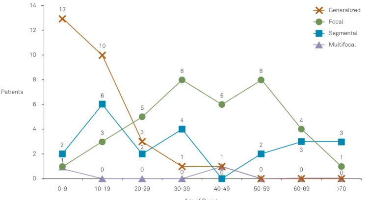
he mean age at onset of the group was 30.47±21.16 years old (range, 5 months to 72 years old). he mean age at onset of focal dystonia (41.05±16.87), and segmental dystonia pa
-tients (35.55±24.13), was higher than the generalized dysto
Table 1. Families with cervical dystonia.
Family Patient Sex Age (years) Age of onset (years) Clinical features and familial history
1 1 M 24 20 GD (CD, Meige syndrome, trunk, left leg).
2 M 18 12 GD (CD, limbs, trunk and cranial dystonia). Several familial members with different forms of dystonia.
2 3 F 13 7 GD (CD, trunk and limbs). Father, a cousin, and an uncle with dystonia.
3 4 M 35 11 GD (CD, limbs and trunk).
5 M 30 10 SD (right arm, left arm and neck). The parents had consanguinity. Two cousins had dystonia.
4 6 M 92 70 SD (Cervical and right arm dystonia). He presented Parkinsonism.
7 M 68 56 FD (CD). No Parkinsonism No other familial members affected.
5 8 M 10 4 GD (CD, trunk and limbs). Two twins brothers with dystonia.
6 9 F 47 30 SD (CD, Meige syndrome and head tremor). Father with dystonia.
7 10 F 40 5 MD (left right and CD). She has a sister with dystonia in legs (most in right leg).
8 11 M 60 57 SD (CD, larynx, right arm and Meige syndrome). Sister and nice with
oromandibular dystonia and CD.
9 12 F 47 39 SD (CD and oromandibular). Several familial members with CD (focal form).
10 13 F 31 9 GD (CD, arms and trunk). A cousin had dystonia.
11 14 F 33 12 GD (CD, limbs and trunk). Her father had CD.
12 15 F 17 2 GD (CD, limbs, cranial region and trunk). She had mental retardation. Her brother had CD without cognition features. The parents had consanguinity.
13 16 M 49 27 GD (CD, larynx, left arm and trunk).
14 17 F 25 18 SD (right arm, cervical and oromandibular dystonia).
18 F 16 6 GD (CD, oromandibular, larynx, limbs and trunk) The grandmother had mild face abnormal movements and speech disorder.
15 19 F 49 37 FD (CD and head tremor).
20 F 27 25 FD (CD and head tremor).
21 F 25 24 FD (CD).
22 M 18 17 FD (CD). Grandmother with CD and head tremor.
16 23 F 67 64 SD (blepharospasm and CD). She had Parkinsonism. Her nice had
blepharospasm.
M: male; F: female; CD: cervical dystonia; GD: generalized dystonia; SD: segmental dystonia; MD: multifocal dystonia; FD: focal dystonia.
0
0-9 10-19 20-29 30-39
Age of Onset
40-49 50-59 60-69
Focal
Segmental
Multifocal Generalized
>70 2
4 6 Patients
8 10 12 14
13
10
8
6 6
5
4 4
3
3
3 3
2
1
0 0 0 0 0 0 0
1 1 1
2 2
8
he main diferences between sporadic and familial cases are in Table 2. he mean age at onset of dystonia in sporad
-ic cases was 32.37±21.09 years old, and 24.78±20.48 years old in familial cases. Although there has not been any statistic diference, cases of generalized dystonia seen in the familial group were higher than in the sporadic group.
here was no diference, between familial and sporad
-ic cases, related to the localization and the spreading of the dystonia. here was also no diference about the site of onset of symptoms among the patients with generalized dystonia (caudal or rostral, limbs or cranial-cervical) even in familial or sporadic cases (p=0.643 to p=1) (Table 3).
Regarding clinical presentations of CD (torticollis, lat
-erocollis, retrocollis and anterocollis), one form was pre
-sented in 46 (52.27%) patients, two forms in 36 (40.9%) patients and three in 4 (4.54%). Among patients with gen
-eralized dystonia, 19 (67.86%) presented more than one form of dystonia. he most of patients with one form was found in a group with focal dystonia, 25 (65.8%) patients. he torticollis was the most common CD presentation, in
64 (72.73%) patients. he laterocollis was observed in 42 (47.73%) patients. he 17 (19.32%) patients with retrocol
-lis, and the 11 (12.5%) patients with anterocollis were found mixed with other forms, no isolated cases were observed. he most common one presented was torticollis plus lat
-erocollis (Table 4).
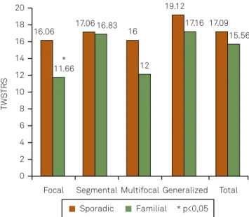
he severity caused by motor neck features, measured for TWSTRS scale, was higher in sporadic cases with focal dys
-tonia (16.06±4.21) than in familial cases with focal dys-tonia
Table 4. Patients with cervical dystonia divided by clinical presentation.
Presentation Sporadic cases (n=65) Familial cases (n=23) Total (n=88) p
1 Type 34 12 46 1
Torticollis 23 9 32
Laterocollis 11 3 14
Retrocollis 0 0 0
Anterocollis 0 0 0
2 Types 27 10 37 1
Torticollis + Laterocollis 10 5 15
Torticollis + Retrocollis 6 2 8
Torticollis + Anterocollis 3 2 5
Laterocollis + Retrocollis 6 0 6
Laterocollis + Anterocollis 2 1 3
3 Types 4 1 5 1
Torticollis + Laterocollis + Retrocollis 2 1 3
Torticollis + Laterocollis + Anterocollis 2 0 2
Table 2. Differences between sporadic cases and familial cases of cervical dystonia
Site of onset Sporadic cases Familial cases Total p
Patients 65 23 88 –
Male:female ratio 1:1.95 1:1.3 1:1.75 –
Age of onset* 32.37±21.09 24.78±20.48 30.47±21.16 p=0.07
Age of onset Focal dystonia
42.45±16.89 (23) 32.2±13.49 (5) 41.05±16.87 (28) p=0.10
Age of onset: segmental dystonia 34±23.58 (15) 42±22.62 (7) 35.55±24.13 (22) p=0.24
Age of onset: multifocal dystonia 42 (1) 5 (1) 24.5±24.75(2) –
Age of onset: generalized dystonia 13.12±9.93 (18) 11±7.16 (10) 12.36±9.10 (28) p=0.28
Focal dystonia: generalized dystonia ratio 1.83:1 1:2.2 1:1 p=0.227
* Years.
Table 3. Site of onset of generalized dystonia (sporadic cases X familial cases).
Site of onset Sporadic cases Familial cases Total
Cranial-cervical 5 4 9
Cranial-facial 1 1 2
Cervical 4 3 7
Limbs 12 7 19
Arms 5 4 9
(11.66±7.56) (p=0.03, Figure 2). here was no diference in se
-verity between CD focal and CD segmental (p=0.13), and be
-tween CD segmental and CD generalized (p=0.19). When the same tool was used, patients with three or two forms of CD presented more severity than patients with one form of dys
-tonia (p<0.001). he generalized cases had more severity than focal cases (p<0.05), mainly in familial patients (p<0.01).
Pain in neck area was related by 53 (60.23%) patients. 26 (49.06%) related a strong or a disability pain. he jerks and spasms, in 13 (14.77%) patients, were an aggravating factor to pain (p<0.001). he sporadic patients had higher pain levels than familial patients (p<0.05). here was more complaint of pain among patients with focal CD than patients with generalized CD (p<0.05). he familial patients with focal CD related less pain than the sporadic patients with focal CD (Figure 3). here was more intensity of the pain in patients with two and three cervical movements than patients with one movement (p<0.05).
Tremor (head tremor and arms postural tremor) was ob
-served in 32 (36.37%) patients. hree patients, one with gen
-eralized and two with segmental dystonia presented parkin
-sonism. he patients with segmental dystonia were familial cases and started the features at 64 and 70 years old. he spo
-radic case started the symptoms at 17 years old by neck area with generalization in two years. No levodopa response was observed in these patients. Other four patients, two with seg
-mental and two with generalized dystonia, had myoclonus. he mean age of onset was 15.5±5.07 (range, 12 to 23 years), without familial history or other movement disorders. A par
-tial response with alcohol was achieved in two patients. A fa
-milial history of movement disorder (tremor, dystonia or par
-kinsonism) was related to 26 (29.55%) patients.
DISCUSSION
he total number of patients with familial history of movement disorders (29.55%, 26.14% with familial history of dystonia) was below the 44% found by Jankovic et al.13
, prob
-ably due to the diiculty of the majority of patients to access their relatives, due to social and economic level conditions, and the presence of unusual ethnic-related genetic transmis
-sion of dystonia in our region.
he relationship of some ethnic groups with speciic types of dystonia is known. he DYT3 dystonia is a movement disorder of adult men from the Philippines. Dystonia DYT3 has only been
diagnosed in the Philippines14
. he frequency of dystonia is esti
-mated at 1/9000 in the Ashkenazi Jewish population15. Among
other ethnic groups the prevalence is likely to be below, about 1/10000 to 1/30000, but has not been deined yet16. he DYT6
gene was described in a study of three Amish-Mennonite fami
-lies. he DYT6 dystonia was initially associated with individu
-als of this ethnic group9
. In 1880, the irst Ashkenazi Jewish im
-migrants arrived in our region, mostly originating from Austrian Galicia. With the First and Second World War, new waves of Jewish immigrants arrived in Paraná17
. he Mennonites formed an important wave of migration from Europe to the South of Brazil in XX century. Despite this fact, we cannot, by clinical his
-tory, establish a relationship of ancestry from any of the 88 pa
-tients evaluated with these ethnic groups.
+ p<0.05 between the total of patients of focal dystonia and the total of patients of generalized dystonia;
# p<0.05 between the sporadic patients with focal dystonia and sporadic patients with generalized dystonia;
* p<0.05 between the familial patients with focal dystonia and sporadic patients with focal dystonia.
VAS: visual analog scale.
Figure 3. Differences of pain level (VAS) in cervical dystonia among patients with several clinical presentations.
Focal
VA
S
0 1 2 3 4 5 6
Generalized 5.06
1.6 *
4.58
3.17 #
3.07 + 2.9
Total Sporadic Familial
Figure 2. Differences of severity (TWSTRS) in cervical dystonia among patients with several clinical presentations.
TWSTRS: Toronto Western Spasmodic Torticollis Rating Scale. Focal
0 2 4 6 8 10 12 14 16 18 20
TW
ST
RS
Segmental Multifocal Generalized Total 16.06
11.66 *
17.06 16.83 16
12 19.12
17.16 17.09 15.56
In agreement with previous studies, we found a predomi
-nance of CD in whites and females (1:1.75)13, 18-21. here was an
earlier onset of symptoms in men than in women at a mean of 5.18 years. Although statistical signiicance was not ob
-served, these values were representative and higher than the diference of 2.2 years observed by Soland et al18.
he onset of symptoms occurred mainly between the fourth and the sixth decades of life (61.2 %) as demon
-strated previously13,21. here was a tendency of earlier onset
when there was a family history, particularly in focal dysto
-nias, however, this inding was not conirmed statistically signiicant. Previous studies also pointed to an earlier on
-set of familial cases than sporadic cases11
. Regardless family history, the generalized form started earlier, and focal and segmental forms later. In our study, patients started gener
-alized dystonia mainly in childhood and adolescence, ac
-cording previous studies11,13,21.
here is a rule in the study of dystonias. he ones which begin in early childhood tend to start in lower limbs and spread to the rest of the body, while the dystonia starting in adults usually begins in the upper half of the body and tend to remain focal22
. With increasing age, there is a caudal-ros
-tral pattern of the site of onset in the following order: low
-er limb dystonia, writ-er’s cramp, CD, spasmodic dyspho
-nia, and blepharospasm/oromandibular dystonia23
. A study comparing the natural history of dystonia between sporadic and familial cases showed that the spread of symptoms to other sites could occur in both groups over time. However, a progression after ive, 10, 15, 20 or 25 years were more pro
-nounced in cases with familial history of dystonia11
. We ob
-served a higher number of patients with generalized dystonia in familial patients and a signiicant number of patients with higher focal dystonia in sporadic group. hese data conirm a higher tendency of earlier onset with faster spreading of dys
-tonia in familial patients than sporadic patients11,23.
he patients in our study who developed generalized dys
-tonia began the symptoms in similar proportion by the limbs or the cranial-cervical region. here was not, unlike the pre
-viously published studies, a tendency of the familial general
-ized cases of starting their symptoms by legs11,23. According to
previous studies, DYT1 dystonia has focal onset in the limbs,
predominantly in the lower limbs, with subsequent general
-ization in childhood7,23. DYT3 dystonia has focal onset with
secondary generalization in 2-5 years14
. DYT5 dystonia, or do
-pa-responsive dystonia, is a generalized dystonia, with onset in the lower limbs, typically in childhood. In DYT7 and DYT13 dystonia, the onset is later, and dystonia remains a focal form. When DYT13 dystonia occurs in childhood, there is a greater tendency to generalize24,25. Although the main presentation
of DYT6 dystonia is the onset in upper limbs in childhood spraying toward generalization, it may occur in the focal or segmental forms, having its onset in cranial-cervical region, and even occurring without a familial history10.
he most common presentation of CD was torticollis, followed by laterocollis, as demonstrated in previous stud
-ies13,20,21. he presence of retrocollis was less common than
the one reported in other series. In our study, patients with tardive dystonia were excluded. he retrocollis and involve
-ment of other regions of the body in CD patients are more prevalent in patients with tardive dystonia. Torticollis and laterocollis, head tremor, geste antagoniste, and familial his
-tory are more common in dystonia “primary”26. In previous
study presented by our group, including patients with mul
-tiple etiologies (8.25% of cases secondary to the use of neuro
-leptics), the presence of retrocollis was higher (37.6%)20.
Patients with two or three types of dystonic movements had severity and pain scores higher than those shown by pa
-tients with a single presentation. Generalized dystonia, with the majority of patients with more than one presentation, showed higher degrees of severity. herefore, we reairm that a higher number of dystonic movements and extra-cervical sites of dystonia are factors of worsening to CD20. Another
important observed datum is when familial patients remain with CD in focal form, tending to have milder features than sporadic patients.
his tendency was repeated when the neck pain was evaluated. he familial patients with focal CD had lower pain scores, which was statistically signiicant for patients with the sporadic form and focal CD. he high incidence of pain con
-tributes signiicantly to the disability of the patients in CD, and distinguishes it from other focal dystonias21
. Diferent degrees of pain in the neck region were reported by 60.23% of patients, similar to 63% and 75% previously described27,28.
CD can be diagnosed among the dystonia with gene loci known or not. Among the known loci with dystonia, CD can have early and familial onset with autosomal dominant in
-heritance (DYT1, DYT6 and DYT13), or early onset with auto
-somal recessive inheritance (DYT2 and DYT17), or late onset
with autosomal dominant inheritance (DYT7, DYT21, DYT23,
DYT24 and DYT25). Currently, despite of the identiication of
several loci related with dystonia, the TOR1A (DYT1), THAP-1 (DYT6), CIZ1 (DYT23), ANO-3 (DYT24) and GNAL (DYT25)
genes are the only ones linked to isolated (“primary”) dysto
-nias. he relationship of CIZ1 (DYT23), ANO-3 (DYT24) and GNAL (DYT25) genes with dystonia was descripted recent
-ly3-7,9, In each monogenetic inheritance dystonia, CD may
be more or less common, and the onset can be part of the spreading even of the generalization3
. he DYT23 dystonia,
for example, has a pure latest onset CD4
. Meanwhile, patients with DYT6 dystonia have a huge range of phenotypes, and
wide range of age of onset8-10. herefore, it is expected that
CD may have several levels of pain and severity within the same etiology, but especially among the diferent etiologies.
Currently, these clinical analyses among diferent etiolo
-gies are not possible due to poor clinical details of the pre
Most likely, with the evolution of genetic studies of dystonia, it will be meaningless to compare clinical symptoms and pain of a familial group versus a sporadic group. In the future, the comparison of these parameters will be among groups with genetically deined etiology.
In conclusion, we descripted diferent clinical features, severity of symptoms and pain, between familial and sporad
-ic patients with CD. his study was part of a continuous proj
-ect. We expect to publish soon the irst data about genetic evaluation of these patients to DYT1 and DYT6 genes.
References
1. Albanese A, Bhatia K, Bressman SB, et al, Phenomenology and classification of dystonia: a consensus update. Mov Disord 2013;28:863-873.
2. Tsui JK. Cervical dystonia. In: Tsui JK, Calne D (Eds). Handbook of distonia. New York: Marcel Dekker, Inc 1995:115-127.
3. Lohmann K, Klein C. Genetics of dystonia: what’s known? What’s new? What’s next? Mov Disord 2013;28:899-905.
4. Xiao J, Uitti RJ, Zhao Y, et al. Mutations in CIZ1 cause adult onset primary cervical dystonia. Ann Neurol 2012;71:458-469.
5. Charlesworth G, Plagnol V, Holmström KM, et al. Mutations in ANO3 cause dominant craniocervical dystonia: ion channel implicated in pathogenesis. Am J Hum Genet 2012;91:1041-1050.
6. Fuchs T, Saunders-Pullman R, Masuho I, et al. Mutations in GNAL cause primary torsion dystonia. Nat Genet 2013;45:88-92.
7. Ozelius LJ, Hewett JW, Page CE, et al. The early-onset torsion dystonia gene (DYT1) encodes an ATP-binding protein. Nat Genet 1997;17:40-48.
8. Saunders-Pullman R, Raymond D, Senthil G, et al. Narrowing the DYT6 dystonia region and evidence for locus heterogeneity in the Amish-Mennonites. Am J Med Genet A 2007;143:2098-2105.
9. Fuchs T, Gavarini S, Saunders-Pullman R, et al. Mutations in the THAP1 gene are responsible for DYT6 primary torsion dystonia. Nat Genet 2009;41:286-288.
10. Bressman SB, Raymond D, Fuchs T, Heiman GA, Ozelius LJ, Saunders-Pullman R. Mutations in THAP1 (DYT6) in early-onset dystonia: a genetic screening study. Lancet Neurol 2009;8:441-446.
11. Elia AE, Filippini G, Bentivoglio AR, Fasano A, Ialongo T, Albanese A. Onset and progression of primary torsion dystonia in sporadic and familial cases. Eur J Neurol 2006;13:1083-1088.
12. Fahn S, Bressman SB, Marsden CD. Classification of dystonia. Adv Neurol 1998;78:1-10.
13. Jankovic J, Leder S, Warner D, Schwartz K. Cervical dystonia: clinical findings and associated movement disorders. Neurology 1991;41:1088-1091.
14. Lee LV, Munoz EL, Tan KT, Reyes MT. Sex linked recessive dystonia parkinsonism of Panay, Philippines (XDP). Mol Pathol 2001;54:362-368.
15. Risch N, de Leon D, Ozelius L, et al. Genetic analysis of idiopathic torsion dystonia in Ashkenazi Jews and their recent descent from a small founder population. Nat Genet 1995;9:152-159.
16. Frédéric M, Lucarz E, Monino C, et al. First determination of the incidence of the unique TOR1A gene mutation, c.907delGAG, in a Mediterranean population. Mov Disord 2007;22:884-888.
17. Gouvêia RR. Comunidade judaica de Curitiba, 1889-1970. Dissertação de Mestrado. Curitiba: PPGH/CNPq/Departamento de História da UFPR, 1980.
18. Soland VL, Bhatia KP, Marsden CD. Sex prevalence of focal dystonias. J Neurol Neurosurg Psychiatry 1996;60:204-205.
19. Almasy L, Bressman SB, Raymond D, et al. Idiopathic torsion dystonia linked to chromosome 8 in two Mennonite families. Ann Neurol 1997;42:670-673.
20. Camargo CH, Teive HA, Becker N, Baran MH, Scola RH, Werneck LC. Cervical dystonia: clinical and therapeutic features in 85 patients. Arq Neuropsiquiatr 2008;66:15-21.
21. Chan J, Brin MF, Fahn S. Idiopathic cervical dystonia: clinical characteristics. Mov Disord 1991;6:119-126.
22. Weiss EM, Hershey T, Karimi M, et al. Relative risk of spread of symptoms among the focal onset primary dystonias. Mov Disord 2006;21:1175-1181.
23. O’Riordan S, Raymond D, Lynch T, et al. Age at onset as a factor in determining the phenotype of primary torsion dystonia. Neurology 2004;63:1423-1426.
24. Bentivoglio AR, Del Grosso N, Albanese A, Cassetta E, Tonali P, Frontali M. Non-DYT1 dystonia in a large Italian family. J Neurol Neurosurg Psychiatry 1997;62:357-360.
25. Bentivoglio AR, Ialongo T, Contarino MF, Valente EM, Albanese A. Phenotypic characterization of DYT13 primary torsion dystonia. Mov Disord 2004;19:200-206.
26. Molho ES, Feustel PJ, Factor SA. Clinical comparison of tardive and idiopathic cervical dystonia. Mov Disord 1998;13:486-489.
27. Lowenstein DH, Aminoff MJ. The clinical course of spasmodic torticollis. Neurology 1988;38:530-532.