he neuroprotective role of therapeutic hypothermia
after cardiac arrest
Papel neuroprotector da hipotermia terapêutica pós paragem
cardio-respiratória
INTRODUCTION
Cardiorespiratory arrest (CRA) is associated with increased morbidity and mortality. he survival rate of in-hospital CRA cases is below 20%, and this rate is reduced by half in out-of-hospital cases.(1)
Notwithstanding the past 50 years of progress in both basic and advanced life support, the mortality rate and severity of neurological sequelae following the recovery of spontaneous circulation (RSC) remain high,(2) jeopardizing long-term quality of life.
In CRA patients with RSC, the mortality rate and neurological sequelae following CRA are primarily ascribed to two pathophysiological mechanisms:
Ana Abreu1, Ana Duque1, Carolina
Paulino1, João Brito1, Joana
Silvestre1,2, João Gonçalves-Pereira1,2,
Vítor Mendes1, Camila Tapadinhas1,
Pedro Póvoa1,2
1. General Intensive Care Unit, Hospital de São Francisco Xavier, Centro Hospitalar de Lisboa Ocidental – CHLO – Lisboa, Portugal. 2. Centro Hospitalar de Lisboa Ocidental – CHLO – Medical College, Universidade Nova de Lisboa – Lisboa, Portugal.
ABSTRACT
Objectives: herapeutic hypothermia following cardiorespiratory arrest has been demonstrated to have cardio- and neuroprotective efects, resulting in improved survival and better neurological outcomes. he objective of this study was to assess the outcomes of patients undergoing therapeutic hypothermia following cardiorespiratory arrest.
Methods: A prospective, 10-month observational study of patients admitted to an intensive care unit and undergoing therapeutic hypothermia after cardiores-piratory arrest was undertaken. hera-peutic hypothermia was induced by cold luid administration and body surface cooling in patients admitted no more than 12 hours after resuscitation from cardiorespiratory arrest. A target tem-perature of 33ºC was maintained for 24 hours.
Results: Overall, 12 patients were included (median age 64 years, 58% male). Half of the cardiorespiratory arrests were in-hospital. he median
irst-day Charlson Index, Sequential Organ Failure Assessment (SOFA) and Acute Physiology and Chronic Health Evaluation II scores were of 2.9, 11 and 24.5, respectively. he intensive care unit mortality rate was 42% (N=5). Five of the 7 surviving patients recovered their pre-cardiorespiratory arrest neurological status. Hypothermia was initiated 120 min (median) after recovery of spontaneous circulation. Most patients (75%) required vasopressor support. During the irst 3 days after cardiorespiratory arrest and therapeutic hypothermia, a progressive SOFA score decrease (median 11 on day 0, 10 on day 1 and 7 on day 2) was observed.
Discussion: In this study, therapeutic hypothermia was applied to all post-cardiorespiratory arrest patients and demonstrated good neurological outcome in surviving patients.
Keywords: Hypothermia, induced; Heart arrest/therapy; Neurological recovery
his study was conducted at the General Intensive Care Unit, Hospital de São Francisco Xavier, Centro Hospitalar de Lisboa Ocidental – CHLO – Lisboa, Portugal.
Conlicts of interest: None.
Submitted on September 8, 2011 Accepted on October 17, 2011
Corresponding author:
Ana Abreu
General Intensive Care Unit Hospital de São Francisco Xavier - Centro Hospitalar Lisboa Ocidental Estrada do Forte do Alto do Duque Zip Code: 1449-005 - Lisboa - Portugal Phone: +351 21 043 1104/5
anoxic encephalopathy resulting from brain circulation impairment and reperfusion syndrome. his syndrome is characterized by a systemic inlammatory response triggered during reperfusion that is a consequence of the activation of several biochemical cascades, leading to release of free oxygen radicals and other molecules that are harmful to brain cell mediators. his inlammatory response may be prolonged for 48-72 hours.(1,3,4)
Moderate therapeutic hypothermia (TH) consists of cooling the body to reach a central temperature of 33 + 0.5ºC, which is aimed to prevent and/or reverse the mechanisms responsible for the reperfusion syndrome and neurological injury.
TH has been used since the mid-1940s, when the irst cases were described(5) and was shown to be efective in animal studies in the mid-1980s.(6,7) However, TH has only recently been validated as an efective CRA therapy. he cardio-protective efects of TH and the reduced mortality rates and neurological sequelae following CRA have been clearly demonstrated in two randomized and controlled trials.(4,8) hese trials have led to the inclusion of TH in the CRA care recommendations by the International Liaison Committee on Resuscitation (ILCOR) since 2003 with an I-B evidence level.(9) Despite the inclusion of TH in this recommendation, the practical use of TH continues to be less frequent than anticipated(3,10) in many European countries, including Portugal.
he most recent ILCOR recommendations state that TH should be used in adult coma patients after CRA due to outpatient ventricular ibrillation and can be beneicial after CRA due to in-hospital, non-shockable rhythms.(11)
his study assessed the feasibility of a TH protocol and outcomes in CRA patients undergoing this therapeutic intervention.
METHODS
A prospective observational study was conducted that included patients admitted to the general intensive care unit (ICU) of the Hospital de São Francisco Xavier, Centro Hospitalar de Lisboa Ocidental (CHLO) undergoing TH after CRA during a 10-month period from August 2009 to May 2010. he study was approved by the hospital’s ethics committee. Informed consent was waived because of the observational nature of this trial, with no additional interventions being made in addition to the usual therapeutic approach for these patients.
Inclusion criteria were spontaneous circulation maintained for > 5 minutes, systolic blood pressure (SBP)
> 80 mmHg, time from CRA < 12 hours, time until advanced life support (ALS) < 10 minutes, ALS time < 45 minutes, time of hypotension after RSC < 30 minutes (SBP < 80 mmHg or mean blood pressure (MBP) < 45 mmHg) and a Glasgow Coma Scale (GCS) score < 9.(12)
Exclusion criteria were primary coagulopathy, terminal disease or indications for non-resuscitation, aorta dissection, intracranial bleeding, signiicant bleeding or myoclonic status epilepticus.
TH was performed according to the local protocol. TH was induced by fast luid infusion, preferably with polyelectrolyte solutions at 4ºC, 30 mL/kg at 100 mL/ min, external cooling by ice packs applied to the axilla and groin areas and eventually wet sheets covering the hands and feed. Additionally, the ventilator’s circuit heating and humidiication was suspended, and a gastric and vesical lavage with 4ºC saline was performed. As this cooling can be performed using peripheral venous access, the process was not delayed by central venous catheter (CVC) placement. he temperature was maintained using wet towels or thermal blankets. Ventilation was maintained on control mode, aiming to maintain peripheral oxygen saturation between 93 and 95%, thereby preventing hypoxemia and hypocapnia. Continuous sedation and analgesia were maintained using midazolam and alfentanil; propofol was avoided, due to its negative inotropic efects. In addition, a neuromuscular blockade was maintained using vecuronium for shivering prevention. he protocol included invasive blood pressure monitoring (arterial line), which also provided access for blood gas control and blood samples for additional laboratory testing. he temperature was continuously monitored using a pharyngeal thermometer.
For maintaining a MBP above 80 mmHg, a venous line was maintained for luid replacement and/or vasopressor drug administration. In cases of renal failure, metabolic acidosis or insuicient cooling with the above described measures (temperature reduction > 1.5ºC or temperature > 35ºC after 90 minutes), continuous renal replacement was started (hemoiltration or continuous veno-venous hemodiailtration) with active extracorporeal circuit cooling with ice. Laboratory panels were performed daily, including speciic neural enolase (SNE) dosage.
Reheating was slow and passively conducted for at least 8 hours at no more than a 0.5ºC/hour pace with hourly blood glucose monitoring, due to the increased risk for hypoglycemia. After the body temperature was > 36ºC, the neuromuscular blockade and then sedation were suspended.
he following clinical and demographic data were collected from the patients undergoing TH: age, gender and Charlson index,(13) Acute Physiology and Chronic Health Evaluation II (APACHE II)(14) and Sequential Organ Failure Assessment (SOFA)(15) scores. CRA was characterized according to the occurrence site, ALS maneuver duration, time until RSC (arrest time) and RSC heart rhythm. he TH parameters assessed were as follows: time from RSC to TH initiation, time from TH initiation until reaching the target temperature (33ºC), TH duration, minimum temperature achieved, volume of cold luid infused, requiring vasopressor drug support, requiring extracorporeal circulation, SNE values, C-reactive protein and white blood cell count. Changes in SOFA scores and neurological outcomes according to the Glasgow Coma Scale (GCS)(16) (1. death; 2. vegetative state; 3. severe disability; 4. moderate disability; 5. good recovery)(12) were also assessed within the 3 days after CRA and TH (D0. TH day; D1. reheating phase; D2. day after reheating), as well as in-hospital mortality.
Descriptive statistical analyses were performed with Microsoft Oice Excel (version 2007) software. Although survival and non-survival group variables were analyzed, we chose to present only descriptive analyses, due to the small sample size. he results are expressed as the median and interquartile interval (IQI) or mean + standard deviation, depending on the variable distribution.
RESULTS
Twelve patients with a median age of 64 years (IQI 29) were included during the trial period. Most patients were male (58.3%). he mean APACHE II scores were 24.5 (IQI 15.25), and the Charlson score was low in ive (41.6%), moderate in three (25%) and high in four (33%) patients. he admission GCS was 5 (IQI 3.75).
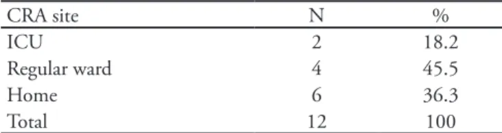
CRA occurred in six patients (50%) in-hospital and two in the ICU (Table 1). he CRA time was 12.5 minutes (IQI 13.3 minutes), and the time to RSC (duration of ALS maneuvers) was 7 minutes (IQI 7.5 minutes). Sinus rhythm was predominant upon RSC in 75% of cases, with atrial ibrillation in 16.6% and other supra-ventricular rhythms being observed in 8.3% of cases.
Table 1 – Cardiorespiratory arrest site
CRA site N %
ICU 2 18.2
Regular ward 4 45.5
Home 6 36.3
Total 12 100
CRA – cardiorespiratory arrest; ICU – intensive care unit.
Hypothermia use
he median time from RSC until initiating TH was 120 minutes (IQI 75 minutes). he time required to reach the target body temperature varied but was 240 minutes (IQI 327.5 minutes) on average, and the duration of hypothermia was 27 hours on average. All patients reached the target temperature, and the minimal temperature was 32.2ºC.
To reach the target temperature, each patient was infused with 5.3 + 2.9 liters of cold luids. In most patients, the target temperature was reached using external cooling and cold luids. In one extreme case, 12.5 liters were required, and in two other patients, continuous renal replacement therapy was necessary.
In most cases (9 patients, 75%), vasopressor support was required during TH. Vasoactive amines selection was conducted under the discretion of the treating physician. he maximal doses given during TH were 5.82 µg/kg/min noradrenalin and 7.5 µg/kg/min dopamine.
SNE was 21.4 (IQI 14 ng/mL) on D1 and 27.15 (IQI 28.9 ng/mL) on D2. However, in some patients (N = 5), SNE was not assessed or was incompletely assessed during TH.
Clinical outcome
Of the included patients, 5 died in the ICU (41.6% mortality rate). he neurological outcome among the survivors was full recovery in 5 and severe disability and vegetative state in the remaining patients. One patient eventually died on the regular ward, resulting in a 50% in-hospital mortality rate.
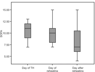
he SOFA scores improved during TH within the irst 3 days in the ICU: 11 (IQI 2.75) on the TH initiation day, 10 (IQI 3) on the reheating day and 7 (IQI 4.5) after reheating (Figure 1).
Age, Charlson index scores, arrest times (CRA + ALS times) and time before starting the TH protocol (median [IQI]) were higher among patients experiencing mortality compared with those with good neurological outcomes (score 5) (66 [33] vs. 56 [43] years; 4.5 [6.8] vs. 1.6 [7]; 30 [25] vs. 24 [8] minutes and 135 [70] vs. 120 [90] minutes, respectively). he time until achieving the target temperature was widely variable both in the good neurological outcome and deceased patient groups.
he SNE value progression could not be assessed during the TH because dosages were partially missing in some patients. he C-reactive protein values increased during TH in all patients (2.3, 2.7 and 4.1 mg/dL on D0, D1 and D2, respectively).(17)
Complications
No evidence of skin injury associated with external cooling was found. hree infective complications were diagnosed after being discharged from the ICU: nosocomial pneumonia in one patient with a good neurological outcome and two patients who died with nosocomial pneumonia and invasive candidiasis.
Figure 1 - Sequential Organ Failure Assessment score progression during therapeutic hypothermia.
D0 – day of therapeutic hypothermia; D1 – time of reheating; D2 – day after reheating; CRA – cardiorespiratory arrest; SOFA - Sequential Organ Failure Assessment.
Table 2 - Clinical and demographic characteristics of patients undergoing therapeutic hypothermia, characterization of cardiorespiratory arrest, hypothermia variables and neurological outcomes
N Age and gender Site of CRA Cause of CRA ALS time (minutes) Initial rhythm Charlson AP
A
CHE II
V
asopr
essors
V
olume of luids (mL) Time of CRA (minutes) Time to star
t TH (minutes)
T
ime to target temperatur
e
(min) TH duration (h) SOF
A D0
SOF
A D1
SOF
A D2
O
utcome
1 70. ♂ ICU Respiratory failure 7 SVT 7 17 Y 6890 10 20 5 28 11 8 4 5
2 74. ♀ ICU Asystole 5 AF 7.4 42 Y 3000 5 30 120 24 12 12 10 5
3 75. ♂ Home Aspiration pneumonia 15 SR 4.5 32 Y 2750 45 135 0 24 12 10 7 1
4 42. ♀ Home Grand mal epilepsy 5 SR 0.2 21 N 7800 15 110 270 35 9 8 7 1
5 66. ♂ Home Asystole 10 SR 7.6 28 N 3200 10 180 420 44 9 9 9 1
6 80. ♂ Hospital Ventricular ibrillation/AMI ? SR 7 42 Y 3550 2 240 10 23 13 NA NA 1
7 62. ♂ Hospital Respiratory failure 3 SR 4.2 19 Y 4700 3 510 420 30 12 12 11 3
8 42. ♂ Hospital Respiratory failure 20 SR 0.2 30 Y 5450 20 75 420 28 13 15 15 1
9 24. ♀ Home Asystole 7 VF 0 17 Y 10150 17 120 180 26 10 7 6 5
10 56. ♀ Home Drug intoxication 10 SR 1.6 6 N 6450 15 120 240 31 7 7 6 5
11 27. ♀ Home Asystole 15 SR 0 16 Y 10700 20 180 1200 19 11 10 6 5
12 70. ♂ Hospital Aspiration pneumonia 2 SR 1.6 33 Y 5800 2 120 240 22 9 10 13 2
DISCUSSION
In the current study, TH was successfully used in CRA patients. The in-hospital mortality rate was 50%, i.e., less than the 71.4% reported by Nolan et
al.(2,9) and 66% reported by Ravetti et al.(18) However,
this finding was influenced by the small sample size, inclusion of both in-hospital and out-of-hospital CRA cases and inclusion of all arrest rhythms. Another aspect that may have influenced this result was the lower number of cardiovascular origin CRA.
In our series, the deceased patients were older, had more comorbidities and had longer CRA times. A full neurological recovery was observed in 41.6% of patients; however, if only the surviving patients are considered, a full recovery was seen in 71.4% of patients. Patients with the best neurological recovery after TH were younger and had fewer comorbidities (higher GCS and lower Charlson index scores at admission).
Similar to other small series, a direct relationship was identified between good neurological outcomes and shorter CRA times and times to initiating TH.(19) Similarly, in our study, the CRA time and time to initating TH were shorter among survivors.
Although evidence for the benefit of TH is clear in out-of-hospial cases of ventricular fibrillation or tachycardia,(20,21) in our series, this therapeutic approach was used independently of the pre-CRA rhythm or arrest site.
The neurological outcome was better for in-ICU CRA patients, which agrees with other studies.(18,22) It is reasonable to assume that continuous hemodynamical monitoring is beneficial for prompt arrest recognition and therapy, the recognition of potentially reversible causes and significantly shortening the time to initiating basic and advanced life support measures and is additionally advantageous for assessing the effectiveness of treatment. After HACA and Bernard(4,8) demonstrated the effectiveness of TH on neurological outcome in CRA patients, TH was included in the 2003 and 2010 ILCOR recommendations.(9,21) However, these recommendations do not state an exact method or protocol for reaching the target temperature. Several external and internal methods are available for achieving the target temperature.(21) One of the most frequently used methods is the perfusion of cold fluids (usually 4ºC saline at 30 mL/kg), which is able to reduce central temperature by 1.5ºC. Other
possible methods for inducing and maintaining TH are ice bags, cool towels, cooling pillows or blankets, gel pillows with circulating water, intravascular heat exchangers and extracorporeal circulation.(21) To the best of our knowledge, no study has compared the effectiveness of these different TH induction methods.
The use of TH in our study was previously defined by a local protocol based on external cooling and cold fluids with no special technology requirements and therefore no incremental costs. It is of note that the target temperature was achieved for all patients. Internal (4ºC fluid infusion) and external (ice bags to the axilla and groin areas and eventually wet sheets, gastric and vesical lavage with cold fluids) methods were used, frequently requiring vasopressor drug support. Only two patients required dialysis to reach the target temperature. In most patients, the hypothermia protocol was both simple and affordable. On average, the target temperature was reached after 5 hours (293 minutes); this finding agrees with those of published literature reports.(4,8)
One case of nosocomial pneumonia was identified among the surviving patients and two cases among the deceased patients. No arrhythmia, coagulopathy, skin ice-burning injury or severe hydroelectrolyctic changes were detected.
Few studies on TH are available in Portugal, suggesting that this therapy is not common. Our study has clearly shown that this proven, beneficial and recommended technique is feasible and safe and can be used in any ICU.
Although our results agree with those of larger series, the limited number of patients, the short follow-up period, this study’s retrospective nature and the inclusion of different CRA rhythms and sites render our findings merely descriptive. Additionally, it was not possible to assess the SNE level progression; therefore, no prognostic assessment based on this parameter could be made.
CONCLUSIONS
RESUMO
Objectivos: A hipotermia terapêutica demonstrou ter efeitos neuro e cardioprotectores, com melhoria da sobrevida e redução das sequelas neurológicas em doentes vítimas de paragem cardio-respira-tória. O objectivo deste estudo foi avaliar a evolução dos doentes sub-metidos a hipotermia terapêutica após paragem cardio-respiratória.
Métodos: Estudo prospectivo observacional dos doentes sub-metidos a hipotermia terapêutica após paragem cardio-respiratória numa unidade de cuidados intensivos polivalente durante 10 meses. Aos doentes admitidos até 12 horas após paragem cardio-respiratória foi induzida a hipotermia terapêutica através da administração de luidos arrefecidos e arrefecimento corporal externo e mantida a tem-peratura alvo, 33°C, durante 24 horas.
Resultados: Foram incluídos 12 doentes, idade (mediana) de 64 anos, 58% do sexo masculino. A paragem cardio-respiratória ocorreu em meio hospitalar em 6 doentes. O índice de Charlson, o Sequential
Organ Failure Assessment (SOFA) e o Acute Physiology and Chronic Health Evaluation II, no primeiro dia, foram 2.9 [IIQ 6.8], 11 [IIQ 2.75], e 24.5 [IIQ 15.25], respectivamente. A taxa de mortalidade na unidade de cuidados intensivos polivalente foi de 42% (N=5). Dos 7 sobreviventes, 5 recuperaram o estado neurológico prévio à paragem cardio-respiratória. A hipotermia terapêutica foi iniciada cerca de 120 minutos [IIQ 78.75], após recuperação de circulação espontânea. A maioria dos doentes (75%) necessitou de suporte vasopressor. Foi constatado, nos 3 dias subsequentes à paragem cardio-respiratória e hipotermia terapêutica, uma diminuição do valor mediano de SOFA (11[IIQ 2.75], no dia 0, 10 [IIQ 3], no dia 1 e 7 [IIQ 4.5], no dia 2).
Conclusão: A aplicação de um protocolo de hipoter-mia terapêutica revelou ser simples e eicaz e permitiu ob-ter em doentes com indicação, boa recuperação neurológica.
Descritores: Hipotermia induzida; Parada cardíaca/terapia; Recuperação neurológica
REFERENCES
1. Pereira JCRG. Care of patient resuscitated from cardiac arrest. Rev Bras Ter Intensiva. 2008;20(2):190-6.
2. Nolan JP, Laver SR, Welch CA, Harrison DA, Gupta V, Rowan K. Outcome following admission to UK intensive care units after cardiac arrest: a secondary analysis of the ICNARC Case Mix Programme Database. Anaesthesia. 2007;62(12):1207-16.
3. Kuiper MA, Spronk PE, Schultz MJ. Use of a standardized treatment protocol for post-cardiac resuscitation care. In: Vincent JL, editor. Yearbook of intensive care and emergency medicine 2009. Berlin: Springer Verlag; 2009. p. 575-88.
4. Hypothermia after Cardiac Arrest Study Group. Mild therapeutic hypothermia to improve the neurologic outcome after cardiac arrest. N Engl J Med. 2002;346(8):549-56. Erratum in N Engl J Med 2002;346(22):1756.
5. Fay T. Observations on generalized refrigeration in cases of severe cerebral trauma. Assoc Res Nerv Ment Dis Proc. 1943;24:611-9.
6. Natale JA, D’Alecy LG. Protection from cerebral ischemia by brain cooling without reduced lactate accumulation in dogs. Stroke. 1989;20(6):770-7.
7. Chopp M, Knight R, Tidwell CD, Helpern JA, Brown E, Welch KM. he metabolic efects of mild hypothermia on global cerebral ischemia and recirculation in the cat: comparison to normothermia and hyperthermia. J Cereb Blood Flow Metab. 1989;9(2):141-8.
8. Bernard SA, Gray TW, Buist MD, Jones BM, Silvester W, Gutteridge G, Smith K. Treatment of comatose survivors of out-of-hospital cardiac arrest with induced hypothermia. N Engl J Med. 2002;346(8):557-63.
9. Nolan JP, Morley PT, Vanden Hoek TL, Hickey RW,
Kloeck WG, Billi J, Böttiger BW, Morley PT, Nolan JP, Okada K, Reyes C, Shuster M, Steen PA, Weil MH, Wenzel V, Hickey RW, Carli P, Vanden Hoek TL, Atkins D; International Liaison Committee on Resuscitation. herapeutic hypothermia after cardiac arrest: an advisory statement by the advanced life support task force of the International Liaison Committee on Resuscitation. Circulation. 2003;108(1):118-21.
10. Brooks SC, Morrison LJ. Implementation of therapeutic hypothermia guidelines for post-cardiac arrest syndrome at a glacial pace: seeking guidance from the knowledge translation literature. Resuscitation. 2008;77(3):286-92. 11. Morrison LJ, Deakin CD, Morley PT, Callaway CW,
Kerber RE, Kronick SL, Lavonas EJ, Link MS, Neumar RW, Otto CW, Parr M, Shuster M, Sunde K, Peberdy MA, Tang W, Hoek TL, Böttiger BW, Drajer S, Lim SH, Nolan JP; Advanced Life Support Chapter Collaborators. Part 8: Advanced life support: 2010 International Consensus on Cardiopulmonary Resuscitation and Emergency Cardiovascular Care Science With Treatment Recommendations. Circulation. 2010;122(16 Suppl 2):S345-421.
12. Teasdale G, Jennett B. Assessment of coma and impaired consciousness. A practical scale. Lancet. 1974;2(7872):81-4. 13. Charlson ME, Pompei P, Ales KL, MacKenzie CR. A
new method of classifying prognostic comorbidity in longitudinal studies: development and validation. J Chronic Dis. 1987;40(5):373-83.
14. Knaus WA, Wagner DP, Draper EA, Zimmerman JE, Bergner M, Bastos PG, et al. he APACHE III prognostic system. Risk prediction of hospital mortality for critically ill hospitalized adults. Chest. 1991;100(6):1619-36. 15. Vincent JL, Moreno R, Takala J, Willatts S, De Mendonça
Failure Assessment) score to describe organ dysfunction/ failure. On behalf of the Working Group on Sepsis-Related Problems of the European Society of Intensive Care Medicine. Intensive Care Med. 1996;22(7):707-10. 16. Jennett B, Bond M. Assessment of outcome after severe
brain damage. Lancet. 1975;1(7905):480-4.
17. Schuetz P, Afolter B, Hunziker S, Winterhalder C, Fischer M, Balestra GM, et al. Serum procalcitonin, C-reactive protein and white blood cell levels following hypothermia after cardiac arrest: a retrospective cohort study. Eur J Clin Invest. 2010;40(4):376-81.
18. Ravetti CG, Silva TO, Moura AD, Carvalho FB. Study of resuscitated in- and out-hospital cardiorespiratory arrest patients undergoing therapeutic hypothermia.
Rev Bras Ter Intensiva. 2009;21(4):369-75.
19. Oddo M, Schaller MD, Feihl F, Ribordy V, Liaudet L. From evidence to clinical practice: efective implementation of therapeutic hypothermia to improve patient outcome after cardiac arrest. Crit Care Med. 2006;34(7):1865-73. 20. Holzer M. Targeted temperature management for
comatose survivors of cardiac arrest. N Engl J Med. 2010;363(13):1256-64.
21. Deakin CD, Nolan JP, Soar J, Sunde K, Koster RW, Smith GB, Perkins GD. European Resuscitation Council Guidelines for Resuscitation 2010 Section 4. Adult advanced life support. Resuscitation. 2010;81(10):1305-52. Erratum in Resuscitation. 2011;82(1):140.