Copyright © 2014 Revista Latino-Americana de Enfermagem This is an Open Access article distributed under the terms of the Creative Commons Attribution Non-Commercial License (CC BY-NC).
This license lets others distribute, remix, tweak, and build upon your work non-commercially, and although their new works must also acknowledge you and be non-commercial, they don’t have to license their derivative works on the same terms.
DOI: 10.1590/0104-1169.3048.2404 www.eerp.usp.br/rlae
Corresponding Author:
Denise de Paula Cerqueira Rua Dr. José de Moura Resende, 111 Vila Tesoura
CEP: 12221-470, São José dos Campos, SP, Brasil E-mail: [email protected]
Denise de Paula Cerqueira
1José Roberto Tavares
2Regimar Carla Machado
21 RN, Specialization student in Cardiology Nursing, Universidade do Vale do Paraíba, São José dos Campos, SP, Brazil. 2 PhD, Professor, Universidade do Vale do Paraíba, São José dos Campos, SP, Brazil.
Objectives: to evaluate the renal function of patients in an intensive care unit, to identify the
predisposing factors for the development of renal failure, and to develop an algorithm to help in
the control of the disease. Method: exploratory, descriptive, prospective study with a quantitative
approach. Results: a total of 30 patients (75.0%) were diagnosed with kidney failure and the
main factors associated with this disease were: advanced age, systemic arterial hypertension,
diabetes mellitus, lung diseases, and antibiotic use. Of these, 23 patients (76.6%) showed a
reduction in creatinine clearance in the first 24 hours of hospitalization. Conclusion: a decline
in renal function was observed in a significant number of subjects, therefore, an algorithm was
developed with the aim of helping in the control of renal failure in a practical and functional way.
Descriptors: Renal Insuficiency; Intensive Care Units; Oliguria.
Rev. Latino-Am. Enfermagem 2014 Mar.-Apr.;22(2):211-7.
Introduction
Renal failure (RF) is a clinical syndrome characterized
by a decrease in renal function with accumulation of
metabolites and electrolytes in the body. Renal failure
can be subdivided into acute renal failure (ARF) and
chronic renal failure (CRF) according to the length of
development of the disease(1-3). Acute renal failure is
deined as the abrupt loss of the glomerular iltration of the kidneys with a subsequent alteration in the
hydroelectrolyte and acid-base balance in the body(4-5).
This imbalance, in turn, leads to the accumulation of
substances in the blood such as urea and creatinine(3).
Recognition of RF in the initial stages is critical to delay
the evolution of the disease, making renal recovery
possible and preventing the individual from needing to
undergo renal replacement therapy(2-3). Some individuals,
in particular, should be monitored carefully in relation to glomerular iltration. Groups that are classiied as at risk for the development of RF include diabetic or
hypertensive patients, those with cardiovascular disease
or a family history of renal failure, patients with other
renal diseases, and those of black race(3-4).
It is noteworthy that patients affected by ARF, in its
early stages, may or may not present a decrease in the
urine volume. However, oliguria is one of the indications that the glomerular iltration of the kidneys is beginning to fail(3,5). In 2002, the Acute Dialysis Quality Initiative
(ADQI) developed the RIFLE system that classiies ARF according to the degree of injury (Risk – class R; Injury
– class I; Failure – Class F; Loss – Class L; and End Stage
– Class E)(4-6). The RIFLE score considers the glomerular
iltration rate from the creatinine clearance (CrCl), which can be obtained using equations such as the Cockcroft-Gault formula – (140 – age in years) × body weight / 72 × serum creatinine, applied to both sexes, with the result multiplied by the constant 0.85(2,7-8).
In Brazil there are few epidemiological studies on
ARF. However, through research conducted in many
Brazilian hospitals, it was found that an increasing
number of patients are developing this disease during
the hospitalization period. It was also observed that it
affects mainly critically ill patients and those with
co-morbidities, making them susceptible to acute renal
disease(4,9-10). Studies conducted in intensive care
units (ICU) have demonstrated the high mortality of
patients with ARF and that many of them developed
the disease after a period of hospitalization in these
units(5,10-11). Given this context, it can be seen that the
nurse has an important role in the prevention of RF, by
actively participating in the care plan. The importance
should be highlighted of conducting precise and swift
nursing actions, based on evidence, in order to provide
appropriate care. Therefore, when considering the
importance of optimizing the work of the nurse through
tools that assist the care, it becomes necessary to
develop instruments to guide and support the decisions
of the nurse in determining the appropriate care for the
RF patient in the ICU.
The aims of this study were to evaluate the renal
function of patients hospitalized in the ICU, to identify
predisposing factors for the development of RF, and
to develop an algorithm to help in the control of the
disease.
Method
This exploratory, descriptive, prospective
study used a quantitative approach. To extract the
information a data collection instrument was prepared, based on the scientiic literature(2-6,8-13). In order to
reine the instrument regarding the coverage, clarity and relevance, a review was performed by four
nurses and one physician, who possessed the title
of intensive care specialist or had a minimum of two
years experience in this area, which was the criteria
for the selection of the experts. The data collection
instrument was divided into Part A and Part B, with the irst composed of the characterization of the subject, including sociodemographic data, personal history and
habits of the patient. The second part consisted of
the clinical data, which considered information on the
diagnosis at admission, clinical progression, treatment,
laboratory examinations, hydric balance, and the
fate of the patient (termination of the participation
of the patient in the study). The research protocol
was submitted to the Research Ethics Committee of
the University of Taubaté and was approved under
protocol No. 563/11.
The study was conducted in a general ICU of a
hospital in the Vale do Paraíba from January to June
2012. The sample consisted of 40 patients hospitalized
in the ICU who did or did not present renal failure.
The inclusion criteria established were, to be patients
18 years of age or over, of either gender, with urinary
catheterization for greater accuracy in the measurement
of the urine volume, with 1.4mg/dL or less of serum
creatinine, and at least 24 hours of hospitalization.
Subjects with chronic renal failure that were undergoing
Data were collected from the patient records
since their hospitalization in the ICU, respecting the
standard established to collect data, such as time and
period of sampling, and manner of collection. It should
be noted that the collection was conducted only by the
researchers. The data collection was terminated from
the time when the bladder catheter was removed, as
it was no longer possible to measure the urine volume;
after the discharge from the ICU; or with the death of the patient. Accordingly, this deined the period of duration of the patients in the study. Daily monitoring of renal
function was conducted in these patients by calculating
creatinine clearance (CrCl) through the Cockcroft-Gault formula and the urine volume. Subsequently, the patients who developed RF, the triggering factors, and the development stage of the disease were identiied, with the use of the RIFLE scale.
Initially, all the variables were analyzed descriptively.
The quantitative variables were described through the
means and standard deviation; and the qualitative
variables were presented as absolute (amount observed)
and relative (percentage) frequencies. For the analysis,
the patients were divided into two groups according to the classiication: with renal failure and without renal
alteration, with a descriptive analysis performed and the
descriptive level of the appropriate tests presented. To
test the association between the categorical variables
and the groups, Fisher’s exact test was used, while the
Mann-Whitney test was used to compare the two groups
regarding the continuous variables. The
Kolmogorov-Smirnov test was also used to test the normality of the variables (whether they were signiicant or not). The level of signiicance for the tests was 5% (α=0.05)(14).
Results
The results, in general, had no relevant statistical signiicance due to the fact that the sample was composed of a small number of subjects (n=40),
however, from the clinical perspective it was possible to
note the differences between the groups analyzed.
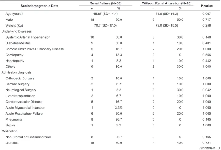
Table 1 shows that RF was present in 30 patients
(75.0%). The groups differed only in terms of age (RF:
65.87 [SD= 14.4] years; Without Renal Alteration: 51.0
[SD=14.2] years; p=0.007) and diagnosis at admission
(Neurological Surgery: RF: 3.3%; Without Renal
Alteration: 30.0%, p=0.042). However, for the other
variables evaluated there were no statistical differences.
Table 1 - Distribution of the patients with Renal Failure acquired during the hospitalization in the ICU and those
without renal alteration. São José dos Campos, SP, Brazil 2012
Sociodemographic Data Renal Failure (N=30) Without Renal Alteration (N=10) P-value
n % n %
Age (years) 65.87 (SD=14.4) 51.0 (SD=14.2) 0.007
Male 18 60.0 5 50.0 0.717
Weight (Kg) 70.7 (SD=17.5) 79.0 (SD=15.5) 0.258
Underlying Diseases
Systemic Arterial Hypertension 18 60.0 3 30.0 0.148
Diabetes Mellitus 9 30.0 1 10.0 0.401
Chronic Obstructive Pulmonary Disease 5 16.7 2 20.0 1.000
Cardiopathy 4 13.3 0 0 0.556
Hepatopathy 1 3.3 1 10.0 0.442
Others 9 30.0 3 30.0 1.000
Admission diagnosis
Orthopedic Surgery 3 10.0 1 10.0 1.000
Cardiac Surgery 2 6.7 1 10.0 1.000
Neurological Surgery 1 3.3 3 30.0 0.042
Liver transplantation 2 6.7 1 10.0 1.000
Cerebrovascular Disease 5 16.7 2 20.0 1.000
Acute Myocardial Infarction 1 3.3% 0 0 1.000
Acute Respiratory Failure 6 20.0 2 20.0 1.000
Pneumonia 8 26.7 0 0 0.165
Sepsis 1 3.3 0 0 1.000
Medication
Non Steroid anti-inlammatories 8 26.7 0 0 0.165
Diuretics 15 50.0 4 40.0 0.721
Rev. Latino-Am. Enfermagem 2014 Mar.-Apr.;22(2):211-7.
Sociodemographic Data Renal Failure (N=30) Without Renal Alteration (N=10) P-value
n % n %
Antihypertensives ACEI* 10 33.3 5 50.0 0.457
Antibiotics 26 86.7 10 100.0 0.556
Immunosuppressives 1 3.3 1 10.0 0.442
Vasoactive drugs 7 23.3 1 10.0 0.653
Outcomes 0.062
Discharge 19 63.3 5 50.0
Death 9 30.0 1 10.0
Bladder Probe Withdrawal 2 6.7 2 20.0
Reversal 0 0 1 10.0
Transfer 0 0 1 10.0
Table 1 - (continuation)
*ACEI=angiotensin converting enzyme inhibitors
CrCl Time n % Mean Standard Deviation
24h 23 76.6 56.47 16.78
48h 4 13.3 67.93 19.79
After 96h 3 10.0 78.53 7.10
SCr Time n % Mean Standard Deviation
24h 2 16.6 1.56 0.18
48h 5 41.6 1.65 0.10
72h 2 16.6 1.81 0.49
After 96h 3 25.0 1.56 0.20
Renal Failure Stages n %
I or class R 00 0
II or class I 14 46.6
III or class F 16 53.3
IV or class L 00 0
V or class E 00 0
The age groups (years) were statistically similar
in both groups. It was also observed that 70% of the
patients with RF were 60 years of age or more.
Table 2 shows the moment when the subjects
presented alterations in creatinine clearance (CrCl). Note
that 76.6% presented CrCl below 90mL/min/1.73m² within the irst 24 hours of hospitalization.
Table 2 - Descriptive measures of CrCl less than or
equal to 90mL/min/1.73m². São José dos Campos, SP,
Brazil 2012 (N=30)
Table 3 - Descriptive Measures of Creatinine greater
than or equal to 1.4 mg/dL. São José dos Campos, SP,
Brazil 2012 (N=12)
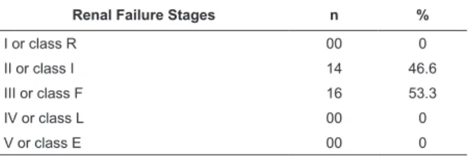
Table 4 - Stages of evolution of Renal Failure, according
to the RIFLE scale. Only the subjects who developed
Renal Failure. São José dos Campos, SP, Brazil 2012
(N=30)
CrCl Time=Creatinine Clearance Time
SCr Time=serum creatinine time – moment at which there was an alteration in the serum creatinine
From the survey data and with the help of the scientiic literature, an algorithm (Figure 1) was elaborated as a manual with guidelines to assist in the prevention, identiication, and control of renal failure, and thus the systematization of the care.
Conversely, in Table 3, the onset of alterations
in serum creatinine (greater than or equal to 1.4mg/ dL) occurred in the subjects within the irst 48 hours, i.e., 24 hours after the onset of the alteration
in CrCl.
The analysis of the stages of evolution of RF in
the patients, according to the RIFLE scale, can be
seen in Table 4, highlighting stages II (46.6%) and
III (53.3%).
Renal Failure Algorithm
Clinical history
Action Description
Identify possible causes loss of extracellular luid (hemorrhage, diarrhea, vomiting);
Identify risk factors elderly (>60 years), arterial hypertension, diabetes mellitus, cardiopathy, lung disease,
kidney disease, recent surgeries.
Calculation of the CrCl Cockcroft & Gault formula
Figure 1 - Renal failure algorithm
Source: NEFROCALC 1.0. Available at: http://www.sbn.org.br/equacoes/link/nefrocalc.htm Renal Failure Algorithm
Physical examination
Action Description
Observe oliguria (volume of urine <400mL in 24 hours) hypovolemia, arterial hypotension, urinary tract obstruction, urinary retention.
Signs and Symptoms back or suprapubic pain; fever; skin rash; dificulty in urinating; mental confusion, agitation or low level of consciousness (uremia)
Laboratory diagnosis
Action Description
Blood urea, creatinine, sodium bicarbonate, sodium, potassium, uric acid;
Urine urinary sediment, sodium, creatinine, and osmolality.
Prevention
Action Description
Establish renal function baseline through the measurement of blood serum creatinine and the CrCl calculation;
Optimize the clinical conditions adequate intravascular volume, maintain blood pressure (mean arterial pressure >80mmHg),
hematocrit above 30%; glucose less than 100mg/dL; adequate tissue oxygenation;
Adjust medication doses according to renal function Tozer equation – NEFROCALC
Maintain adequate hydration and monitor renal function Hydric balance
Discussion
Renal failure is responsible for the high rate of
hospital mortality, especially in intensive care units
(ICU). From this information, this study sought to
monitor the renal function of patients admitted to an
ICU to identify those who developed renal failure, the
stages of evolution, and the factors that may have
triggered the disease. The study sample was composed
of a small number of patients (40), due to the high rate
of hospitalization of patients with CRF with creatinine
greater than 1.4mg/dL, the high turnover of patients,
i.e., patients recovering post-surgery that remained
in the ICU for less than 24 hours, and those who had
incomplete medical records.
In the data analysis, the subjects were analyzed
regarding the presence of RF when they presented
CrCl <90mL/min/1.73m² during the hospitalization
in the ICU and those who already had CrCl <90mL/
min/1.73m² at the time of admission. However,
the subjects without RF who did not present renal
alterations were characterized as having a CrCl >90mL/
min/1.73m². Next, a comparative analysis between the
RF and without renal alteration groups was performed.
According to the literature, the individual with CrCl
lower than 90mL/min/1.73m² is in the early stage of
loss of kidney function. At this stage, the levels of urea
and plasma creatinine are still normal and there are no signiicant clinical signs or symptoms of renal failure(7,15).
In the sociodemographic analysis of this study,
it was noted that the group over 60 years of age
had a higher incidence of renal failure. This can be
explained by the fact that with advancing age, the glomerular iltration rate decreases as part of the body aging process(9,16-17). The analysis of the associations
between comorbidities and acute renal failure in the
literature is scarce. Studies show the association of a particular co-morbidity that may or may not inluence the onset of ARF in critically ill patients(18-19). In this
study, there were high percentages of comorbidities
present in the RF patients. The comorbidities described in the scientiic literature, such as diabetes mellitus, arterial hypertension, cardiopathy, and vascular
disorders, are factors that predispose individuals
to develop RF(10).
The underlying diseases more frequently presented
in this study were systemic arterial hypertension
(SAH), diabetes mellitus (DM), chronic obstructive
pulmonary disease (COPD), and cardiopathy. In many
studies these diseases are also highlighted as a risk
factors for the development of RF(9-10,19-21). However,
during hospitalization, complications presented by
patients such as sepsis, septic and hypovolemic shock,
hypotension, and pulmonary and metabolic diseases,
may be associated with renal dysfunction(10,17). One study
found that sepsis was the main triggering factor for ARF
among patients during hospitalization in the ICU(19).
The admission diagnosis variable generally included
clinical factors, such as pneumonia (pneumonia), acute
respiratory failure (ARP) and cerebrovascular disease
Rev. Latino-Am. Enfermagem 2014 Mar.-Apr.;22(2):211-7.
predispose patients to the development of RF(9-10,19).
The medications used in the treatment of the
diseases with higher prevalence in this study were
antibiotics, diuretics and antihypertensives and
ACEI inhibitors. According to the literature, these
drugs can enhance renal alterations in patients
with RF, or may even lead to the development of
the disease(3,12).
By analyzing the RF, it was found that 75% of
the patients developed this disease. According to the
development stage of the RF (from the RIFLE scale),
47% of the patients had class I of the disease and 53% (16) class F. These indings corroborate another study which showed that, when analyzing the renal function
of patients in an ICU, who presented ARF, classes R, I
and F of the RIFLE scale were more prevalent(5). In Table
3, the onset of changes in serum creatinine (greater
than or equal to 1.4mg/dL) occurred in the subjects within the irst 48 hours, i.e., 24 hours after the onset of the change in CrCl. However, the scientiic literature emphasizes that creatinine can not be used as a
parameter for the detection of changes in the glomerular iltration rate, since, when this is elevated the kidney is already suffering(7-8).
A mortality rate of 25% was observed, of which
30% corresponded to subjects who presented RF.
The mortality of patients with RF is highly variable,
with it depending on the development stage of the
disease, the clinical treatment, aggravating health
factors, the physical condition of the patient, and
other factors that are not directly related to kidney
disease(10,22). However, the mortality of these patients
still remains at high levels, which reinforces the need
for effective and careful prevention of RF(10,23). Recent
study analyzed 564 patients with and without ARF in an
ICU, comparing the clinical features and development, as well as the identiication of risk factors associated with the development of ARF and with the mortality,
showing that 25% of the patients developed ARF
during the hospitalization period and that the mortality
was 62%(22).
Studies on the role of nursing in the prevention
of nosocomial renal disease are scarce. However, this
fact does not make the issue less important, because
nurses, through resources that guide them in the
prevention and progression of RF, will be able to perform
their actions in a safer and more effective manner.
Therefore, the development of an action plan to identify signs of renal alterations qualiies and systematizes the care.
Conclusions
It was observed that the majority of the patients
presented RF, highlighted by the change in serum
creatinine (SCr) after 48 hours of ICU admission and
by alterations in creatinine clearance (CrCl) in the irst 24 hours. This shows that SCr is not a reliable parameter for the early diagnosis of IR. The main factors
associated with the development of RF were advanced
age, hypertension, diabetes mellitus, cardiopathy, lung
disease, and prolonged use of antibiotics. The daily
evaluation of renal function in critically ill patients can
minimize renal injury during hospitalization. Therefore,
an algorithm was developed in order to serve as a
guide to assist in the prevention and control of RF. The
algorithm was developed considering the practicality of
its use, thus serving as a quick tool to support healthcare
professionals in hospital inpatient units.
References
1. Stevens LA, Levey AS. Measurement of kidney
function. Med Clin North Am. 2005;89: 457-73.
2. Sociedade Brasileira de Nefrologia. Diretrizes de
insuficiência renal aguda [Internet]. 2007. [acesso 28
julho 2011]; Disponível em: http://www.jbn.org.br/
diretrizes.asp
3. Silva VTC, Yu L. Consulta nefrológica em 10 minutos:
abordagem clínica da oligúria. Serviço de Nefrologia
do Hospital das Clínicas da USP. J Bras Nefrol.
2009;31(3):173-4.
4. Santos ER. Associação do RIFLE com letalidade
e tempo de internação em pacientes críticos com
lesão renal aguda. Rev Bras Ter Intensiva. 2009;
21(4):359-68.
5. Young WP, Eun Ah H, Jang MH, Park SB, Chul-hyun
K. The Risk Factors and Outcome of Acute Kidney
Injury in the Intensive Care Units. Korean J Intern Med.
2010;25(2):181-7.
6. Mehta RL, Pascual MT,Gruta CG, Zhuang S, Chertow GM. Refining predictive models in critically ill patients with acute renal failure. J Am Soc Nephrol. 2002;
13(5):1350-7.
7. Sodré FL, Costa JCB, Lima JCC. Avaliação da função e
da lesão renal: um desafio laboratorial. J Bras Patol Med
Lab. 2007;43(5):329-37.
8. Pecoits-Filho R. Diagnóstico de doença renal
crônica: avaliação da função renal. J Bras Nefrol. 2004;
26(3 supl 1):4-5.
Received: Nov. 30th 2012 Accepted: Dec. 19th 2013
insuficiência renal em unidade de terapia intensiva. Acta
Paul Enferm. 2008;21:174-8.
10. Garcia TPR, Romero MP, Poletti NAA, Cesarino CB, Ribeiro RCHM. Principais motivos de internação
do paciente com insuficiência renal aguda na unidade
de terapia intensiva. Arq Ciênc Saúde. 2005;
12(3):146-50.
11. Carmo PAV, Amaral CF, Paiva ARB, Ribeiro CCOS, Ramalho GT, Bastos MG, et al. Insuficiência renal aguda dialítica: experiência em hospital universitário. J Bras
Nefrol. 2006; 28(1):7-14.
12. Pinto PS, Carminatti M, Lacet T, Rodrigues DF, Nogueira LO, Bastos MG, et al. Insuficiência renal aguda nefrotóxica: prevalência, evolução clínica e desfecho. J
Bras Nefrol. 2009;31(3):183-9.
13. Vukusich AC, Alvear FM, Villanueva PA, González CT, Olivari FP, Alvarado NA, et al. Epidemiología de la
insuficiencia renal aguda grave: un estudio prospectivo
multicéntrico en la Región Metropolitana. Rev Méd Chile.
2004;132(11):1355-61.
14. Bussab WO, Morettin P. Estatística básica. 6ª ed. São
Paulo: Saraiva; 2010.
15. Koyner JL, Vaidya VS, Bennett MR, Worcester E,
Akhter SA, Raman J, et al. Urinary biomarkers in the
clinical prognosis and early detection of acute kidney
injury. Section of Nephrology, Department of Medicine,
University of Chicago, Chicago, Illinois, USA. Clin J Am
Soc Nephrol. 2010;5(12):2154-65.
16. Romão JE Júnior, Haiashi ARM, Vidonho AF Júnior,
Abensur H, Quintaes PSL, Araújo MRT, et al. Causas
e prognóstico da insuficiência renal aguda hospitalar
em pacientes idosos. Rev Assoc Med Bras. 2000;
46(3):212-7.
17. Kusumota L, Rodrigues RAP, Marques S.
Elderly persons with chronic kidney failure: health
status alterations. Rev. Latino-Am. Enfermagem.
2004;12(3):525-32.
18. Murugan R, Karajala-Subramanyam V, Lee M, Yende
S, Kong L, Carter M, et al. Acute kidney injury in
non-severe pneumonia is associated with an increased
immune response and lower survival. Kidney Int.
2010;77(6):527-35.
19. Medve L, Antek C, Paloczi B, Kocsi S, Gartner B. Epidemiology of acute kidney injury in Hungarian
intensive care units: a multicenter, prospective,
observational study. BMC Nephrol. 2011; 12:43.
20. Bezerra KV, Santos JLF. Daily life of patients
with chronic renal failure receiving hemodialysis
treatment. Rev. Latino-Am. Enfermagem. 2008;
16(4):686-91.
21. Ponce D, Zorzenon CPF, Santos NY, Teixeira UA, Balbi AL. Injúria renal aguda em unidade de terapia
intensiva: estudo prospectivo sobre a incidência, fatores
de risco e mortalidade. Rev Bras Ter Intensiva. 2011;
23(3):321-6.
22. Chertow GM, Burdick E, Honour M, Bonventre JV, Bates DW. Acute Kidney Injury, Mortality, Length of Stay,
and Costs in Hospitalized Patients. J Am Soc Nephrol.
2005;16: 3365-70.
23. Camerini FG, Cruz I. Cuidados de enfermagem na prevenção da insuficiência renal provocada por
contraste após cateterismo. Acta Paul Enferm. 2008;