Microcirculatory assessment: a new weapon in the
treatment of sepsis?
Avaliação da microcirculação: uma nova arma no manejo da
sepse?
INTRODUCTION
In spite of the signiicant advances in the study of the pathophysiology and treatment of sepsis, mortality remains high.(1) he progression into
multi-organ failure is a common feature of sepsis, even after hemodynamic stability is achieved. Multi-organ failure is apparently related to the redistribution of blood low to the organs, as has been shown in studies on microcirculation and morphologic or functional cell changes, such as mitochondrial dysfunction.
During the 1990s, microcirculation studies that used invasive techniques in experimental sepsis models described microcirculatory changes that showed a marked reduction in capillary density.(2-4) During
this period of research, human studies were dependent upon the large microscopes that were used for periungual microcirculation assessments. These assessments showed a reduced rate of capillary flow in normotensive patients with fever.(5)
Orthogonal polarization spectral imaging (OPS) has the capacity to assess microcirculation in the sublingual mucosa and in other tissues covered by thin epithelial layers (such as the conjunctiva, the rectal mucosa, the vagina and the newborn axilla). he use of OPS as a real-time non-invasive tool for observation has been validated in several trials.(6,7) he technology used in
OPS is based on Beer’s law, which states that the constant found by dividing
Guilherme Loures Penna1,3,
Diamantino Ribeiro Salgado2,
André Miguel Japiassú1, Marcelo
Kalichsztein1, Gustavo Freitas
Nobre1, Nivaldo Villela2, Eliete
Bouskela3
1. Intensive Care Unit, Casa de Saúde São José – Rio de Janeiro (RJ), Brazil. 2. Hospital Universitário Clementino Fraga Filho – Rio de Janeiro (RJ), Brazil. 3. Vascular Biology Clinical and Experimental Research Laboratory (BioVasc), Biomedical Center, Universidade do Estado do Rio de Janeiro – UERJ – Rio de Janeiro (RJ), Brazil.
ABSTRACT
he progression into multi-organ failure continues to be a common feature of sepsis and is directly related to microcirculatory dysfunction. Based on a PubMed database search using the key words microcirculation and sepsis, twenty-six articles were selected for this review. he relevant references from these articles were also selected and included in this analysis. Orthogonal polarization spectral imaging allows for the bedside assessment of the microcirculation of critically ill patients. Such imaging
has established a correlation between microvascular dysfunction and patient outcomes, which allows practitioners to directly assess the efects of therapeutic interventions. However, the causal relationships between microcirculatory dysfunction, adverse outcomes, and the efects of therapies aimed at these microcirculatory changes in sepsis, are not clear.
Keywords: Microcirculation/ physiology; Sepsis/physiopathology; O r g a n d y s f u n c t i o n ; Mo r t a l i t y ; Hemodynamics; Shock, septic/therapy
Conlicts of interest: None.
Submitted on June 21, 2011 Accepted on August 2, 2011
Corresponding author:
Guilherme Loures Penna
Rua Dona Mariana, 143 - Sala B 31 - Botafogo
Zip Code: 22280-020 - Rio de Janeiro (RJ), Brazil.
the object’s optic density by the wave length is directly proportional to the concentration of the constituents causing the absorbance. In the case of microcirculation, the constituent is primarily hemoglobin. OPS imaging has the capacity to evaluate microcirculation up to 3 millimeters under the examined surface if the light intensity is suicient, the light sources are orthogonally situated, and an appropriate objective is used. he OPS device is easy to use and is portable. More recently, the sidestream dark ield (SDF) technique was also developed, based on the same principles.(8)
he analysis of microcirculation can provide relevant information. he total capillary density corresponds to all ield visible capillary vessels, which is proportional to the number of capillary vessels crossing three horizontal and three vertical screen lines. he capillary vessels are categorized as perfused in the following manner: the vessels show continued low, no low, any low that can be identiied, or intermittent
low if no low is seen for at least 50% of the time. he density and percentage of perfused vessels can be calculated with the use of particular mathematical formulas. Capillary vessels, which are less than 20 micrometers in diameter, can also be diferentiated from venules. Microvascular low heterogeneity, which is present in sepsis, is assessed by the comparison of the four quadrants.(9) In an experimental trial, Verdant
et al. showed that septic microcirculatory changes were similar in both the intestinal and sublingual mucosa, perhaps because of their shared embryonic origin.(10)
Due to its accessibility, the sublingual mucosa was selected for microcirculatory studies in septic patients.
Evidence of septic microcirculatory changes
Several trials in which OPS was used to evaluate microcirculation in sepsis were recently published (Table 1). De Backer et al. showed evidence of reduced functional capillary density, reduced proportion of
Table 1 – he main set of published articles on microcirculation and sepsis
Author Year Journal Method Title
Lam et al.(2) 1994 J Clin Invest. Experimental Microvascular perfusion is impaired in a rat model of normotensive sepsis.
Piper et al.(3) 1996 Am J Respir Crit Care Med. Experimental Microcirculatory changes in rat skeletal muscle in sepsis.
Farquhar et al.(4) 1996 J Surg Res. Experimental Decreased capillary density in vivo in bowel mucosa of rats with
normotensive sepsis.
De Backer et al.(11) 2002 Am J Respir Crit Care Med.* Clinical Microvascular blood low is altered in patients with sepsis.
Sakr et al.(12) 2004 Crit Care Med.* Clinical Persistent microcirculatory alterations are associated with organ failure
and death in patients with septic shock.
De Backer et al.(24) 2006 Crit Care Med.* Clinical Efects of drotrecogin alfa activated on microcirculatory alterations in
patients with severe sepsis.
Sakr et al.(20) 2007 Crit Care Med.* Clinical Microvascular response to red blood cell transfusion in patients with
severe sepsis.
Trzeciak et al.(13) 2008 Intensive Care Med.* Clinical Early increases in microcirculatory perfusion during protocol-directed
resuscitation are associated with reduced multi-organ failure at 24 h in patients with sepsis.
Verdant et al.(10) 2009 Crit Care Med.* Experimental Evaluation of sublingual and gut mucosal microcirculation in sepsis: a
quantitative analysis.
Dubin et al.(19) 2009 Crit Care Med.* Clinical Increasing arterial blood pressure with norepinephrine does not improve
microcirculatory blood low: a prospective study.
Büchele et al.(23) 2009 Crit Care Med.* Clinical Efects of hydrocortisone on microcirculatory alterations in patients
with septic shock.
Pottecher et al.(21) 2010 Intensive Care Med.* Clinical Both passive leg raising and intravascular volume expansion improve sublingual
microcirculatory perfusion in severe sepsis and septic shock patients.
Boerma et al.(26) 2010 Crit Care Med.* Clinical Efects of nitroglycerin on sublingual microcirculatory blood low
in patients with severe sepsis/septic shock after a strict resuscitation protocol: a double-blind randomized placebo controlled trial.
Su et al.(30) 2010 Shock.* Experimental Efects of a novel anticoagulant compound (TV7130) in an ovine
model of septic shock.
Salgado et al.(29) 2011 Shock.* Experimental Sublingual microcirculatory efects of enalaprilat in an ovine model of
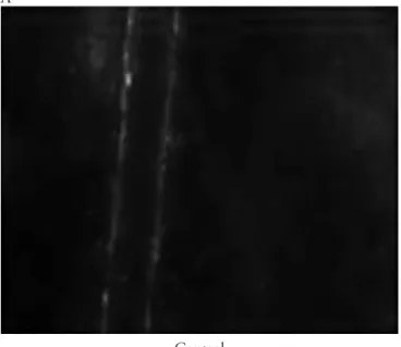
perfused capillary and low heterogeneity with no low capillary vessels in severe sepsis patients as compared to healthy subjects (Figure 1).(11)
A
B
Figure 1 – Orthogonal polarization spectral imaging (OPS) of a patient admitted to the intensive Care Unit of Casa de Saúde São José: A) during septic shock; B) during recovery after shock just before extubation.
In another study, persistent microcirculatory changes in septic patients were shown to be associated with poorer outcomes.(12) OPS was used to assess the
microcirculatory status of patients each day from the irst day of septic shock until the shock was resolved or the patient had died. During the irst rounds of microcirculatory assessment, surviving patients had shown a capillary density that was similar to the non-surviving patients. However, patients who maintained a reduced capillary density, even if the initial septic shock was reversed, did not survive. Even with improved hemodynamic stability, a microcirculatory assessment was able to discriminate between patients
who would survive and those who would progress into multi-organ failure. he relevance of microcirculatory oxygenation is that, even when global oxygen supply is preserved, microcirculatory impairment may lead to tissue hypoxia. In another trial, Trzeciak et al. have shown that the rate of capillary low is closely correlated to the patient’s outcome.(13) In this trial, a
microcirculatory assessment was completed during the irst 3 hours of early goal directed therapy (EGDT)(14)
and repeated 3 to 6 hours later. he Sequential Organ Failure Assessment (SOFA) score was calculated at the start of EGDT and after 24 hours. Out of 33 patients, 48% had at least a 2 point drop in their SOFA score. he Age, Acute Physiology and Chronic Health Disease Classiication System (APACHE II) score, along with overall hemodynamics, were not signiicantly diferent between the groups. Among patients who had a decrease in their SOFA score, 88% had a mean microcirculatory low improvement of 23%. hose with no SOFA score improvement had a mean decrease in microcirculatory low of 5%. An improvement in microcirculation would apparently reduce the likelihood of organ failure.
he hemodynamic parameters used for EGDT, such as central venous pressure, central venous saturation, blood pressure and urinary output should always be optimized during the management of sepsis, especially in the early stages of treatment, as hemodynamic stability reduces mortality. However these parameters are not directly related to the prognostic relevancy of microcirculatory changes.(15)
Possible pathophysiologic explanations
Septic microcirculatory changes can broadly be understood as the impaired balance between vasoconstrictor and vasodilator production and release, which leads to a reduction in microvascular low. here is also an increase in the production of free oxygen radicals. hese radicals react with nitric oxide (NO) and reduce its intracellular levels. Adequate levels of NO are needed to activate the enzyme guanylate cyclase, which is responsible for the production of cyclic GMP (a mediator of smooth muscle cell relaxation).(16) In
addition, the endothelium normally acts as an anti-adhesive barrier for blood components. However, septic endotoxins create pro-adhesive and pro-thrombotic efects on red and white blood cells and platelets that then alter the capillary low.(17) Free oxygen radicals
A
Control
B
Sepsis
Figure 2 – Intravital microscopy images from experimental animals studies from the Vascular Biology Clinical and Experimental Research Laboratory of Universidade do Estado do Rio de Janeiro. he images show the adhesion of white blood cells to the vessel wall during septic shock. A) Control; B) During sepsis.
Efect of diferent therapeutic interventions
he therapeutic interventions initiated during sepsis afect the microcirculation. Although it is known that the microcirculation will be impaired during severe hypotension, it is diicult to establish the ideal mean blood pressure (MBP) that will optimize microcirculatory low. Dubin et al. evaluated the microcirculation of 20 septic shock patients with a mean blood pressure (MBP) of 65
mmHg. After an increase in the noradrenalin infusion, which increased the MBP to 75-85 mmHg, they reassessed the microcirculatory status of the patients and found that microvascular perfusion was not signiicantly changed across the entire set of patients, in spite of any inter-subject variation. However, there was an increase in capillary density for those patients with baseline microcirculatory changes. Capillary density worsened for those patients with normal microcirculatory baselines.(19) Similarly, in a series
that included 35 severely septic patients, Sakr et al. showed that the transfusion of 1 to 2 units of packed red blood cells did not improve microcirculatory low. here was, however, a considerable degree of inter-subject variability. Patients with worse baseline microcirculatory perfusion presented with a remarkable improvement in capillary perfusion.(20)
To optimize microcirculation, it must be monitored.
Another recent trial focused on the inluence of rheological factors on microcirculation. A group of patients underwent passive leg raising maneuvers and a later infusion of a crystalloid solution. he patients showed similar increases in cardiac output, functional capillary density, microcirculatory low rate and the number of perfused capillary vessels for both interventions. here appeared to be no rheological inluence, at least with the limited volume of infusion.(21) In a recent study, Ospina-Tascon
et al. compared a volume infusion of either crystalloids or colloids in two groups of patients: a group with severe sepsis for less than 24 hours and a group with severe sepsis for more than 48 hours. A signiicant increase in small vessel density and perfusion was shown only in the former group, which demonstrates the relevance of early optimization of microcirculation. he observed microcirculatory efects were not related to the overall hemodynamic parameters or the type of volume expansion solution.(22)
Other non-vasopressor drugs afect microcirculation in the setting of severe sepsis. Hydrocortisone and activated protein C can improve capillary perfusion, although the modes of action are not clear.(23,24) Hypothetically,
nitroglycerin, a NO donor vasodilator could improve microcirculatory low heterogeneity and provide capillary recruitment, as has been described in the literature.
(25) However, a randomized double-blind trial infusing
either nitroglycerin or a placebo failed to show these beneits in septic patients after volume resuscitation and hemodynamic stabilization.(26) Microcirculatory
parameters were considerably improved during the initial clinical stabilization, but the efects did not last later in the clinical course of treatment.(27) he sedative agent propofol
reduced capillary density by 9.1%,(28) but the drug’s efect
studies have explored the possible therapeutic efects of drugs such as enalapril(29) or the anticoagulant TV7130(30)
on microcirculatory low improvement. hese studies show the possible range of experimental and clinical trials over the next few years.
CLOSING REMARKS
Microcirculatory dysfunction is directly related to poorer patient outcomes. he impairment of microcirculation in sepsis is heterogeneous with some capillary areas showing very slow low or even no low, while in neighboring areas, capillary perfusion is preserved. An organ, then, may be ischemic even when the global oxygen supply for this organ is preserved. It is not known if any causal relationship exists, or if microcirculatory changes can be more than a marker. Future studies are expected to test the hypothesis that improvements in microcirculation may serve as efective intervention in the treatment of sepsis.
RESUMO
A evolução para disfunção de múltiplos órgãos ainda é co-mum na sepse e está relacionada diretamente com a disfunção microcirculatória. Através de pesquisa nas bases de dados Pub-Med, empregando-se os unitermos microcirculação e sepse, vinte e seis artigos foram selecionados para esta revisão, bem como citações consideradas relevantes extraídas de artigos de revisão. Com o advento da técnica de imagem obtida através de polarização ortogonal, que permite a observação à beira do leito da microcirculação em pacientes críticos, é possível estabe-lecer uma relação entre disfunção microvascular e prognóstico, além de observar diretamente o efeito de diferentes interven-ções terapêuticas. No entanto, a relação causal entre disfunção microcirculatória e prognóstico adverso na sepse, bem como os efeitos de terapias dirigidas para correção destas anormalidades microcirculatórias ainda precisam ser melhor deinidos.
Descritores: Microcirculação/isiologia; Sepse/isiopatologia; Disfunção orgânica; Mortalidade; Hemodinâmica; Choque séptico/terapia
REFERENCES
1. Wenzel RP. Treating sepsis. N Engl J Med. 2002;347(13):966-7.
2. Lam C, Tyml K, Martin C, Sibbald W. Microvascular perfusion is impaired in a rat model of normotensive sepsis. J Clin Invest. 1994;94(5):2077-83.
3. Piper RD, Pitt-Hyde M, Li F, Sibbald WJ, Potter RF. Microcirculatory changes in rat skeletal muscle in sepsis. Am J Respir Crit Care Med. 1996;154(4 Pt 1):931-7. 4. Farquhar I, Martin CM, Lam C, Potter R, Ellis CG,
Sibbald WJ. Decreased capillary density in vivo in bowel mucosa of rats with normotensive sepsis. J Surg Res. 1996;61(1):190-6.
5. Weinberg JR, Boyle P, homas K, Murphy K, Tooke JE, Guz A. Capillary blood cell velocity is reduced in fever without hypotension. Int J Microcirc Clin Exp. 1991;10(1):13-9.
6. Harris AG, Sinitsina I, Messmer K. he Cytoscan Model E-II, a new relectance microscope for intravital microscopy: comparison with the standard luorescence method. J Vasc Res. 2000;37(6):469-76.
7. Mathura KR, Vollebregt KC, Boer K, De Graaf JC, Ubbink DT, Ince C. Comparison of OPS imaging and conventional capillary microscopy to study the human microcirculation. J Appl Physiol. 2001;91(1):74-8. 8. Ince C. he microcirculation is the motor of sepsis. Crit
Care. 2005;9 Suppl 4:S13-9.
9. De Backer D, Hollenberg S, Boerma C, Goedhart P, Büchele G, Ospina-Tascon G, et al. How to evaluate the
microcirculation: report of a round table conference. Crit Care. 2007;11(5):R101.
10. Verdant CL, De Backer D, Bruhn A, Clausi CM, Su F, Wang Z, et al. Evaluation of sublingual and gut mucosal microcirculation in sepsis: a quantitative analysis. Crit Care Med. 2009;37(11):2875-81.
11. De Backer D, Creteur J, Preiser JC, Dubois MJ, Vincent JL. Microvascular blood low is altered in patients with sepsis. Am J Respir Crit Care Med. 2002;166(1):98-104. 12. Sakr Y, Dubois MJ, De Backer D, Creteur J, Vincent JL.
Persistent microcirculatory alterations are associated with organ failure and death in patients with septic shock. Crit Care Med. 2004;32(9):1825-31.
13. Trzeciak S, McCoy JV, Phillip Dellinger R, Arnold RC, Rizzuto M, Abate NL, Shapiro NI, Parrillo JE, Hollenberg SM; Microcirculatory Alterations in Resuscitation and Shock (MARS) investigators. Early increases in microcirculatory perfusion during protocol-directed resuscitation are associated with reduced multi-organ failure at 24 h in patients with sepsis. Intensive Care Med. 2008;34(12):2210-7.
14. Rivers E, Nguyen B, Havstad S, Ressler J, Muzzin A, Knoblich B, Peterson E, Tomlanovich M; Early Goal-Directed herapy Collaborative Group. Early goal-directed therapy in the treatment of severe sepsis and septic shock. N Engl J Med. 2001;345(19):1368-77.
15. De Backer D, Ortiz JA, Salgado D. Coupling microcirculation to systemic hemodynamics. Curr Opin Crit Care. 2010;16(3):250-4.
microvascular endothelium in sepsis: role of oxidants. J Clin Biochem Nutr. 2008;42(3):175-84.
17. Eichelbrönner O, Sielenkämper A, Cepinskas G, Sibbald WJ, Chin-Yee IH. Endotoxin promotes adhesion of human erythrocytes to human vascular endothelial cells under conditions of low. Crit Care Med. 2000;28(6):1865-70. 18. Singer G, Urakami H, Specian RD, Stokes KY, Granger
DN. Platelet recruitment in the murine hepatic microvasculature during experimental sepsis: role of neutrophils. Microcirculation. 2006;13(2):89-97.
19. Dubin A, Pozo MO, Casabella CA, Pálizas F Jr, Murias G, Moseinco MC, et al. Increasing arterial blood pressure with norepinephrine does not improve microcirculatory blood low: a prospective study. Crit Care. 2009;13(3):R92. 20. Sakr Y, Chierego M, Piagnerelli M, Verdant C, Dubois
MJ, Koch M, et al. Microvascular response to red blood cell transfusion in patients with severe sepsis. Crit Care Med. 2007;35(7):1639-44.
21. Pottecher J, Deruddre S, Teboul JL, Georger JF, Laplace C, Benhamou D, et al. Both passive leg raising and intravascular volume expansion improve sublingual microcirculatory perfusion in severe sepsis and septic shock patients. Intensive Care Med. 2010;36(11):1867-74. 22. Ospina-Tascón G, Neves AP, Occhipinti G, Donadello
K, Büchele G, Simion D, et al. Efects of luids on microvascular perfusion in patients with severe sepsis. Intensive Care Med. 2010;36(6):949-55.
23. Büchele GL, Silva E, Ospina-Tascón GA, Vincent JL, De Backer D. Efects of hydrocortisone on microcirculatory
alterations in patients with septic shock. Crit Care Med. 2009;37(4):1341-7.
24. De Backer D, Verdant C, Chierego M, Koch M, Gullo A, Vincent JL. Efects of drotrecogin alfa activated on microcirculatory alterations in patients with severe sepsis. Crit Care Med. 2006;34(7):1918-24.
25. Spronk PE, Ince C, Gardien MJ, Mathura KR, Oudemans-van Straaten HM, Zandstra DF. Nitroglycerin in septic shock after intravascular volume resuscitation. Lancet. 2002;360(9343):1395-6.
26. Boerma EC, Koopmans M, Konijn A, Kaiferova K, Bakker AJ, van Roon EN, et al. Efects of nitroglycerin on sublingual microcirculatory blood low in patients with severe sepsis/septic shock after a strict resuscitation protocol: a double-blind randomized placebo controlled trial. Crit Care Med. 2010;38(1):93-100.
27. Favory R, Salgado D, Vincent JL, De Backer D. Can normal be more normal than normal? Crit Care Med. 2010;38(2):737-8; author reply 738.
28. Koch M, De Backer D, Vincent JL, Barvais L, Hennart D, Schmartz D. Efects of propofol on human microcirculation. Br J Anaesth. 2008;101(4):473-8. 29. Salgado DR, He X, Su F, de Sousa DB, Penaccini L,
Maciel LK, et al. Sublingual microcirculatory efects of enalaprilat in an ovine model of septic shock. Shock. 2011;35(6):542-9.