(1) Universidade Federal de Santa Maria,
Santa Maria, RS, Brasil.
(2) Departamento de Fisioterapia e
Reabilitação, Universidade Federal de Santa Maria, Santa Maria, RS, Brasil. Source: FIPE and CAPES
Conlict of interest: non-existent
Implications of mouth breathing on the pulmonary function
and respiratory muscles
Implicações da respiração oral na função pulmonar
e músculos respiratórios
Helenize Lopes Veron(1) Ana Gabrieli Antunes(1) Jovana de Moura Milanesi(1) Eliane Castilhos Rodrigues Corrêa(2)
Received on: July 24, 2015 Accepted on: November 10, 2015 Mailing address:
Helenize Veron Lopes
Rua Professor Braga, 79, Apto. 61,
Bairro Centro
Santa Maria – RS – Brasil
CEP: 97015-530
E-mail: [email protected]
doi: 10.1590/1982-0216201618111915
ABSTRACT
The mouth breathing syndrome is characterized by a set of signs and symptoms, which may be present in subjects who replace an adequate and eficient nasal breathing mode by the mouth or mist breathing mode, for a period equal or superior of six months. The mouth or mist breathing mode may be associated to changes in the ventilatory function and mechanics. This review has the objective of investigating more deeply the consequences of the mouth breathing in the lung function and respiratory muscles, empha-sizing the development of such changes since childhood to adult age. Eighteen articles were selected through data basis PubMed and Web of Science and they were grouped in the text, comprehending the follow topics:1) Implications of mouth breathing on the lung function and 2)Implications of mouth bre-athing on the respiratory muscles. From the information derivate of the analyzed articles, it may reali-zed that few studies reject or did not ind some relation between pulmonary changes and mouth brea-thing. It was suggested that the muscular unbalance produced by these changes may contribute for the mechanic disadvantage of the diaphragm muscle and increase of accessory inspiratory muscles work. Nevertheless, studies with more selective method, including objective and reproducible evaluation of the
respiratory muscles are still need.
Keywords: Mouth Breathing; Respiratory System; Pulmonary Ventilation; Respiratory Muscles
RESUMO
A Síndrome do Respirador Oral é caracterizada por um conjunto de sinais e sintomas que podem estar presentes em indivíduos que substituem o modo adequado e eiciente da respiração nasal pelo modo respiratório oral ou misto, por um período igual ou superior a seis meses. O modo respiratório oral ou misto pode estar associado a mudanças na função e mecânica ventilatória. Esta revisão tem como obje-tivo investigar mais profundamente as consequências da respiração oral na função pulmonar e músculos respiratórios, ressaltando o desenvolvimento de tais alterações desde a infância até a idade adulta. Foram selecionados 18 artigos por meio das bases de dados Pubmed e Web of Science e foram agrupados no texto, compreendendo os seguintes tópicos: 1) Implicações da respiração oral sobre a função pulmonar e 2) Implicações da respiração oral sobre os músculos respiratórios. A partir das informações oriundas dos resultados dos artigos analisados, percebe-se que poucos estudos refutam ou não encontram alguma relação entre as alterações pulmonares e a respiração oral. Sugere-se que, o desequilíbrio muscular produzido por estas alterações pode contribuir para a desvantagem mecânica do músculo diafragma e aumento do trabalho dos músculos acessórios da inspiração. Entretanto, são necessários estudos com métodos mais criteriosos, incluindo avaliações objetivas e reprodutíveis dos músculos respiratórios.
Descritores: Respiração Bucal; Sistema Respiratório; Ventilação Pulmonar; Músculos Respiratórios
Revision articles
INTRODUCTION
Breathing is a vital function and is strongly dependent on the adequate permeability of the nasal route, establishing itself as the main function of the body. The physiologic breathing mode in the human being is nasal, regardless of age1.2. The nasal cavity has a fundamental role in the physiology of respiration. It promotes iltering, heating and humidiication of the inhaled air, causing it to reach the lungs at the ideal temperature and favoring the adequate oxygenation of the body2.
Any factor leading to the upper airway (UA) obstruction causes nasal breathing to be replaced by mouth breathing, among which mechanical events, allergic and nonallergic inlammatory diseases, congenital malformation and tumoral lesions1.
Mouth breathing has been studied since the beginning of the twentieth century, with scientiic publi -cations directed to the scope of dentistry emphasizing the occlusal consequences3. This condition, considered as a public health problem, is attracting growing scien-tiic interest in recent years, and greater coverage in the multidisciplinary aspects surrounding it.
Mouth Breathing Syndrome is deined as a set of signs and symptoms that may be completely or incom-pletely present in subjects who, for several reasons, replace the correct pattern of nasal breathing by an oral or mixed pattern4 for mor than six months5.
Among these signs and symptoms are included daytime sleepiness, headache, agitation and nocturnal enuresis, frequent fatigue, poor appetite, bruxism, school problems and even learning deicits and behav -ioral problems6. Abreu et al.7 when studying mouth breathing children, detected the following: sleeping with mouth open (86%), snoring (79%), nasal itching (77%), drooling on the pillow (62%), nocturnal breathing dificulty or restless sleep (62%) nasal obstruction (49%) and irritability during the day (43%). Felcar et al.8 concluded that children under seven years old who drooled and snored at night were more prone to mouth breathing occurrence. Menezes et al.9 , when verifying the socioeconomic and demographic inluences in determining mouth breathing mode, obtained higher prevalence of mouth breathing in public than private schools.
In addition, this syndrome has as main character-istics the lack of lip seal, ogival or high-arched palate, Angle’s Class II malocclusion, unilateral or bilateral crossbite, open bite, sleep apnea, everted lower lip, retracted upper lip, generalized facial hypotonia,
stomatognathic malfunction and postural changes10,11. Other characteristics include the presence of dark
circles12, elongated face13, lowered mandible and
dental alterations14, abnormal speech15, habitual tongue position in the oral loor and mentual muscle hyper -function during lip-closing16.
Mouth breathing may be associated with genetic factors, inadequate oral habits (paciier and inger sucking, baby bottle) and nasal obstruction with variable intensity and duration 7. Rhinitis is one of the main causes of nasal obstruction with a high preva-lence in the population17. Mouth breathing in children is a frequent complaint in the pediatrician, allergist and otorhinolaryngologist’s practice18.
The altered breathing mode can also be associated with changes in the ventilatory function and mechanics. The respiratory tract can be considered a single morphofunctional entity extending from nose to the alveoli and any change can affect the rest19. It is well-known the coexistence between mouth breathing and asthma, highlighting the high prevalence of mouth
breathing among asthmatic children20. However,
studies explaining the complex relationship between the mouth breathing mode and changes in the venti-latory function and mechanics are still scarce.
According to Correa and Bérzin21, the persistence of mouth breathing even after the resolution of the initial functional abnormality (increased nasal resistance) has been described in the literature. This can be attributed to neural adaptations, changes in the central control of the upper airway and the muscular function and skeletal changes. Therefore, it is important to emphasize that the effects of mouth breathing can be perpetuated into the adulthood 22,23.
Currently, most studies regarding the mouth breathing mode address ENT, dental and of orofacial motricity aspects23. A few studies focus on the respi-ratory changes, the main ones being targeted to mouth breathing in children. In addition, some of these studies are inconsistent and present controversies. Based on the abovementioned, this review aims to inves-tigate further the consequences of mouth breathing for pulmonary function and respiratory muscles, emphasizing the development of such alterations from childhood to adulthood.
METHODS
measures? After this stage, from December 2014 to March 2015, work began with the search and selection of published studies, without restriction regarding the publication period, in PubMed and Web of Science databases. In both databases, the survey was conducted by crossing the keywords “oral breathing” or “mouth breathing” and “spirometry” or “ventilatory function” or “respiratory muscles” or “respiratory mechanics” or “respiratory muscular strength” or “ventilatory muscular strength” or “maximal respi-ratory pressure” or “maximal inspirespi-ratory pressure” or “maximal expiratory pressure” or “accessory inspiratory muscles” or “diaphragm excursion”.
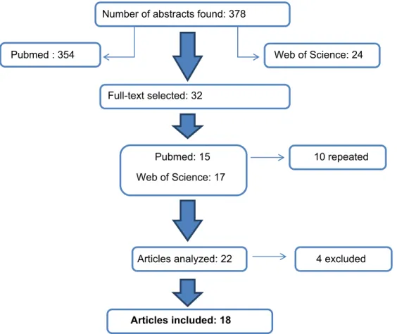
The survey resulted in a total of 378 abstracts, with 354 of them referring to PubMed and 24 related to Web of Science databases. The titles and abstracts were analyzed and all articles written in Portuguese, English or Spanish and with potential relevance to achieve the proposed objectives, as well as for resolving the study problem were selected. Thus, articles analysing some measure of pulmonary function and/or ventilatory mechanics in mouth breathers were included. The articles found in both databases were counted only once. Twenty-two articles were selected to read the full text and of these, 4 were excluded for not having information compatible with the purpose of this review. (Figure 1)
Number of abstracts found: 378
Pubmed : 354 Web of Science: 24
Full-text selected: 32
Pubmed: 15 10 repeated
Web of Science: 17
Articles analyzed: 22 4 excluded
Articles included: 18
LITERATURE REVIEW
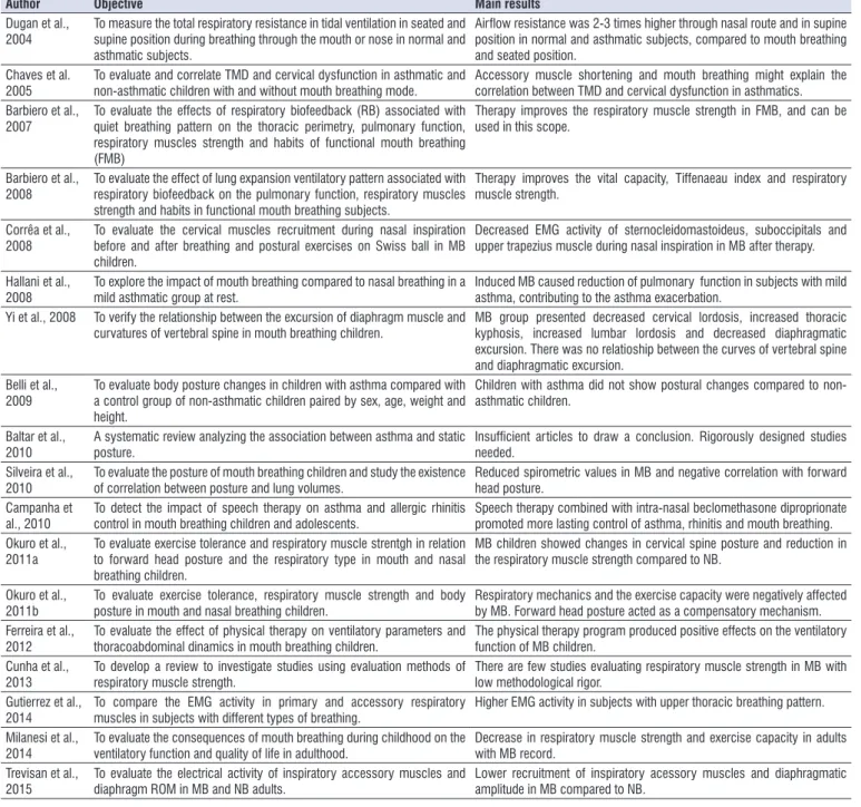
Table 1 shows the articles included in this review, in chronological order, describing the author and date, objectives and key results. It is observed that the articles found date from the last 11 years, with higher concentration of publications in 2008. In recent decades, there has been a growing interest in under-standing the pathophysiological mechanisms involved
in mouth breathing, however the relationship between pulmonary function and ventilatory mechanics seems to be recent and have not yet been fully elucidated.
The discussion of the articles in the text was organized in chronological order and divided into topics as follows: 1) Implications of the mouth breathing for the pulmonary function and 2) Implication of the mouth breathing for the respiratory muscles.
Table 1. Objectives description and main results of the studies selected for the review.
Author Objective Main results
Dugan et al.,
2004 To measure the total respiratory resistance in tidal ventilation in seated and supine position during breathing through the mouth or nose in normal and
asthmatic subjects.
Airlow resistance was 2-3 times higher through nasal route and in supine position in normal and asthmatic subjects, compared to mouth breathing
and seated position.
Chaves et al.
2005
To evaluate and correlate TMD and cervical dysfunction in asthmatic and
non-asthmatic children with and without mouth breathing mode. Accessory muscle shortening and mouth breathing might explain the correlation between TMD and cervical dysfunction in asthmatics.
Barbiero et al.,
2007 To evaluate the effects of respiratory biofeedback (RB) associated with quiet breathing pattern on the thoracic perimetry, pulmonary function, respiratory muscles strength and habits of functional mouth breathing
(FMB)
Therapy improves the respiratory muscle strength in FMB, and can be used in this scope.
Barbiero et al., 2008
To evaluate the effect of lung expansion ventilatory pattern associated with respiratory biofeedback on the pulmonary function, respiratory muscles strength and habits in functional mouth breathing subjects.
Therapy improves the vital capacity, Tiffenaeau index and respiratory muscle strength.
Corrêa et al.,
2008
To evaluate the cervical muscles recruitment during nasal inspiration before and after breathing and postural exercises on Swiss ball in MB children.
Decreased EMG activity of sternocleidomastoideus, suboccipitals and upper trapezius muscle during nasal inspiration in MB after therapy.
Hallani et al., 2008
To explore the impact of mouth breathing compared to nasal breathing in a
mild asthmatic group at rest. Induced MB caused reduction of pulmonary function in subjects with mild asthma, contributing to the asthma exacerbation.
Yi et al., 2008 To verify the relationship between the excursion of diaphragm muscle and
curvatures of vertebral spine in mouth breathing children. MB group presented decreased cervical lordosis, increased thoracic kyphosis, increased lumbar lordosis and decreased diaphragmatic excursion. There was no relatioship between the curves of vertebral spine and diaphragmatic excursion.
Belli et al.,
2009 To evaluate body posture changes in children with asthma compared with a control group of non-asthmatic children paired by sex, age, weight and height.
Children with asthma did not show postural changes compared to non-asthmatic children.
Baltar et al., 2010
A systematic review analyzing the association between asthma and static
posture.
Insuficient articles to draw a conclusion. Rigorously designed studies
needed. Silveira et al.,
2010
To evaluate the posture of mouth breathing children and study the existence
of correlation between posture and lung volumes. Reduced spirometric values in MB and negative correlation with forward head posture.
Campanha et
al., 2010
To detect the impact of speech therapy on asthma and allergic rhinitis
control in mouth breathing children and adolescents. Speech therapy combined with intra-nasal beclomethasone diproprionate promoted more lasting control of asthma, rhinitis and mouth breathing. Okuro et al.,
2011a
To evaluate exercise tolerance and respiratory muscle strentgh in relation to forward head posture and the respiratory type in mouth and nasal breathing children.
MB children showed changes in cervical spine posture and reduction in the respiratory muscle strength compared to NB.
Okuro et al.,
2011b To evaluate exercise tolerance, respiratory muscle strength and body posture in mouth and nasal breathing children. Respiratory mechanics and the exercise capacity were negatively affected by MB. Forward head posture acted as a compensatory mechanism. Ferreira et al.,
2012 To evaluate the effect of physical therapy on ventilatory parameters and thoracoabdominal dinamics in mouth breathing children. The physical therapy program produced positive effects on the ventilatory function of MB children.
Cunha et al.,
2013 To develop a review to investigate studies using evaluation methods of respiratory muscle strength. There are few studies evaluating respiratory muscle strength in MB with low methodological rigor.
Gutierrez et al.,
2014 To compare the EMG activity in primary and accessory respiratory muscles in subjects with different types of breathing. Higher EMG activity in subjects with upper thoracic breathing pattern. Milanesi et al.,
2014 To evaluate the consequences of mouth breathing during childhood on the ventilatory function and quality of life in adulthood. Decrease in respiratory muscle strength and exercise capacity in adults with MB record. Trevisan et al.,
2015 To evaluate the electrical activity of inspiratory accessory muscles and diaphragm ROM in MB and NB adults. Lower recruitment of inspiratory acessory muscles and diaphragmatic amplitude in MB compared to NB.
breathing may play a role in the pathogenesis of acute asthma exacerbations.
In 2008, Barbiero et al.29 conducted a randomized clinical study with 60 functional mouth breathing children to evaluate the eficacy of respiratory therapy by means of reexpansive ventilatory exercises and respi-ratory biofeedback (RBF). Considering the possibility of restrictive ventilatory alterations in mouth breathers, the authors utilized measures of pulmonary function and maximal respiratory pressures. The results showed a signiicant increase of the forced vital capacity (FVC), maximal expiratory and inspiratory pressures (PEMáx and PIMáx), besides the reduction in the FEV1/ FVC ratio in subjects with functional mouth breathing under-going reexpansive ventilatory exercises associated with respiratory biofeedback. The reexpansive venti-latory exercises promote an increase of inspiratory reserve volume, expiratory reserve volume, functional residual capacity and total lung capacity, also producing an increase of FVC. Respiratory muscular training performed with RBF associated with breathing exercises generated an increase of respiratory muscles strength evidenced by alterations in the respiratory pattern of children from control and study group, inducing changes in the dynamics of the respiratory movements and consequent improvement of respi-ratory mechanics.
In order to analyze the posture of mouth breathing children and to study the possible correlations between posture and lung volumes, Silveira et al. 30 showed a signiicant reduction of the pulmonary function values in mouth breathers compared to nasal breathers. They also found a negative correlation between the forced vital capacity and the anterior projection of the head in the mouth breathing group, explained by the fact that this anterior projection of the head acts with the purpose of facilitating the air entry through the mouth, resulting in postural changes that determine the worsening of pulmonary function. Therefore, the researchers report that postural changes (especially the anterior projection of the head) may contribute to the worsening of the respiratory dysfunction criating a feedback system that generates a progressive respi-ratory and musculoskeletal worsening .
Campanha et al.31 utilized peak expiratory low (PEF) and forced expiratory volume in one second (FEV1) measures, among other variables, to analyze the eficacy of speech therapy in children presenting with mouth breathing, asthma andallergic rhinitis. Therapy to achieve nasal breathing awareness and automation,
Implications of mouth breathing for the pulmonary
function
The respiratory system is an assembly of tubular and alveolar organs situated in the head, neck and chest cavity. Under the command of the Central Nervous System, it performs functions such as gas exchange, acid-basic balance and phonation. The primary function of the respiratory system is diffusion, which is the gas exchange between the alveolar air and the pulmonary capillary blood, culminating with the oxygen supply required for the tissue metabolism24.
The upper airways are the most responsible for the increased resistance with increasing airlow, therefore factors that modify the airways diameter (as nasal obstruction) may alter their resistance25. The failure in the iltration, humidiication and heating of the inhaled air stimulates increased presence of leukocytes in the blood, increasing the lung hypersensitivity and decreasing its volumes and capacity. In addition, there is evidence that the nasal or upper airways obstruction determines disturbances in the afferent nerves with profound effects on breathing and airway caliber of the lungs, negatively affecting chest expansion and alveolo-pulmonary ventilation21.
The relationship between asthma and rhinitis does not seem to be fully established, since both disorders may represent two distinct entities or a disease involving both airways 17. Chaves et al. 26 reported that the association between both diseases can lead to the development of a series of postural changes and in the primary and accessory muscles of inspiration.
Implications of mouth breathing on the respiratory
muscles
Breathing is a process that involves neural, chemical and muscular components and its main agents are the diaphragm, intercostal and abdominal muscles33. The breathing process occurs due to movements that increase and decrease the chest size causing air to be inspired into the lungs and subsequently expired. The thoracic movement only becomes possible when there is suficient effort to overcome the elastic retraction and airlow resistance25. The diaphragm, the main muscle of breathing, contracts during inspiration together with the accessory muscles, including external intercostal, sternocleidomastoid and scalene. This contraction promotes chest expansion and reduction of intratho-racic pressure, thus allowing the air to enter the lungs. The expiration occurs by relaxing the diaphragm and other activated muscles and, predominantly, by the lung elastic recoil34.
The effectiveness of diaphragm depends, above all, on the stability of the abdominal wall, which promotes the visceral support during inspiration and also depends on the stability of the lumbar paraspinal muscles, the site of vertebral insertion of the diaphragm. Thus, these muscles prevent en bloc elevation of thoracic cage, characterizing the synergistic antagonistic relationship2.
The prominent inspiratory movement on the upper chest inluences the thoracoabdominal mechanics by changing the diaphragm muscle position and its apposition zone due to reduced intra-abdominal pressure. This fact could lead to the development of thoracic deformities, such as elevation of last ribs, upper displacement of thoracic cage and increased lumbar lordosis2.
Nasal obstruction can lead to decreased olfactory stimuli, increased pulmonary hyperresponsiveness and nasal congestion5,7. Therefore, the upper airway obstruction can bring as consequence mouth breathing, impaired ventilation and chest expansion, subsequently resulting in developmental disorders of the thoracic cage. Changing the breathing pattern by MBS also implies adaptative postural needs.
In 2005, Chaves et al.35 compared and correlated the clinical signs of Temporomandibular Disorder (TMD) and cervical dysfunction in asthmatic and non-asthmatic children with and without mouth breathing mode. The authors speculated that the increased resistance in the respiratory tract could lead to changes in head posture, dysfunction of respiratory mechanics associated with hyperactivity of the neck muscles and the development in conjunction with intranasal beclomethasone
dipro-pionate, improved signiicantly the respiratory capacity as compared to the use of medication alone. Thus, speech therapy contributed for the respiratory pattern adequacy and facilitated early and lasting control with favorable impact on functional and clinical management of asthma and allergic rhinitis in the mouth breathers studied.
In order to evaluate the effect of physical therapy on ventilatory parameters and on the thoracoabdominal dynamics, Ferreira et al.32 veriied the maximal inspi -ratory (MIP) and expi-ratory (MEP) pressures, inspi-ratory capacity (IC), peak expiinspi-ratory low (PEF) and thoracoabdominal mobility in mouth breathing children. The authors observed a considerable increase in lung volumes, evidenced by the signiicant increment of the IC. This inding was explained by the increasedinspi -ratory muscle strength, also obtained with treatment. Higher values of MIP were also observed after treatment and may indicate, according to the authors, that children developed better use of diaphragm, which may have favored its strengthening. After treatment, they showed better distribution of the ventilatory pattern in upper chest and abdomen regions, with preference of costo-diaphragmatic pattern. Furthermore, there was an increase in the Charpy angle, atributted by the authors to the release of the chest cavity through manual diaphragmatic stimulation and by stretching the inspiratory accessory muscles, since the diaphrag-matic muscle recruitment provides greater mobility in the lower ribs and the increase of lower transverse diameter of the rib cage.
speculated that the obstruction, present in asthmatic subjects, might cause muscle shortening that, by compensation, would promote postural changes and the consequent impairment of respiratory mechanics. Nevertheless, after analyzing the studies found in the literature, they concluded that data were still insuficient to draw a conclusion.
Based on the assumption of a possible relationship between body posture and breathing muscles, Okuro et al.37 compared the maximal respiratory pressures and head posture among mouth and nasal breathing children. The authors observed a decrease in maximal inspiratory pressure (MIP) and maximal expiratory pressure (MEP) in mouth breathers. Another important and surprising inding relates to the fact that forward head posture (lexion of the lower cervical spine and extension of the upper cervical spine) acted in this case as a compensation mechanism for better performance of the respiratory muscles strength37.
Cunha et al.38 point out that further studies are needed to assess respiratory muscle strength in mouth breathers, including a comparison with other instru-ments with the same purpose, as these studies are scarce in the literature.
The recruitment of the diaphragm and sternoclei-domastoid muscles appears to signiicantly increase during breathing with resistence39. However, with the maintenance of large loads, the contribution of these muscles to the respiratory effort varies over time, so that the diaphragm decreases its activity and increases the sternocleidomastoid muscle recruitment. In addition, it is assumed that the diaphragm does not have the sensory receptors required to mediate the sensation of dyspnea, which reinforces the theory that the receptors present in the accessory muscles of inspiration may be involved in generating the sensation of dyspnea39.
Gutierrez et al.40 showed a signiicantly higher muscle activation in the diaphragm and external inter-costal muscles by comparing individuals with upper thoracic breathing and costo-diaphragmatic patterns during tasks such as tidal and deep breathing, speech, swallowing and clenching. The research team demon-strated the participation of respiratory muscles during other stomatognathic functions noting that the upper thoracic breathing pattern can be a decisive factor in the capacity of differentiated muscle adaptation. More recently, Trevisan et al.23 evaluated the activity of the sternocleidomastoid and upper trapezius muscles, using surface electromyography and the amplitude of diaphragmatic movement, by means of ultrasound, of cervical abnormalities. A positive correlation between
the scores of TMD and cervical dysfunction was found only in the asthmatic children group and 90% of these were mouth breathers.
Yi et al.2 analyzed, by means of luoroscopy, the diaphragmatic excursion in nasal and mouth breathing children and found a decrease in the diaphragm amplitude caused by mouth breathing. The authors also report that, when there is a signiicant nasal obstruction (as in mouth breathing mode), an attempt to overcome this obstruction occurs through conscious effort, by increasing the inspiratory effort through the accessory muscles of inspiration.
In 2008, Corrêa and Bérzin21 evaluated the cervical muscles recruitment during nasal inspiration, by means of electromyography, before and after a physical therapy program with breathing and postural exercises using Swiss ball in children presenting with Mouth Breathing Syndrome. Suboccipitals muscles showed the highest levels of electromyographic activity, probably due to their function as extensors of the upper cervical spine in the forward head posture, induced by the nasal obstruction. However, the biggest difference observed after physical therapy program was found in the sterno-cleiodomastoid muscle, which is justiied by its action as an accessory muscle of inspiration, because 70% of inspiratory capacity is obtained with no activity of this muscle, but its recruitment increases with the decrease of diaphragm activity due to low mechanical advantage. The lower activity obtained for this muscle with the intervention suggests advantageous postural changes, with restriction in mouth breathing due to forward head posture and the excessive use of accessory muscles of breathing. The indings of this study may be the result of a better postural alignment and muscular balance and, consequently, a reduced recruitment of cervical muscles in these children during nasal inspiration.
of the pulmonary function 30. In the long run, the hyperactivity of the neck muscles may be associated with cervical changes that, as a result, can cause temporomandibular disorders (TMD) and spine cervical disorders21. Considering all these aspects, a cycle seems to be established where mouth breathing alters the respiratory function and mechanics and produces postural compensations, which in turn perpetuate the respiratory changes.
When analyzing these studies from the method -ological point of view, many differences remain regarding not only the diagnosis of oral breathing, but also the variables related to respiratory mechanics. When addressing mouth breathing, the utilization of a uniform classiication, including the same terminology and the same laboratory tests, is desirable. Further studies are needed with more detailed methods, including objective and reproducible parameters in the evaluation of the respiratory muscles.
REFERENCES
1. Brant TCS, Parreira VF, Mancini MC, Becker HMG, Reis AFC, Brito RR. Breathing pattern and thoracoabdominal motion in mouth-breathing children. Rev Bras Fisioter. 2008;12(6):495-501. 2. Yi LC, Jardim JR, Inoue DP, Pignatari SSN. The
relationship between excursion of the diaphragm and curvatures of the spinal column in mouth breathing children. J Pediatr. 2008;84(2):171-7. 3. Hartsook JT. Mouth breathing as a primary etiologic
factor in the production of malocclusion. J Dent Child. 1946;13(4):91-4.
4. Barbiero EF, Vanderlei LCM, Nascimento PC, Costa MM, Neto AC. Inluência do biofeedback respiratório associado ao padrão Quiet Breathing sobre a função pulmonar e hábitos
de respiradores bucais funcionais. Rev Bras
Fisioter. 2007;11(5):347-53.
5. Conti PBM, Sakano E, Ribeiro MAGO, Schivinski CIS, Ribeiro JD. Assessment of the body posture of mouth-breathing children and adolescents. J Pediatr. 2011;87(4):357-63.
6. Menezes VA, Tavares RLO, Granville-Garcia AF. Síndrome da respiração oral: alterações clínicas e comportamentais. Arq Odontol. 2009;45(3):160-5. 7. Abreu RR, Rocha RL, Lamounier JA, Guerra AFM.
Etiology, clinical manifestations and concurrent indings in mouth-breathing children. J Pediatr. 2008;84(6): 529-35.
in mouth and nasal breathing adults. The authors observed lower recruitment of accessory muscles of inspiration during fast inspiration andsmaller amplitude of the diaphragmatic movement in mouth breathers. The increased workload required during rapid inspi-ration and the burden imposed by a possible transient edema of the nasal mucosa were the reasons attributed by the authors to the electromyographic indings. The lower diaphragmatic amplitude was explained by the biomechanical disadvantage of the diaphragm due to the excessive use of accessory muscles in mouth breathers.
FINAL CONSIDERATIONS
From this analysis perspective, it seems to be important to consider pulmonary function and respi-ratory mechanics in the approach of the mouth breather. Mouth breathing leads to musculoskeletal impairment requiring a comprehensive intervention to prevent pathological compensatory mechanisms that can be perpetuated into adulthood33.
It can be seen that the articles found in the databases used for research on oral breathing and respiratory changes were written mainly in the last 10 years. In recent decades there has been a growing interest in understanding the etiological and patho-physiological mechanisms involved in mouth breathing, although controversies remain related to their deinition and diagnosis. Regarding respiratory muscle strength, Cunha et al.38 point out the absence of studies on this topic, while it is necessary to evaluate the respiratory muscles and the consequences of mouth breathing on the respiratory system. Another relevant observation, also conirmed by the authors above mentioned, refers to the fact that most of these studies were conducted by Brazilian researchers. It seems that there are still many gaps to be illed regarding this topic.
airway obstruction. Int J Pediatr Otorhinolaryngol. 2010;74(8):860-3.
20. Belli JFC, Chaves TC, Oliveira AS, Grossi DB. Analysis of body posture in children with mild to moderate asthma. Euro J Pediatr. 2009;68(10):1207-16.
21. Corrêa ECR, Bérzin F. Mouth Breathing Syndrome: cervical muscles recruitment during nasal inspiration before and after respiratory and postural exercises on swiss ball. Int J Pediatr Otorhinolaryngol. 2008;72(9):1335-43.
22. Milanesi JM, Borin G, Correa ECR, Silva AMT, Bortoluzzi DC, Souza JÁ. Impact of the mouth breathing occurred during childhood in the adult age: biophotogrammetric postural analysis. Int J Pediatr Otorhinolaryngol. 2011; 75(8):999-1004. 23. Trevisan ME, Bouleur J, Soares JC, Haygert CJP,
Ries LGK, Correa ECR. Diaphragmatic amplitude and accessory inspiratory muscle activity in nasal and mouth-breathing adults: a cross-sectional study. J Electromyogr Kinesiol. 2015;25(3):463-8. 24. West JB. Fisiologia respiratória. 8ª ed. Porto
Alegre: Artmed; 2010.
25. Pires MG, Di Francesco RC, Junior JFM, Grumach AS. Alterações Torácicas Secundárias ao Aumento de Volume de Tonsilas Palatinas e Faríngeas. Arq Int Otorrinolaringol. 2007;11(2):99-105.
26. Chaves TC, Silva TSA, Monteiro SAC, Watanabe PCA, Oliveira AS, Grossi DB. Craniocervical posture and hyoid bone position in children with mild and moderate asthma and mouth breathing. Int J Pediatr Otorhinolaryngol. 2010;74(9):1021-7.
27. Duggan CJ, Watson RA, Pride NB. Postural Changes in Nasal and Pulmonary Resistance in Subjects with Asthma. J Asthma. 2004;41(7):695-701.
28. Hallani M, Wheatley JR, Amis TC. Enforced mouth breathing decreases lung function in mild asthmatics. Respirol. 2008;13:553-8.
29. Barbiero EF, Vanderlei LCM, Neto AC, Nascimento PC. Inluence of respiratory biofeedback associated to re-expansive ventilation patterns in individuals with functional mouth breathing. Int J Pediatr Otorhinolaryngol. 2008;72:1683-91.
30. Silveira W, Mello FCQ, Guimarães FS, Menezes SLS. Postural alterations and pulmonary function of mouth-breathing children. Braz J Otorhinolaryngol. 2010;76(6):683-6.
31. Campanha SMA, Fontes MJF, Camargos PAM, Freire LMS. The impact of speech therapy on asthma and allergic rhinitis control in mouth 8. Felcar JM, Bueno IR, Massan ACS, Torezan RP,
Cardoso JR. Prevalence of mouth breathing in children from an elementary school. Ciênc Saúde Coletiva. 2010;15(2):437-44.
9. Menezes VA, Leal RB, Moura MM, Granville-Garcia AF. Inluence of socio-economic and demographic factors in determining breathing patterns: a pilot study. Rev Bras Otorrinolaringol. 2007;73(6):826-34. 10. Cattoni DM, Fernandes FDM, Di Francesco
RC, Latorre MRDO. Quantitative evaluation of the orofacial morphology: anthropometric measurements in healthy and mouth-breathing children.Int J Orofacial Myology. 2009;35:44-54. 11. Posnick JC, Agnihotri N. Consequences and
management of nasal airway obstruction in the dentofacial deformity patient.Curr Opin Otolaryngol Head Neck Surg. 2010;18(4):323-31.
12. Menezes VA, Leal RB, Pessoa RS, Pontes RMES. Prevalência e fatores associados à respiração oral em escolares participantes do projeto Santo Amaro-Recife. Rev Bras Otorrinolaringol. 2006;72(3):394-9. 13. Falcão DA, Grinfeld S, Grinfeld A, Melo MVR.
Oral breathers clinically diagnosed and by autodiagnosed. Body posture consequences. Int J Dent. 2003;2(2):250-6.
14. Motonaga SM, Berte LC, Anselmo-Lima WT. Respiração bucal: causas e alterações no sistema estomatognático. Rev Bras Otorrinolaringol. 2000;66(4):373-9.
15. Bicalho GP, Motta AR, Vicente LCC. Evaluation of Swallowing in Mouth Breathing Children. Rev CEFAC. 2006;8(1):50-5.
16. Cattoni DM, Fernandes FDM, Di Francesco RC, Latorre MRDO. Características do sistema estomatognático de crianças respiradoras orais: enfoque antroposcópico. Pró-Fono R Atual Cient. 2007;19(4):347-51.
17. Menezes VA, Barbosa AMF, Souza RMS, Freire CVC, Granville-Garcia AF. Ocorrência de rinite, respiração oral e alterações orofaciais em adolescentes asmáticos. Rev CEFAC. 2013;15(3):663-71.
18. Costa Jr EC, Sabino HAC, Miura CS, Azevedo CB, Menezes UP, Valera FCP et al. Atopia e hipertroia adenoamigdaliana em pacientes respiradores bucais em um centro de referência. Braz J Otorhinolaryngol. 2013;79(6):663-7.
breathing children and adolescents. J Pediatr. 2010;86(3):203-8.
32. Ferreira FS, Weber P, Correa ECR, Milanesi JM, Borin GS, Dias MF. Efeito da isioterapia sobre os parâmetros ventilatórios e a dinâmica tóraco-abdominal de crianças respiradoras bucais. Fisioter Pesq. 2012;19(1):8-13.
33. Milanesi JM, Weber P, Berwig LC, Ritzel RA, Silva AMT, Correa ECR. Childhood mouth-breathing consequences at adult age: entilator function and quality of life. Fisioter Mov. 2014;27(2) 211-8. 34. Nason LK, Walker CM, McNeeley MF, Burivong
W, Fligner CL, Godwin JD. Imaging of the diaphragm: anatomy and function. Radiographics. 2012;32(2):E51–E70.
35. Chaves TC, Grossi DB, Oliveira AS, Bertolli F, Holtz A, Costa D. Correlation between signs of temporomandibular (TMD) and cervical spine (CSD) disorders in asthmatic children.J Clin Pediatr Dent. 2005;29(4):287-92.
36. Baltar JÁ, Santos MSB, Silva HJ. Does asthma promote changes in static posture? – Systematic review. Rev Port Pneumol. 2010;16(3):471-6.
37. Okuro RT, Morcillo AM, Ribeiro MAGO, Sakano E, Conti PBM, Ribeiro JD. Mouth breathing and forward head posture: effects on respiratory biomechanics and exercise capacity in children. J Bras Pneumol. 2011;37(4):471-9.
38. Cunha RA, Cunha DA, Assis RB, Bezerra LA, Silva HJ. Evaluation of Respiratory Muscle Strength in Mouth Breathers: Clinical Evidences. Int Arch Otorhinolaryngol. 2014;18(3):289-93.
39. Breslin EH, Garoutte BC, Kohlman-Carrieri V, Celli BR. Correlations between dyspnea, diaphragm and sternomastoid recruitment during inspiratory resistance breathing in normal subjects. Chest. 1990;98(2):298-302.