PULMONARY KAPOSI’SSARCOMAINAFEMALEPATIENT: CASEREPORT
REV ASSOC MED BRAS 2016; 62(5):395-398 395
IMAGE IN MEDICINE
Pulmonary Kaposi’s sarcoma in a female patient: Case report
NATACHA CALHEIROSDE LIMA PETRIBU1*, MAYANA SILVA CISNEIROS2, GLAUBER BARBOSADE CARVALHO3, LUCYANADE MELO BAPTISTA4
1MSc -- MD and Professor of Radiology and Diagnostic Imaging, Hospital Barão de Lucena, Recife, PE, Brazil 2Graduate degree – MD, Radiologist, Hospital Barão de Lucena, Recife, PE, Brazil
3Graduate student – Resident Physician in Radiology and Diagnostic Imaging, Hospital Barão de Lucena, Recife, PE, Brazil 4Graduate degree – MD, Medical Clinic and Cardiology Resident, Hospital Barão de Lucena, Recife, PE, Brazil
S
UMMARYStudy conducted at Hospital Barão de Lucena, Recife, PE, Brazil
Article received: 7/5/2015 Accepted for publication: 7/6/2015
*Correspondence: Address: Av. Caxangá, 3860
Iputinga Recife, PE – Brazil Postal code: 50731-005 Phone: +55 81 3184-6400 [email protected]
http://dx.doi.org/10.1590/1806-9282.62.05.395
Kaposi’s sarcoma (KS) is a multicentric lymphoproliferative malignancy. Most of the time this tumor is conined to the skin and subcutaneous tissue, but it can present with widespread visceral involvement, such as in the lung. Pulmo-nary KS is the most frequent form in young adult males, in a ratio of 15:1. The disease usually affects individuals with low CD4 lymphocyte counts (<150-200 cells/mm³). We report a case of a female patient aged 35 years, with the presence of skin lesions, self-limiting episodes of diarrhea and weight loss of 15 kg for nearly 9 months, progressing to persistent fever. AIDS was diagnosed and biop-sy of the lesions revealed Kaposi’s sarcoma. Computed tomography of the chest showed peribronchovascular thickening, areas of ground glass opacity, conden-sations with air bronchograms surrounded by ground glass opacity (halo sign) and bilateral pleural effusion. The diagnosis of pulmonary KS is still a challenge, especially due to the occurrence of other opportunistic diseases that may also occur concurrently. Therefore, suspecting this diagnosis based on clinical and laboratory manifestations, and even more with CT indings, is fundamental, es-pecially in patients who already have the cutaneous form of the disease.
Keywords: Kaposi’s sarcoma, HIV, acquired immunodeiciency syndrome, lung.
I
NTRODUCTIONKaposi’s sarcoma (KS) was irst described in 1872 by Moritz Kaposi as “idiopathic multiple pigmented sarco-ma of the skin”. It is a multicentric lymphoproliferative malignancy macroscopically characterized by the devel-opment of grape-like tumors, often raised. Most of the time these tumors are limited to the skin and subcuta-neous tissue, but they may present with widespread vis-ceral involvement.1
The disease usually affects individuals with low CD4 lymphocyte counts (<150-200 cells/mm³). The sites most affected by visceral involvement in AIDS-related KS are lymph nodes (72% of cases), lung (51%), gastrointestinal tract (48%), liver (34%) and spleen (27%). Thoracic involve-ment may include trachea, lymph vessels, pleura and lung parenchyma.2,3
C
ASE REPORTJ.M.S., 35, female, admitted into Hospital Barão de Luce-na due to the presence of purplish lesions on the face, trunk, limbs, palate and right external ear, episodes of
self-limiting diarrhea, and weight loss of approximately 15 kg in the course of approximately 9 months. AIDS diagno-sis was made based on rapid HIV testing with serologic conirmation by ELISA. The patient’s CD4 lymphocyte count was 64 cells/mm³. Biopsy of the lesions with histo-pathological examination revealed Kaposi’s sarcoma.
She had to be hospitalized due to fever and the pres-ence of bullous impetigo and cellulitis in her left foot, treated with empiric antibiotic therapy with cephalothin. At the time, screening for other sites of infection was
neg-ative. There was no clinical improvement and therefore we chose to broaden the antibiotic therapy with cipro-loxacin and clindamycin. However, the patient contin-ued to have daily fever peaks.
The culture of secretion from the impetigoid lesions showed growth of ciproloxacin-resistant Pseudomonas
pseu-doalcaligenes, and thus cefepime was started with
can-PETRIBU NCL ETAL.
396 REV ASSOC MED BRAS 2016; 62(5):395-398
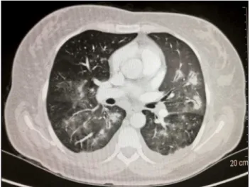
didiasis, then treated with luconazole. Computed to-mography of the chest showed peribronchovascular thick-ening, areas of ground glass opacity, condensations with air bronchograms surrounded by ground glass opacity (halo sign) and bilateral pleural effusion (Figures 1, 2 and 3). Following guidance from the oncology team, only an-tiretroviral therapy was maintained for immune recovery, with subsequent evaluation regarding chemotherapy.
Two weeks after admission, the patient progressed with disorientation, hallucinations, worsening of respira-tory pattern, and oliguria. She had worsening of renal func-tion, and strong metabolic acidosis. The patient was re-ferred to the ICU, and after central venous access puncture
she progressed with respiratory failure requiring mechan-ical ventilation. As chest X-ray showed extensive pneumo-thorax, chest tube drainage was performed. Increased ni-trogenous compounds and metabolic acidosis were treated with dialysis requiring vasoactive drug due to he-modynamic instability.
Due to the possible association of hallucinogenic symp-toms with efavirenz and the risk of nephrotoxicity of te-nofovir, the team chose to replace the Antiretroviral Ther-apy (ART), introducing lopinavir/ritonavir and abacavir with lamivudine. The antibiotic coverage was broadened and tuberculostatic medication was started to treat a pos-sible disseminated tuberculosis, later discarded after a neg-ative result of PCR tests for the disease in the blood, CSF, and urine. Cranial CT scan showed no changes, and study of CSF ruled out any association with opportunistic dis-eases of the central nervous system. A new CT scan of the chest revealed consolidations at the base of both lungs within air bronchograms and residual pneumothorax. The patient progressed with clinical improvement, and the tra-cheal tube was removed. Given that control chest X-ray showed complete lung expansion, we opted for the remov-al of the chest drain.
The patient continued with hallucinations and psy-chomotor agitation, which were explained after psychi-atric evaluation as sepsis-associated delirium, then treat-ed with quetiapine.
During hemodialysis, the patient had an episode of hypotension, followed by massive aspiration and cardio-respiratory arrest with asystole. There was no reversal af-ter cardiopulmonary resuscitation maneuvers.
FIGURE 3 Chest CT scan, axial mediastinal window, arterial
phase. See small bilateral pleural effusion and mediastinal lymph nodes with preserved size.
FIGURE 2 Chest CT scan, axial lung window. Some areas of
consolidation with irregular margins, and a ground glass halo giving the appearance of flame. There is bilateral pleural effusion and peribronchovascular infiltrate.
FIGURE 1 Chest CT scan, axial lung window. See areas of ground
PULMONARY KAPOSI’SSARCOMAINAFEMALEPATIENT: CASEREPORT
REV ASSOC MED BRAS 2016; 62(5):395-398 397
D
ISCUSSIONKS associated with infection by the human immunode-iciency virus (HIV) is common in adult homosexual and bisexual young men.4-6
Men are more affected than women in a ratio of 15:1. The annual incidence is between 0.02 and 0.06% of all ma-lignant tumors.7 KS rarely affects American or European heterosexuals, but it often occurs in African or Caribbe-an heterosexuals.8
Most patients with pulmonary KS already present with cutaneous lesions at the time of diagnosis; however, visceral disease may occasionally precede the skin. The most affected skin sites are the lower limbs, head and neck, and the lesions may have a varied appearance (spots, pap-ules, nodules or plaques), all palpable and non-pruritic, with sizes ranging from millimeters to centimeters, and brownish, pink or violet color. These lesions may also be conluent and are generally symmetrical in distribution.16
The precise incidence of pulmonary KS is still not well known. Some clinical studies have shown a higher prevalence in individuals with AIDS who already had the cutaneous form of the disease (over 32%) compared to those without it (3-13%).3,9-11 In autopsies, however, bron-chopulmonary KS was found in 47% of patients with AIDS who had the cutaneous form, which reveals a
pos-sible underdiagnosis of pulmonary involvement.12 Since the variety of opportunistic diseases is great, pulmonary involvement in SK can be dificult to diag-nose. Symptoms are unspeciic (cough, dyspnea, hemop-tysis, chest pain) and may represent several other diseas-es, including tuberculosis. Pleural effusion, hypoxemia and acute respiratory failure requiring mechanical venti-lation have also been reported in some cases.13-15
Kaposi’s sarcoma may involve the tracheobronchial tree, the lung parenchyma and pleura. Pleural involvement occurs only in the presence of parenchymal abnormalities, just as the presence of lesions in the bronchial tree below the tracheal carina is, as a rule, accompanied by parenchy-mal lesions. In the lung, Kaposi’s sarcoma grows along the lymph node routes, i.e. around bronchovascular sheaths, especially in the axial interstitium, interlobular septa, and pleura. The cellular iniltrate advances in the interstitium along the peribronchovascular sheaths, the pulmonary veins, and the pleural surface. From these regions, the cells proliferate as “tentacles” to the adjacent alveolar septa and ill the alveoli, forming solid nodules.17,18
The diagnosis of pulmonary KS (PKS) is still a chal-lenge, mainly because the clinical, laboratory and radio-graphic indings are non-speciic and cannot differenti-ate PKS from other lung diseases that may occur in AIDS.
Therefore, these indings should be combined with those obtained by CT scan, bronchoscopy and transbronchial biopsy.19
Differential diagnoses include: lymphoma, lung tu-mors, infections (such as tuberculosis, viruses and fun-gi), infection with Pneumocystis jirovecii and bacillary angiomatosis.18,20,21
Histologically, the tumor consists of vascular spac-es interspersed with spindle-shaped reticuloendothelial cell, containing atypical nuclei and occasional mitoses. The vascular gaps contain erythrocytes and hemosider-in deposits.18
The indings on chest radiography are many, usual-ly bilateral and diffuse. Reticulonodular iniltrates with predilection for hilar areas and the base of the lungs, con-solidations, multiple ill-deined nodules, bilateral pleu-ral effusion in 30% of cases, hilar and mediastinal lymph node enlargement in 10% of cases, and even normal x-rays have also been described.2,19-25 The presence of other as-sociated lung infections may complicate the interpreta-tion of the indings. Due to the above and the fact that the indings are not speciic of PKS, radiography has the main role to dismiss lung involvement and/or monitor disease progression.26
High-resolution CT (HRCT) has higher sensitivity and speciicity compared to radiography in the event of sus-pected diagnosis of PKS. Early indings include thicken-ing of peribronchovascular interstitium, especially in the base of the lungs, which can even simulate other oppor-tunistic infections of the airways. Late changes include ill-deined nodules/consolidations (“candle lame” appear-ance) with peribronchovascular distribution, symmetri-cal and bilateral, which may be surrounded by ground glass opacity (halo sign), thickening of peribronchovas-cular axial interstitium, interlobular septal thickening and pleural effusion. These changes were observed in our pa-tient (Figure 3). In addition to these indings, there is also nodular thickening of issures, subpleural nodules, hilar and mediastinal lymph node enlargement, absent in this case, which presented some mediastinal lymph nodes with preserved size. In most patients, the presence of typical nodules in CT and perihilar distribution of the aforemen-tioned abnormalities justify KS being considered togeth-er with othtogeth-er thoracic affections in AIDS.2,15,18-20,25,27-32
C
ONCLUSIONPETRIBU NCL ETAL.
398 REV ASSOC MED BRAS 2016; 62(5):395-398
manifestations, and even more with CT indings, is fun-damental, especially in patients who already have the cu-taneous form of the disease, so that proper treatment can be initiated as soon as possible.
R
ESUMOSarcoma de Kaposi pulmonar em paciente do sexo femi-nino: Relato de caso
O sarcoma de Kaposi (SK) é uma neoplasia angioprolifera-tiva maligna multicêntrica. Na maioria das vezes, esse tu-mor restringe-se à pele e ao tecido subcutâneo, mas pode cursar com comprometimento visceral amplamente disse-minado, como é o caso do pulmão. O SK pulmonar é a for-ma for-mais frequente em homens adultos jovens, em ufor-ma pro-porção de 15:1. A doença geralmente acomete indivíduos com baixa contagem de linfócitos CD4 (<150-200 células/ mm³). Relatamos um caso de uma paciente do sexo femi-nino, 35 anos, com presença de lesões cutâneas, episódios de diarreia autolimitada e perda de peso de 15 kg havia aproximadamente 9 meses, evoluindo para quadro febril persistente. Foi dado o diagnóstico de aids e foi realizada biópsia das lesões evidenciando sarcoma de Kaposi. A to-mograia computadorizada do tórax evidenciou imagens de espessamento peribroncovascular, áreas de vidro fosco, condensações com broncogramas aéreos circundados por área de vidro fosco (sinal do halo) e derrame pleural bilate-ral. O diagnóstico da forma pulmonar de SK ainda é um desaio, principalmente pela ocorrência de outras doenças oportunistas que, inclusive, podem ocorrer de forma con-comitante. Portanto, a suspeição desse diagnóstico pelas manifestações clínicas, laboratoriais e, principalmente, to-mográicas é de fundamental importância, sobretudo em pacientes que já apresentam a forma cutânea da doença.
Palavras-chave: sarcoma de Kaposi, HIV, síndrome de imunodeiciência adquirida, pulmão.
R
EFERENCES1. Kalil JA, Jovino MAC, Papaiordanou F, Arriaga M, Ribeiro Jr MAF. Sarcoma de Kaposi em membros inferiores: relato de caso. J Vasc Bras. 2010; 9(4):261-5. 2. Restrepo CS, Martínez S, Lemos JA, Carrillo JA, Lemos DF, Ojeda P, et al. Imaging
manifestations of Kaposi sarcoma. Radiographics. 2006; 26(4):1169-85. 3. Zibrak JD, Silvestri RC, Costello P, Marlink R, Jensen WA, Robins A, et al.
Bronchoscopic and radiographic features of Kapsosi’s sarcoma involving the respiratory tract. Chest. 1986; 90(4):476-9.
4. Fonseca BAL, Bollela, VR, da Justa Neto P. Sarcoma de Kaposi e síndrome da imunodeiciência adquirida: características desta associação, incluindo novos conceitos sobre patogênese e tratamento. Medicina, Ribeirão Preto. 1999; 32:26-39.
5. Patrikidou A, Vahtsevanos K, Charalambidou M, Valeri RM, Xirou P, Antoniades K. Non-AIDS Kaposi’s sarcoma in the head and neck area. Head Neck. 2009; 31(2):260-8.
6. Friedman-Kien AE, Saltzman BR. Clinical manifestations of classical, endemic African, and epidemic AIDS-associated Kaposi’s sarcoma. J Am Acad Dermatol. 1990; 22(6 Pt 2):1237-50.
7. Antman K, Chang Y. Kaposi’s sarcoma. N Engl J Med. 2000; 342(14):1027-38. 8. Sitas F, Newton R. Kaposi’s sarcoma in South Africa. J Natl Cancer Inst
Monogr. 2001; (28):1-4.
9. Murray JF, Felton CP, Garay SM, Gottlieb MS, Hopewell PC, Stover DE, et al. Pulmonary complications of the acquired immunodeiciency syndrome. Report of a National Heart, Lung, and Blood Institute workshop. N Engl J Med. 1984; 310(25):1682-8.
10. Garay SM, Belenko M, Fazzini E, Schinella R. Pulmonary manifestations of Kaposi’s sarcoma. Chest. 1987; 91(1):39-43.
11. Gill PS, Akil B, Colletti P, Rarick M, Loureiro C, Bernstein-Singer M, et al. Pulmonary Kaposi’s sarcoma: clinical indings and results of therapy. Am J Med. 1989; 87(1):57-61.
12. Meduri GU, Stover DE, Lee M, Myskowski PL, Carvelli JF, Zaman MB. Pulmonary Kaposi’s sarcoma in the acquired immunodeiciency syndrome. Clinical, radiographic, and pathologic manifestations. Am J Med. 1986; 81(1):11-8.
13. Miller RF, Tomlinson MC, Cottrill CP, Donald JJ, Spittle MF, Semple SJ. Bronchopulmonary Kaposi’s sarcoma in patients with AIDS. Thorax. 1992; 47(9):721-5.
14. Arruda E, Jacome AA, Toscano AL, Silvestrini AA, Rêgo AS, Wiermann EG, et al. Consensus of the Brazilian Society of Infectious Diseases and Brazilian Society of Clinical Oncology on the management and treatment of Kaposi’s sarcoma. Braz J Infect Dis. 2014; 18(3):315-26.
15. Cadranel J, Naccache J, Wislez M, Mayaud C. Pulmonary malignancies in the immunocompromised patient. Respiration. 1999, 66(4):289-309. 16. Pantanowitz L, Dezube BJ. Kaposi sarcoma in unusual locations. BMC
Cancer. 2008; 8:190-199.
17. Marchiori E, Valiante PM, Sales AR, Capone D, Moraes HP, Santos MLO. Sarcoma de Kaposi pulmonar: correlação da tomograia computadorizada de alta resolução com a anatomopatologia. Rev Imagem. 2000; 22:63-7. 18. McGuinness G. Changing trends in the pulmonary manifestations of AIDS.
Radiol Clin North Am. 1997; 35(5):1029-82.
19. Kang EY, Staples CA, McGuinness G, Primack SL, Müller NL. Detection and differential diagnosis of pulmonary infections and tumors in patients with AIDS: value of chest radiography versus CT. AJR Am J Roentgenol. 1996; 166(1):15-9.
20. Hartman TE, Primack SL, Muller NL, Staples CA. Diagnosis of thoracic complications in AIDS: accuracy of CT. AJR Am J Roentgenol. 1994; 162(3):547-53.
21. Moore EH, Russell LA, Klein JS, White CS, McGuinness G, Davis LG, et al. Bacillary angiomatosis in patients with AIDS: multiorgan imaging indings. Radiology. 1995; 197(1):67-72.
22. Mann SG. Kaposi’s sarcoma. AJR Am J Roentgenol. 1974; 121(4):793-9. 23. Leibowitz MR, Dagliotti M, Smith E, Murray TF. Rapidly fatal
lymphangioma-like Kaposi’s sarcoma. Histopathology. 1980; 4(5):559-66.
24. Davis SD, Henschke CI, Chamides BK, Westcott JL. Intrathoracic Kaposi sarcoma in AIDS patients: radiographic-pathologic correlation. Radiology. 1987; 163(2):495-500.
25. McGuinness G, Gruden JF, Bhalla M, Harkin TJ, Jagirdar JS, Naidich DP. AIDS-related airway disease. AJR Am J Roentgenol. 1997; 168(1):67-77. 26. O’Mahony D, Gandjbakhche AH, Hassan M, Vogel A, Yarchoan R. Imaging
techniques for Kaposi’s sarcoma. J HIV Ther. 2008; 13(3):65-71. 27. Huang L, Schnapp LM, Gruden JF, Hopewell PC, Stansell JD. Presentation
of AIDS-related pulmonary Kaposi’s sarcoma diagnosed by bronchoscopy. Am J Respir Crit Care Med. 1996; 153(4 Pt 1):1385-90.
28. Gruden JF, Huang L, Webb WR, Gamsu G, Hopewell PC, Sides DM. AIDS-related Kaposi sarcoma of the lung: radiographic indings and staging system with bronchoscopic correlation. Radiology. 1995; 195(2):545-52. 29. Khalil AM, Carette MF, Cadranel JL, Mayaud CM, Bigot JM. Intrathoracic
Kaposi’s sarcoma. CT indings. Chest. 1995; 108(6):1622-6.
30. Marchiori E, Müller NL, Soares Souza A Jr, Escuissato DL, Gasparetto EL, Franquet T. Pulmonary disease in patients with AIDS: high resolution CT and pathologic indings. AJR Am J Roentgenol. 2005; 184(3):757-64. 31. da Silva Filho FP, Marchiori E, Valiante PM, Escuissato DL, Gasparetto TD.
AIDS-related Kaposi sarcoma of the lung presenting with a “crazy-paving” pattern on high-resolution CT: imaging and pathologic indings. J Thorac Imaging. 2008; 23(2):135-7.