Treatment of brain metastases
TRATAMENTO DAS METÁSTASES ENCEFÁLICAS
Authorship: Sociedade Brasileira de Neurocirurgia
Participants: Frederico de Melo Tavares de Lima1, Ana Lucia Mello de Carvalho1, Joacil Carlos da Silva Jr.1,
Robson Amorim1, Marcos Dellaretti1, Leonardo Augusto Wendling-Henriques1, Marcos Maldaun1,
Daniel Kirchhoff1, Ricardo Vieira Botelho1
Conflict of interest:none
1Sociedade Brasileira de Neurocirurgia
http://dx.doi.org/10.1590/1806-9282.62.05.389
The Guidelines Project, an initiative of the Brazilian Medical Association, aims to combine information from the medical ield in order to standard-ize procedures to assist the reasoning and decision-making of doctors.
The information provided through this project must be assessed and criticized by the physician responsible for the conduct that will be adopted, de-pending on the conditions and the clinical status of each patient.
I
NTRODUCTIONMetastases are the most common cancers that affect the in-tracranial space. They have an incidence of 6 to 10 times that of primary brain tumors. The estimate is that they oc-cur in 20 to 40% of all malignant neoplasms affecting the human body, being symptomatic in 10 to 20% of these pa-tients. Prevalence is growing due to aging of the population, greater access to neuroimaging techniques and increased survival of patients with malignancies. Brain metastases (BM) occur with equal frequency in men and women, and have the same effect in all races. Any form of cancer can pro-mote secondary spread in the brain. Primary tumors that contribute the most to BMs are, in descending order: lung, breast, melanoma, kidney and colorectal carcinoma. How-ever, in over 15% of cases the primary site is unknown. Brain metastases are most often diagnosed in patients with a known malignancy (metachronous presentation). Less fre-quently, the lesions are diagnosed simultaneously with the primary tumor (synchronous presentation), or even before the discovery of the primary disease. About 85% of BMs are located in the supratentorial compartment; 10 to 15% are in the infratentorial space; and 1 to 3%, in the brainstem. The most common symptoms in BMs are: headache, sei-zures, behavioral changes, hemiparesis, vomiting, dyspha-sia, and consciousness disorders. Most metastases spread in the brain through the blood but, less often, through the venous plexus. Without treatment, the median survival is less than 2 months.
In Brazil, based on the estimate for 2012 by the Na-tional Cancer Institute, and considering the probable 90,300 new cases of breast, lung and colon cancer, and melanoma, it is possible to estimate the occurrence of 13,545 new cases of BM per year. That number places BMs
as the fourth most common form of cancer in Brazil, re-gardless of gender. Furthermore, it is an incidence rough-ly three times higher than all tumors of the central ner-vous system taken together.
Treatments include surgery, radiosurgery and radio-therapy, and in some cases chemotherapy. Treatment can include one method alone, or a combination of therapies.
O
BJECTIVEThe aim is to clarify the effect of surgery, radiotherapy and radiosurgery in patients with brain metastases.
G
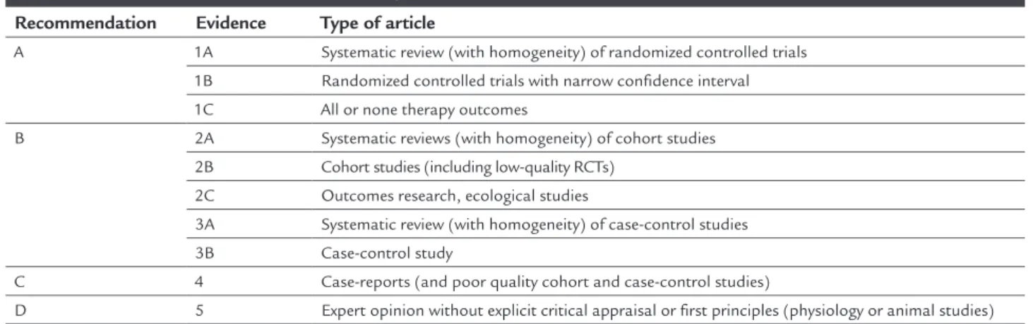
RADE OFRECOMMENDATION AND STRENGTH OF EVIDENCEThe grade of recommendation and strength of evidence were deined in accordance with the Brazilian Medical As-sociation, as displayed in the Table 1.
T
YPES OF STUDYThe primary study for deinition of this guideline is the randomized clinical trial. Clinical studies of lower meth-odological quality were used in the absence of the irst.
1) C
LINICAL QUESTIONWhat is the beneit of surgery in the treatment of brain metastases compared to radiotherapy?
Description of the evidence collection method
The following terms were searched as “MeSH” and words in the text:
• 1# (((“brain”[MeSH Terms] OR “brain”[All Fields]) AND (“neoplasm metastasis”[MeSH Terms] OR (“neoplasm”[All Fields] AND “metastasis”[All Fields]) OR “neoplasm metastasis”[All Fields] OR “metastasis”[All Fields])) • 2# “brain neoplasms/secondary”[MeSH Terms] OR
“supratentorial neoplasms/secondary”[MeSH Terms] AND ((“surgery”[Subheading] OR “surgery”[All Fields] OR “surgical procedures, operative”[MeSH Terms] OR (“surgical”[All Fields] AND “procedures”[All Fields] AND “operative”[All Fields]) OR “operative surgical procedures”[All Fields] OR “surgery”[All Fields] AND “surgery”[All Fields]) OR “microsurgery”[MeSH Terms]
Related articles were searched from the citations in the primary texts.
• Inclusion criteria: Only randomized controlled trials were evaluated, including those published in English, Spanish and Portuguese.
• Clinical outcomes included were: Functional indepen-dence, survival, tumor control, cost-effectiveness, qual-ity of life, cognitive decline and other adverse effects. • Combined results of searches: 7,963 articles were
ini-tially retrieved. 366 studies were considered clinical tri-als, of which only three were randomized and selected for critical evaluation of the strength of evidence. The remaining non-randomized were excluded.
Methodological quality analysis
The three works were classiied according to the Jadad scale as ≥ 3. Considering the size of the samples in the three studies, the one by Patchel1(1B) had a strength of 91.93%, while the studies by Vecht2 (1B) and Mintch3 (1B) had the same strength, 16.96%.
The study by Patchell1(1B) (1990) was randomized at a single center, and included 48 patients to compare surgery, followed by radiotherapy and biopsy plus radio-therapy. Patients had mean age of 60 years; the mean Kar-nofsky score was 90, conirming the good functional sta-tus of patients. Randomization was done by computer but the assessment of outcomes was not made by inde-pendent observers or blinded to the treatment. The out-comes studied were survival, functional independence, tumor size progression, time to recurrence and cause of death. Statistical analysis was performed with survival study (Kaplan-Meier and log rank test).
The study by Vecht2 (1B) (1993) was a Dutch multi-center randomized clinical trial of surgical resection, fol-lowed by whole brain radiotherapy versus radiotherapy alone. 63 patients were randomized (mean age 60 years). Randomization was done in blocks, controlled by call center, but outcome assessors were not blinded to treat-ment. Assessment measures included survival, function-ally independent survival and cause of death.
The third3 (1B) (Mintz, 1996) was a Canadian multi-center study, comparing surgical resection followed by whole brain radiotherapy versus whole brain radiothera-py alone. The authors randomized 84 patients (mean age 59 years). Randomization was based on call center after stratiication for prognostic factors. Outcome assessors were not blinded for treatment type. Outcomes included survival (percentage), cause of death, functional status (Karnofsky) and quality of life (using the Spitzer scale) and surgical complications after 30 days.
Outcome data extraction
Three types of outcomes were extracted and evaluated as clus-ters in the three randomized trials: Survival time, percentage of lesion recurrence, and time of functional independence.
TABLE 1 Grade of recommendation and strenght of evidence.
Recommendation Evidence Type of article
A 1A Systematic review (with homogeneity) of randomized controlled trials 1B Randomized controlled trials with narrow confidence interval
1C All or none therapy outcomes
B 2A Systematic reviews (with homogeneity) of cohort studies 2B Cohort studies (including low-quality RCTs)
2C Outcomes research, ecological studies
3A Systematic review (with homogeneity) of case-control studies 3B Case-control study
C 4 Case-reports (and poor quality cohort and case-control studies)
Survival time
The study by Patchel (1B) showed 40 weeks of survival in the surgical group and 15 weeks in the group undergoing radiotherapy (RT) (p<0.01), favoring surgical treatment.
Vecht (1B) found a longer survival (10 months) in the surgical group compared with the RT group (6 months) (p=0.04).
In the study by Mintz (1B), there was no difference regarding survival: 5.62 months in the surgical group, 6.28 months in the RT group (p=0.24).
The analysis of randomized trials based on survival curves has several limitations.4,5 The survival analysis did not show a statistically signiicant difference between the two treatments (HR=0.72, 95CI 0.34-1.53, p=0.40).6 The studies by Patchell and Vecht reported longer survival in patients undergoing surgery plus radiotherapy, while the study by Mintz revealed longer survival in patients treat-ed with radiotherapy alone.
Outcome: Lesion recurrence
The study by Patchel (1B) was the only one to properly describe the results in this outcome. Recurrence in the surgical group totaled 20% and in the RT group, 52%. The difference was statistically signiicant (p<0.02).
Methodological quality analysis
This trial was classiied as Jadad = 3. The study’s strength reached 64.77%.
Evidence summary
Surgery decreases the recurrence of the lesion as single metastases, compared to radiotherapy.
Outcome: Functional independence
The three studies examined functional independence. Patchel and Mitch used the Karnofsky scale. Vecht used the performance status level of the World Health Organization (between 0 and 4).
In the study by Patchel, the surgical group maintained Karnofsky score > 70 in up to 38 weeks of follow-up (on average), and the radiotherapy group maintained this score only up to 8 weeks (p<0.0005).
In the study by Vecht, there was no difference in func-tional independence between the groups.
The study by Mitch also revealed no difference in functional independence (Karnofsky) (p=0.98).
Evidence summary
There is no evidence that surgical treatment preserves the functional independence of patients, compared with ra-diotherapy.
2) C
LINICAL QUESTIONIs radiosurgery combined with holocranial radiotherapy more effective than radiosurgery or radiotherapy alone (1 to 3 metastases)?
Description of the evidence collection method
The search strategy was conducted in the MedLine (PubMed) database to identify articles published from 1964 to 2013. The objective was to identify studies com-paring radiotherapy (combined with radiosurgery) and radiosurgery or radiotherapy alone.
The following terms were searched as “Mesh” and words in the text: “Brain Neoplasms”, “Radiosurgery”, “Radiotherapy”, “Brain Neoplasms/radiotherapy”,
“Ra-diotherapy, Adjuvant”, “Radiosurgery/methods”, “Treat-ment Outcome”, “Radiosurgery/adverse effects”.
Related articles were searched from the citations in the primary texts.
• Inclusion criteria: Randomized clinical trials, includ-ing those published in English, Spanish and Portu-guese.
• Clinical outcomes included were: Functional indepen-dence, survival, tumor control, cost-effectiveness, qual-ity of life, cognitive decline and other adverse effects.
Results
In all, 2,638 articles were identiied in the initial search strategy and 29 articles were retrieved based on inclusion criteria. Of these, 19 articles were excluded because they were narrative reviews, evaluation of radiosurgery alone, or treatment of high-grade gliomas.
Another article was manually selected. In the end, nine randomized trials were analyzed to answer this question.
Methodological quality analysis
Andrews et al.7(1B) (2004) studied, between January 1996 and June 2001, 333 patients in 55 participating centers – 167 underwent whole brain radiotherapy and
previous-ly calculated considering 124 patients per group and eventually adjusted to 326 patients. The analysis of loss to follow-up was done based on intention-to-treat. There is no data on extraction of results.
Kondziolka8(1B)(1999) was a single-center trial dis-continued after the interim analysis of 27 patients that revealed signiicant beneit in the rate of local metastat-ic control with whole brain radiotherapy plus radiosur-gery. This study included patients with 2 to 4 brain me-tastases sized 25 mm or less. Local tumor control was also assessed as a primary outcome. No other results were presented. MRI scans were read by an independent ob-server blinded for treatments.
Aoyama et al.9(1B) (2006) studied patients aged 18 years or older with 1 to 4 brain metastases measuring a maximum of 3 cm in diameter on MRI divided into two groups: stereotactic radiosurgery alone versus stereotac-tic radiosurgery combined with whole brain radiothera-py. Eligible patients should have a Karnofsky performance score of 70 or more. The study was conducted at the Hok-kaido University (Japan) and ten other centers. Random-ization was centralized at the Hokkaido University, in blocks of four. The patients were stratiied based on the number of brain metastases (single vs. 2-4), extent of ex-tracranial disease (active vs. stable), and location of the primary tumor (lung vs. other sites).
The study by Chang et al.10(1B) (2009) considered el-igible patients who were treated at the Departments of Ra-diation Oncology and Neurosurgery of MD Anderson Can-cer Center, Houston, aged 18 years or older, Karnofsky = 70 or more, with 1-3 brain metastases. Randomization was done by computer (1:1) between the group of stereotactic radiosurgery combined with radiotherapy and stereotac-tic radiosurgery alone, in blocks of 2, 4, 6, or 8 patients. The sequence was hidden until all interventions were des-ignated. Intention-to-treat analysis was conducted.
Kocher et al.11(1B) studies the role of whole brain ra-diotherapy after surgery or radiosurgery in a limited num-ber of brain metastases in patients in good conditions with stable systemic cancer. The trial was randomized by center, organized by the European Organisation for Re-search and Treatment of Cancer. The study was designed to detect a difference of 11% in the proportion of live pa-tients, with strength of 80% and 5% of two-tailed signii-cance. 340 patients were planned to be recruited.
Sofietti R et al.12(1B): This was a multicenter, ran-domized, international, phase III trial comparing patients undergoing radiosurgery or surgery with whole brain ra-diotherapy as adjuvant treatment or monitoring.
Results
Outcome: Survival
Andrews et al.7(1B) (2004) studied 333 patients evaluat-ing the effects of the “boost” of radiosurgery in the tu-mor bed after radiotherapy. There was no beneit in sur-vival between the groups. However, in patients with single metastasis, combined treatment proved to be superior (6.5 months vs. 4.9 months, p=0.03) (1B).
Kondziolka et al.8(1B) (1997) studied 27 patients and found no signiicant differences with respect to sur-vival. The mean survival in the group undergoing com-bination treatment was 11 months, while the group treat-ed with radiotherapy alone had a survival of 7.5 months (p=0.22). This study was terminated early because an in-terim analysis showed signiicant beneit in terms of lo-cal tumor control in favor of the combined treatment (ev-idence level 2B).
Aoyama et al. (2006), in 132 patients, obtained a 7.5 month survival with the combined treatment, and 8 months in the group undergoing radiosurgery alone (p=0.42) (level of evidence 1B).
Chang et al. (2009) compared the combined treat-ment with radiosurgery alone in 58 patients. In the ra-diosurgery group, survival was 15.2 months versus 5.7 in the combined treatment group (p=0.003) (level of evi-dence 1B). This study was terminated at 4 months after an interim analysis showed cognitive decline in the com-bined treatment group.
Evidence summary
Compared to radiosurgery alone, it is not possible to de-termine whether the combination treatment is superior or not in terms of survival (A).
O
UTCOME: F
UNCTIONAL INDEPENDENCE3) C
LINICAL QUESTIONDoes radiosurgery combined with holocranial radiother-apy increase the time of functional independence com-pared with radiosurgery or holocranial radiotherapy alone?
Results
Andrews et al. (2004) found that combination treatment provides less possibility of functional worsening at 6 months (RR=0.78, 95CI 0.61-1, p=0.05) compared with holocranial radiotherapy alone (level of evidence 2B).
for those not treated with radiotherapy vs. 9.5 months for patients undergoing RT (95CI 7.8-11.9), p=0.71 (level of evidence 1B).
Aoyama et al. (1996) did not ind beneit in function-al prognosis (p=0.53) or preservation of neurologicfunction-al func-tion (p=0.99) at 12 months with combined treatment (lev-el of evidence 1B).
Evidence summary
Radiotherapy combined with holocranial radiotherapy does not increase the time of functional independence com-pared with radiosurgery (A). However, compared to patients treated with radiotherapy, the combined treatment is supe-rior in terms of functional independence at 6 months (B).
O
UTCOME: T
UMOR CONTROL4) C
LINICAL QUESTIONDoes radiosurgery combined with holocranial radiother-apy improve tumor control compared with radiosurgery or holocranial radiotherapy alone?
Results
The study by Kondziolka et al. (1997) ended prematurely after an interim analysis identiied considerable beneit in terms of local tumor control with combined treatment compared to radiotherapy alone. The median time to lo-cal recurrence was 36 months for the irst and 6 months for radiotherapy alone (p=0.005) (level of evidence 2B).
In the study by Kocher et al. (2004), combined treat-ment led to better local control of tumors at 2 years com-pared with radiosurgery alone: reduction from 31% (95CI 22-40%) to 19% (95CI 11-27%; p=0.04). There was also bet-ter control of tumors in other locations: a reduction from 48% (95CI 38-58%) to 33% (95CI 24-43%; p=0.023).
In the study by Chang et al. (2009), tumor control at 1 year occurred in 100% of the patients undergoing com-bined treatment versus 67% of tumor control with radio-surgery alone (p=0.012).
In the study by Aoyama et al. (1996), tumor control at 12 months reached 88.7% (95CI 80.1-97.3%) in the group of combined treatment versus 72.5% (95CI 60.3-84.7%) in the group undergoing radiosurgery alone (p=0.002). Tumor control at 1 year in other sites of the brain was also more favorable than under combined treat-ment. There was a decline from 73% to 45% (p=0.02).
Evidence summary
Radiosurgery combined with holocranial radiotherapy reduces the likelihood of local tumor recurrence and the development of new brain lesions (A).
O
UTCOME: C
OGNITIVE DECLINE5) C
LINICAL QUESTIONIs radiosurgery combined with holocranial radiotherapy associated with greater cognitive decline compared with radiosurgery or holocranial radiotherapy alone?
Results
The study by Chang et al. (2009) had cognitive function as the primary endpoint, which was evaluated by a set of neu-ropsychological tests covering various domains. This study was terminated early after an interim analysis demonstrat-ed that the possibility of deterioration of learning and mem-ory at 4 months was signiicantly higher in patients under-going combined treatment (52%) than in patients treated with radiosurgery alone (24%) (level of evidence 1B).
Aoyama et al.13 (2007) used the mini-mental state ex-amination to assess cognitive function, and found that the main factor related to cognitive function is tumor control. Cognitive worsening was earlier the single treat-ment group (7.6 months vs. 16.5 months, p=0.05). How-ever, after 36 months, only 14.7% of patients who under-went combined treatment had no cognitive worsening (level of evidence 2B).
Evidence summary
Radiosurgery combined with holocranial radiotherapy leads to signiicant worsening of cognition compared with radiosurgery alone (A).
O
UTCOME: Q
UALITY OF LIFE6) C
LINICAL QUESTIONIs radiosurgery combined with holocranial radiotherapy associated with poorer quality of life compared to radio-surgery or holocranial radiotherapy alone?
Results
Sofietti et al. (2013) analyzed the aspects of quality of life based on the EORTC QLQ-C30 scale in patients un-dergoing radiosurgery/surgery alone or combined treat-ment. Patients who underwent combined treatment had poorer overall quality of life at 9 months (p=0.014) (lev-el of evidence 2B).
Evidence summary
Radiosurgery combined with holocranial radiotherapy is associated with poorer quality of life (B).
N
OTErecommendations were formulated for each outcome, and the best type of treatment for each type of manifes-tation should be chosen at the discretion of the attend-ing physician.
R
EFERENCES1. Patchell RA, Tibbs PA, Walsh JW, Dempsey RJ, Maruyama Y, Kryscio RJ, et al. A randomized trial of surgery in the treatment of single metastases to the brain. N Engl J Med. 1990; 322(8):494-500.
2. Vecht CJ, Haaxma-Reiche H, Noordijk EM, Padberg GW, Voormolen JH, Hoekstra FH, et al. Treatment of single brain metastasis: radiotherapy alone or combined with neurosurgery? Ann Neurol. 1993; 33(6):583-90. 3. Mintz AH, Kestle J, Rathbone MP, Gaspar L, Hugenholtz H, Fisher B, et al.
A randomized trial to assess the efficacy of surgery in addition to radiotherapy in patients with a single cerebral metastasis. Cancer. 1996; 78(7):1470-6. 4. Pamar MKB, Torri V, Stewart L. Extracting summary statistics to perform
meta-analysis of the published literature for survival endpoints. Stat Med. 1998; 17(24):2815-34,.
5. Williamson PR, Smith CT, Hutton JL, Marson AG. Aggregate data meta-analysis with time-to-event outcomes. Stat Med. 2002; 21(22):3337-51. 6. Hart MG, Grant R, Walker M, Dickinson H. Surgical resection and whole
brain radiation therapy versus whole brain radiation therapy alone for single brain metastases. Cochrane Database Syst Rev. 2005; (1):CD003292. 7. Andrews DW, Scott CB, Sperduto PW, Flanders AE, Gaspar LE, Schell
MC, et al. Whole brain radiation therapy with or without stereotactic
radiosurgery boost for patients with one to three brain metastases: phase I I I results of the RTOG 9508 ra ndomised tria l. La ncet. 20 04; 363(9422):1665-72.
8. Kondziolka D, Patel A, Lunsford LD, Kassam A, Flickinger JC. Stereotactic radiosurgery plus whole brain radiotherapy versus radiotherapy alone for patients with multiple brain metastases. Int J Radiat Oncol Biol Phys. 1999; 45(2):427-34.
9. Aoyama H, Shirato H, Tago M, Nakagawa K, Toyoda T, Hatano K, et al. Stereotactic radiosurgery plus whole-brain radiation therapy vs stereotactic radiosurgery alone for treatment of brain metastases: a randomized controlled trial. JAMA. 2006; 295(21):2483-91.
10. Chang EL, Wefel JS, Hess KR, Allen PK, Lang FF, Kornguth DG, et al. Neurocognition in patients with brain metastases treated with radiosurgery or radiosurgery plus whole-brain irradiation: a randomised controlled trial. Lancet Oncol. 2009; 10(11):1037-44.
11. Kocher M, Sofietti R, Abacioglu U, Villà S, Fauchon F, Baumert BG, et al. Adjuvant whole-brain radiotherapy versus observation after radiosurgery or surgical resection of one to three cerebral metastases: results of the EORTC 22952-26001 study. J Clin Oncol. 2011; 29(2):134-41.
12. Sofietti R, Kocher M, Abacioglu UM, Villa S, Fauchon F, Baumert BG, et al. A European Organization for Research and Treatment of Cancer phase III trial of adjuvant whole-brain radiotherapy versus observation in patients with one to three brain metastases from solid tumors after surgical resection or radiosurgery: quality-of-life results. J Clin Oncol. 2013; 31(1):65-72.