(1) Universidade Federal do Rio Grande do Norte – UFRN – Natal (RN), Brasil. (2) Universidade de Fortaleza – UNIFOR –
Fortaleza (CE), Brasil.
(3) Universidade Estadual do Ceará – UECE – Fortaleza (CE), Brasil.
Source: Fundação Cearense de Apoio ao Desenvolvimento Cientíico e Tecnológico – FUNCAP
Conlict of interest: non-existent
Audiologic characteristics of patients
with diabetes mellitus type 2
Características audiológicas de pacientes com diabetes mellitus tipo 2
Juliana Mota Ferreira(1)
Marília Fontenele e Silva Câmara(2)
Paulo César de Almeida(3)
José Brandão Neto(1)
Carlos Antonio Bruno da Silva(2)
Received on: December 23, 2015 Accepted on: June 10, 2016
Mailing address:
Carlos Antonio Bruno da Silva Universidade de Fortaleza, Programa de Pós Graduação em Saúde Coletiva Av. Washington Soares, 1321, Bloco S, Sala S-11, Edson Queiroz
Fortaleza – CE – Brasil CEP: 60811-904
E-mail: [email protected]
ABSTRACT
Purpose: to identify the audiological characteristics of patients with type 2 diabetes mellitus.
Methods: Cross-sectional study in secondary care unit for diabetes in Fortaleza, April-July 2010. The sample comprised 152 patients with type 2 diabetes, regardless of sex, between 36 and 60 years of age. Held pure tone audiometry (PTA), transient-evoked otoacoustic emissions (TEOAE) and distortion product
otoacoustic emissions (DPOAE).
Results: association of age and time of diagnosis with hearing loss, and the time of diagnosis with the absence of DPOAE. It was observed sensorineural hearing loss in 63.2% of patients, which 71.9% were bilateral and 75% symmetric. Approximately 50% of hearing loss had lat coniguration. TOAEs were absent in 75% of the patients and DPOAE in 78.9%. There were no otoacoustic emissions in the presence of normal PTA, on average, 32% of the patients on the right and 48% on the left. The presence of otoa-coustic emissions on the occurrence of hearing loss was observed in approximately 30% of patients for TEOAE and 14% for DPOAE on the right; and 25% for TEOAE and 11% for DPOAE on the left.
Conclusions: Prevalence of symmetrical bilateral sensorineural hearing loss with a lat coniguration, and absence of TEOAE and DPOAE. The association analysis of the results of PTA and OAE suggests loss of outer hair cells of the cochlea or possible auditory neuropathy. These indings justify the hearing monito-ring of these patients, as well as conducting speciic tests to evaluate the central auditory system. Keywords: Diabetes Mellitus, Type 2; Hearing; Evaluation; Audiometry; Cochlea
RESUMO
Objetivo: identiicar as características audiológicas de pacientes com diabetes mellitus tipo 2.
Métodos: estudo transversal, realizado em unidade de atenção secundária para diabetes em Fortaleza,
de abril a julho de 2010. Amostra composta por 152 pacientes diabéticos tipo 2, independente do sexo, entre 36 e 60 anos. Realizou-se audiometria tonal liminar (ATL), emissões otoacústicas evocadas por estímulo transiente (EOAT) e emissões otoacústicas evocadas-produto de distorção (EOAPD).
Resultados: associação da idade e do tempo de diagnóstico com a presença de perda auditiva, e do tempo de diagnóstico com a ausência de EOAPD. Observou-se perda auditiva sensorioneural em 63,2% dos pacientes, das quais 71,9% eram bilaterais e 75% simétricas. Aproximadamente, 50% das perdas auditivas apresentaram coniguração plana. As EOAT estavam ausentes em 75% dos pacientes e as EOAPD em 78,9%. Houve ausência de emissões otoacústicas na presença de ATL normal, em média, em 32% dos pacientes à direita e 48% à esquerda. Já a presença de emissões otoacústicas na ocorrência de perda auditiva foi observada em, aproximadamente, 30% dos pacientes para EOAT e 14% para EOAPD à direita; e 25% para EOAT e 11% para EOAPD à esquerda.
Conclusão: predomínio de perda auditiva sensorioneural bilateral simétrica com coniguração plana, e ausência de EOAT e EOAPD. A análise da associação dos resultados da ATL e das emissões otoacústicas sugere prejuízo das células ciliadas externas da cóclea ou possível neuropatia auditiva. Tais achados justiicariam o monitoramento da audição destes pacientes, bem como a realização de testes especíicos
para avaliação do sistema auditivo central.
INTRODUCTION
Diabetes mellitus (DM) is a metabolic disorder characterized by hyperglycemic resulting from defects of the secretion and/or action of insulin, which can cause a variety of metabolic, neurological and vascular complications1,2.
It is estimated that 1out of every 11 individuals worldwide has diabetes, and Brazil is the fourth country in number of people affected between 20-79 years old, with 14.3 millions3. The type 2 DM is the most common,
representing approximately 90% of all cases of the disease, affecting mainly middle-aged and elderly individuals4.
People with diabetes are at increased risk of devel-oping chronic health complications. Constant blood glucose levels can affect the heart and blood vessels, eyes, kidneys and nerves3. Changes in the inner ear are
also identiied in some studies, in which they observed changes in the basement membrane of the capillaries of the vascular stria and the basilar membrane, remarkably thickened, giving rise to diabetic microangiopathy5.
It is believed that one of the causes of hearing loss in individuals with DM is a microangiopathy, which can interfere with the supply of nutrients and oxygen from the cochlea. And can be directly on the reduction of transport caused by the thickening of the walls of the capillaries, and indirectly, by reducing the low due to vascular narrowing, leading to the death of cells and biological tissues. In addition to the cochlear, changes the DM can also cause secondary degeneration of the eighth cranial nerve, provoking neural hearing loss6.
The association between DM and hearing alter-ation is pointed to in several studies7,8 that found high
audiometric thresholds2,9 and change in response of
otoacoustic emissions (OAEs) 2,10 in diabetic patients.
The DM is also associated with increased risk of devel-oping sensorioneural sudden hearing loss11.
Even though there is vast literature that points to the DM with auditory alterations, there is still no consensus among the related indings to the audio -logic proile. Some studies indicate sensorioneural hearing loss at high frequencies12,13, while others have
found low and medium frequencies deicit14. Studies
involving the analysis of the OAE are also diverging to indings, while some showed reduction of amplitude of responses in diabetic patients2,15; others found no
signiicant difference between the groups with DM and the control16.
Speciic tests for hearing are not part of routine tests for diabetic patient care1,17. Whereas the hearing loss
caused by the type 2 DM can be prevented17, the
devel-opment of research related to auditory function in DM would assist in raising awareness among health profes-sionals about the necessary referrals.
On the above, the present study sought to identify the characteristics of patients with DM type audio-logic 2, through the pure tone audiometry (PTA) and transient-evoked otoacoustic emission (TEOAE) and distortion product otoacoustic emissions (DPOAE).
METHODS
The study was approved by the Ethics and Research Committee of the Universidade de Fortaleza and registered with the mien n° 384/08. All subjects involved in research have signed the “informed consent free and cleared”, allowing, thus, with the completion and dissemination of this research and its results as Resolution 196/96.
Study, analytical, quantitative approach, carried out in secondary attention unit for the care in diabetes in Fortaleza. The sample was composed of 152 patients with type 2 DM, selected randomly, regardless of sex, with ages varying between 36 and 60 years of age, in the period from April to July 2010. All patients were evaluated with clinical monitoring of the disease in the institution, being in treatment with oral hypoglycemics or insulin.
Excluded from the research were patients with altered meatoscopy at the time of evaluation; with a history of middle ear infections; and who have other risk factors for hearing loss, among them: exposure to noise and/or ototoxic chemicals; severe cranial injury; history of infectious diseases; radiotherapy in head and neck and chemotherapy; family history of hearing loss.
Considering the prevalence of hearing loss increases with age18 were excluded patients above 60
years old, thus seeking to reduce the chances of natural aging of hearing (presbycusis) in the sample selected.
Selected patients were referred to the rehabilitation of the institution, to the achievement of the audiologic evaluation, being composed of PTA, TEOAE and DPOAE, preceded by the inspection of the external acoustic meatus, for investigation of possible changes that could interfere with the result of the evaluation.
cochlea (sub-clinical), sensory changes even before these were veriied onaudiometry2.
Instruments for data collection used were the form of an audiologic anamnesis and the chart of the health unit, being analyzed the following variables: sex, age, time of diabetes diagnosis, presence of hearing complaints and complaint type.
Variables related to hearing-the presence of hearing loss, type and degree of hearing loss audiogram conig -uration, and the presence of otoacoustic emissions, were analyzed according to the audiological evaluation results.
Audiological evaluation
For the audiologic evaluation the following instru-ments were used: Otoscope Heine brand, mini model 2000; audiometer Vibrasom brand, model AVS 500; otoacoustic emissions Vivosonic brand, model vivo 200DPS; audiometric cabin Redusom brand, all properly calibrated.
In the PTA it was searched the air passage thresholds, in the frequencies of 0.25; 0.5; 1; 2; 3; 4; 6 and 8 kHz, and in bone via, in the frequencies of 0.5; 1; 2; 3 and 4 kHz, being considered normal thresholds to 25dBNA. The results were classiied according to the presence of hearing loss, type and degree of loss, the presence of hearing loss in isolated frequency, laterality19,20 audiogram coniguration and symmetry of
alteration21.
The hearing classiication was held by the ear, considering normal when all the frequencies presented values less than or equal to 25dBNA; and by individual, using the same criteria and considering normal when both ears showed values within the normal range.
The TEOAE were performed with click stimulus, which occurs predominantly in the range of 0.5 to 4 kHz, in the intensity of 84dB NPS, 11ms window, in the frequency bands of 1, 2, 3 and 4 kHz. The general reproducibility was observed, considered normal when more than 50%, and the relation signal/noise, whereas emissions present when the result was greater than or equal to 3dB22.
For the DPOAE two pure tones of different frequencies, presented simultaneously (F1 and F2, F2/F1 = 1.22) were used, with intensities of F1 and F2, respectively, 65dB NPS (L1) and 55dB NPS (L2). Frequencies 1.5; 2; 2.5; 3; 3.2; 3.5; 4; 4.5; 5; 5.5; 6; 7 and 8 kHz were analyzed, being considered present emissions when the signal/noise was greater than or equal to 6dB22.
Otoacoustic emissions classiication was realized per ear, considering present when all the frequencies presented normal values, according to the criteria previ-ously described; and per individual, using the same criteria and considering the presence of emissions when both ears showed values within the normal range.
Analysis of the data
Data were analyzed according to averages, standard deviation and proportions. To compare the averages used the student t-test, and to verify the binding between the averages, we used the Chi-square test. To verify the association between the variables used the Chi-square test, the calculation of the ratio of chance and the conidence interval. For all the testing it was settled the signiicance level of 5%.
The data was tabulated and analyzed in the program SPSS (Statistical Package for the Social Sciences
-SPSS version 15.0).
RESULTS
152 patients were evaluated with DM, of which 57 (37.5%) were male and 95 (62.5%) female. The average age was 53.4 years of age (SD = 6.02), with predominance of aged 51 to 60 years, with 109 (71.7%) evaluated patients, and 43 (28.3%) aged 36 to 50 years.
The average time from diagnosis of DM was 11.2 years (SD = 6.3), with 97 (64.2%) patients diagnosed between 1 and 12 years, and 54 (35.8%) patients with between 13 and 33 years of age.
The indings related to the hearing showed that 89 (58.6%) patients presented auditory complaints, among them: 18 (20.2%) reported tinnitus; 27 (30.3%), hypoacusis and 44 (49.4%) reported both hypoacusis and tinnitus.
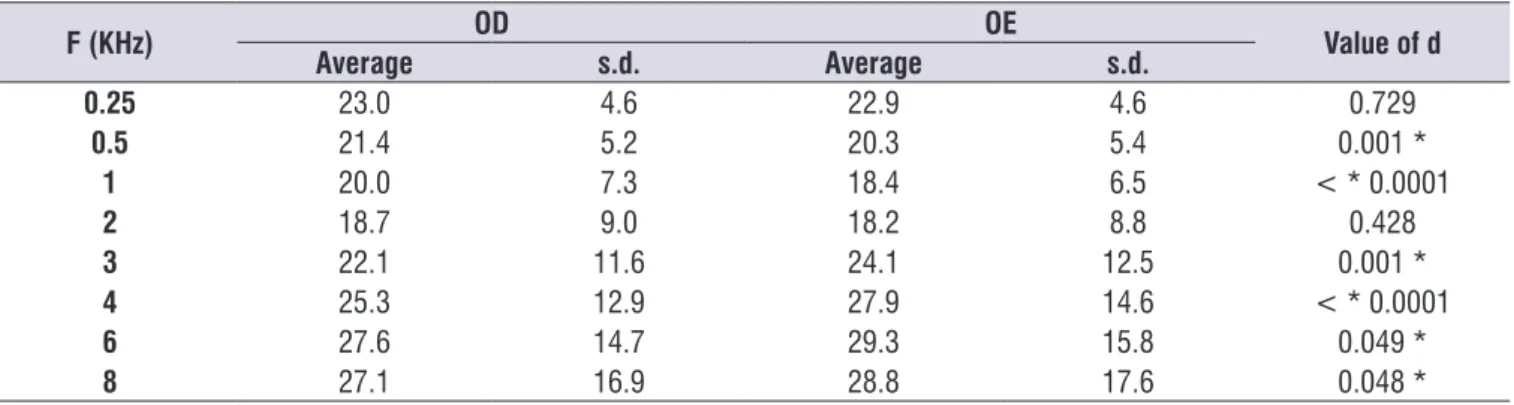
The analysis of the results of the tonal threshold audiometry demonstrated the presence of hearing loss in 96 (63.2%) individuals. All losses found were the sensorioneural type, of which 69 (71.9%) were bilateral, 27 (28.1%) unilateral, 72 (75%) symmetrical and 24 (25%) asymmetrical. Analyzing the ears separately, hearing loss in the right ear (RE) in 80 (52.6%) patients, and in left ear (LE) in 85 (55.9%).
in most frequencies, with the exception of 250 Hz and
2 kHz (p > 0.05), noting that the low frequencies the
right ear has higher averages, and the high frequencies
higher averages are in left ear (table 2). presence of isolated hearing loss in the frequencies of
6 and/or 8 kHz at 24 (30.0%) patients in the RE and 20 (23.5%) in LE (table 1).
Comparing the ears, is a statistically signiicant difference between the average loudness thresholds
Table 1. Distribution of the number of patients according to the characteristics of hearing loss in the right ear and left ear
Features
Hearing loss
RE (n = 80) LE (n = 85)
n % n %
Degree (average 0.5 to 4 kHz)
Normal 53 66.3 48 56.5
Light 21 26.3 32 37.6
Moderated 6 7.5 5 5.9
Isolated loss in 6 kHz and 8 kHz
Yes 24 30.0 20 23.5
No 56 70.0 65 76.5
Audiogram coniguration
Flat 44 55.0 44 51.8
High frequencies slightly demoted 24 30.0 26 30.6
Descendant 11 13.8 15 17.6
U-shaped average frequency 1 1.3 0 0
Caption: RE = right ear; LE = left ear
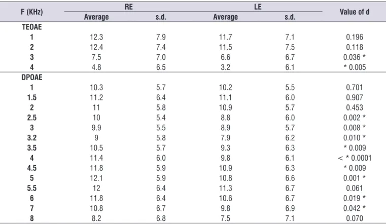
Otoacoustic emissions analysis was found in 114 (75%) patients with alterations in TEOAE and in DPOAE 120 (78.9%). For the RE, TEOAE and DPOAE absence in 86 (56.6%) and in 110 (72.4%) patients, respectively. For the LE, TEOAE and DPOAE absence in 95 (65.2%) and in 108 (71.1%) patients, respectively.
The average signal/noise relation of frequency bands in the TEOAE and in frequency DPOAE revealed lower values in RE in all tested frequencies, showing greater commitment on the left side. However, statisti-cally, in the frequency bands 1 and 2 kHz the TEOAE,
and in the frequencies of 1; 1.5; 2; 5.5 and 8 kHz in DPOAE, there was no signiicant difference between the two ears (p > 0.05) (table 3).
To analyze the association between PTA and results of the TEOAE, absence of OAE in presence of normal PTA, on average, in 32% of patients at 48% RE and LE. Already the presence of OAE in occurrence of hearing loss was observed in approximately 30% of patients to TEOAE and DPOAE in RE 14%; and 25% for TEOAE and DPOAE 11% on LE (table 4).
Table 2. Comparison of the averages of the tonal airway thresholds of the right ear and the left ear
F (KHz) OD OE Value of d
Average s.d. Average s.d.
0.25 23.0 4.6 22.9 4.6 0.729
0.5 21.4 5.2 20.3 5.4 0.001 *
1 20.0 7.3 18.4 6.5 < * 0.0001
2 18.7 9.0 18.2 8.8 0.428
3 22.1 11.6 24.1 12.5 0.001 *
4 25.3 12.9 27.9 14.6 < * 0.0001
6 27.6 14.7 29.3 15.8 0.049 *
8 27.1 16.9 28.8 17.6 0.048 *
* Signiicant Values (p < 0.05) – Chi-square Test
Table 3. Comparison of the averages of the signal/noise of the right ear and the left ear
F (KHz) RE LE Value of d
Average s.d. Average s.d.
TEOAE
1 12.3 7.9 11.7 7.1 0.196
2 12.4 7.4 11.5 7.5 0.118
3 7.5 7.0 6.6 6.7 0.036 *
4 4.8 6.5 3.2 6.1 * 0.005
DPOAE
1 10.3 5.7 10.2 5.5 0.701
1.5 11.2 6.4 11.1 6.0 0.907
2 11 5.8 10.9 5.7 0.453
2.5 10 5.4 8.8 6.0 0.002 *
3 9.9 5.5 8.9 5.7 0.008 *
3.2 9 5.8 7.9 6.2 0.010 *
3.5 10.5 5.7 9.3 6.3 * 0.009
4 11.4 6.0 9.8 6.1 < * 0.0001
4.5 11.8 5.9 10.9 6.3 * 0.009
5 12.1 5.9 10.8 6.6 0.001 *
5.5 12 6.4 11.3 6.7 0.061
6 11.8 6.4 10.6 6.7 0.019 *
7 10.8 6.7 9.8 6.9 0.042 *
8 8.2 6.8 7.5 7.1 0.070
* Signiicant Values (p < 0.05) – Chi-square Test
Caption: RE = right ear; LE = left ear; s.d. = standard deviation; DPOAE = distortion product otoacoustic emissions; TEOA = transient-evoked otoacoustic
emissions.
Also, analyses were performed in order to verify
a possible association between the variables age
and time of diagnosis and the presence of auditory
changes. With regard to the age group, it was observed
that patients in the range of 51 to 60 years old showed
2.6 times more chance of hearing loss compared to the
36 to 50 years of age. Relating to the time of diagnosis,
it was found that patients who were between 13 and 33
years of age DM presented 2.1 times more chance of
hearing loss than those who had a diagnosis less than 13 years old (table 5).
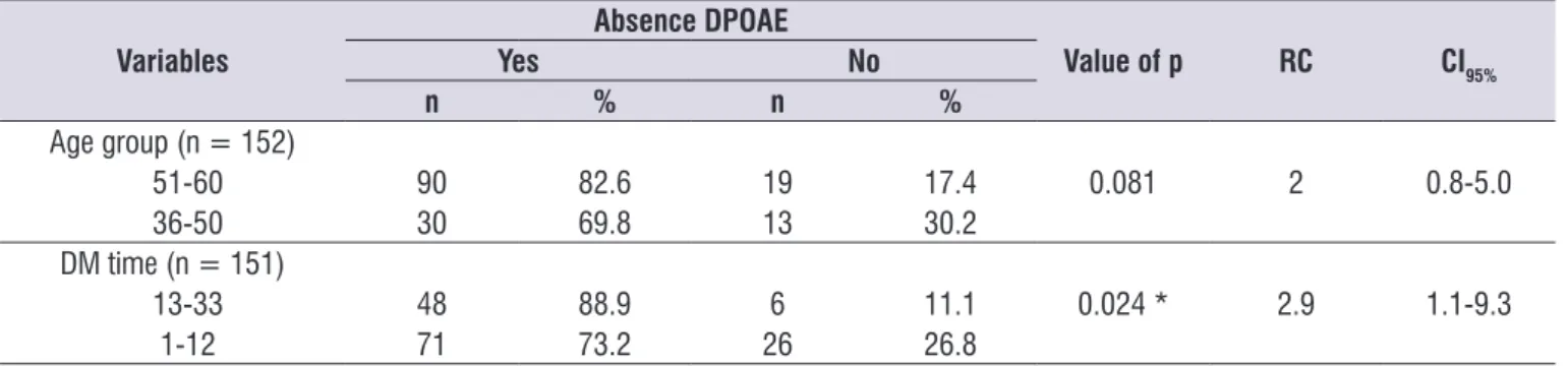
Finally, analyses were performed in order to verify a possible association between the variables age group and time of diagnosis and the absence of OAE. A relationship was not observed between the age group or the time of diagnosis and the absence of TEOAE (table 6). For the DPOAE, patients with diagnostic time between 13 and 33 years of age presented 2.9 times more likely to have a lack of answers (Table 7).
Table 4. Distribution of the number of patients according to the association between the presence of hearing loss and the response of
TEOAE and DPOAE in right ear and on the left
HEARING LOSS RE
p
LE
p
Yes No Yes No
n % n % n % n %
TEOAE PresentAbsent 2159 26.373.7 4527 62.537.5 < * 0.0001 6322 25.974.1 3235 52.247.8 0.001 *
DPOAE Present 6 7.5 36 50.0 < * 0.0001 10 11.8 34 50.7 < * 0.0001
Absent 74 92.5 36 50.0 75 88.2 33 49.3
* Signiicant Values (p < 0.05) – Chi-square Test
Table 7. Distribution of the number of patients according to the absence of DPOAE related to age group and time of diagnosis
Variables
Absence DPOAE
Value of p RC CI95%
Yes No
n % n %
Age group (n = 152)
51-60 90 82.6 19 17.4 0.081 2 0.8-5.0
36-50 30 69.8 13 30.2
DM time (n = 151)
13-33 48 88.9 6 11.1 0.024 * 2.9 1.1-9.3
1-12 71 73.2 26 26.8
* Signiicant Values (p < 0.05) – Chi-square Test
Caption: DPOAE = distortion product otoacoustic Emissions; RC = right of chance; CI = conidence interval; DM = Diabetes Mellitus.
Table 5. Distribution of the number of patients according to age-related hearing loss and time of diagnosis
Variables
Hearing loss
Value of p RC CI95%
Yes No
n % n %
Age group (n = 152)
51-60 76 69.7 33 30.3 0.008 * 2.6 1.3-5.4
36-50 20 46.5 23 53.5
DM time (n = 151)
13-33 40 74.1 14 25.9 0.045 * 2.1 1.0-4.3
1-12 56 57.7 41 42.3
* Signiicant Values (p < 0.05) – Chi-square Test
Caption: RC = right of chance; CI = conidence interval; DM = Diabetes Mellitus
Table 6. Distribution of the number of patients according to the absence of TEOAE related to age and time of diagnosis
Variables
Absence TEOAE
Value of p RC CI95%
Yes No
n % n %
Age group (n = 152)
51-60 83 76.1 26 23.9 0.603 1.2 0.5-2.9
36-50 31 72.1 12 27.9
DM time (n = 151)
13-33 42 77.8 12 22.2 0.534 1.3 0.5-3.1
1-12 71 73.2 26 26.8
* Signiicant Values (p < 0.05) – Chi-square Test
Caption: TEOAE = transient-evoked otoacoustic emissions; RC = right of chance; CI = conidence interval; Dm = Diabetes Mellitus.
DISCUSSION
In the present study we could observe the preva-lence of diabetic patients ranging in age from 51 to 60 years, with the age-associated hearing loss. There was no association of age with the absence of responses in the OAE. Some studies involving individuals with older age, observed the relation of DM with the hearing loss14, 18,23 and with the absence of OAE2, while others found
no association15,24.
Considering the prevalence and severity of hearing loss increases signiicantly with age18, the positive
association could be related to presbycusis, however, the association was found with auditory changes even in a sample of diabetic patients, with up to 50 years of age25.
Sunkum et al. 9 Other authors showed no association
between the duration of the illness and the alterations in PTA or OAE2,14,25,26, however, it should be noted that in
two of these studies2,25 the sample evaluated presented
recent diagnosis, up to 7 years.
Mitchell et al.23 results found in a consistent cross
association between the duration of diabetes and hearing loss, revealing that in 5 years, there was an accelerated progression of deafness in patients early diagnosed with DM. However, in the same study, people with previously diagnosed diabetes showed a non-signiicant inverse association with the progression of hearing loss. The authors suggest that the early diagnosis and treatment for the DM can avoid potential fall in the hearing, giving a protective effect.
Hearing complaints such as tinnitus and hypocusis, present in 89 (58.8%) patients, seem common in diabetics, being cited, to a greater or lesser frequency in many studies relating hearing and DM12,13,25. According
to Gibrin et al. 27, changes in the inner ear, causing
Vertigo and/or tinnitus, hearing loss, can be related to a microcirculatory failure due to a vascular occlusion by emboli, hemorrhage or Vasospasm, and these, in turn, would be due to hyper viscosity or microangiopathy by DM or hypertension.
Relating to the audiometric indings, the hearing loss was observed in 63.2% of patients evaluated, being all of the sensorioneural type, with a predominance of bilateral and symmetrical losses. Similar results were found in other studies25,28.
Sunkum et al.9 found prevalence of hearing loss in
58.3% of patients with controlled glucose and 85.2% of diabetic patients with uncontrolled glucose levels, corroborating with the hypothesis that the glucose control can protect against hearing deiciency23,29.
Study realized in diabetic patients with early diagnosis (before 40 years old) showed prevalence of 21.7% sensorioneural uni or bilateral hearing loss, stating that the type 2 DM early start and with poor glycemic control leads to an increased prevalence of subclinical hearing loss and prejudice in the responses of the brain stem13.
In the present study the glycemic control of patients was not evaluated, so it is not possible to infer about its relation with the hearing loss.
On the audiogram characterization of this study a greater number of ears with degree considered normal were found, however, the degree classiication only takes into consideration the frequencies of 0.5 to 4 kHz 19, and were found in isolated hearing loss in the
frequencies of 6 and/or 8 kHz. This fact explains why
many losses were classiied as normal hearing degree. The losses encountered, lightweight degree prevailed lat coniguration.
The audiometric coniguration of patients with DM offers great variation between the authors. Some studies point out the loss attributed to the DM as the sensorioneural bilateral type, affecting mainly high frequencies9,12,13,15,25,28; others have found worse
thresholds at low frequencies30, low and medium-sized14
or low and high2; and some refer to predominant14
lat coniguration. As for the degree, most research concerns take hearing loss on the DM12,13,18,25,28.
Analyzing the frequencies speciically, it was observed that the frequencies above 4 kHz were most affected in patients evaluated. Other authors also reported worse thresholds in 4 kHz26 and in 4 and
8 kHz10.
The predominance of high frequency alterations can be related to presbycusis, characterized by high-frequency symmetric bilateral sensorioneural loss27,
whereas studies involving elderly diabetic patients18,23.
Some authors suggest that the DM mechanisms could act synergistically with the underlying processes related hearing loss with age, resulting in worsening of hearing2,23. However, other studies9,13 evaluated
younger diabetic patients (up to 50 years of age) and found the same characteristics of hearing, contradicting the theory that the loss could be merely a phenomenon attributed to age.
The results are quite contradictory, as there are studies with individuals with up to 69 years old who have a predominance of latconiguration14, similar to
what was found in the results of this study. There was no consensus in the literature study on the characteristics of hearing loss associated with the DM; it is believed that the variations found could be related to inner ear affected region. Although many studies point to the existence of histopathological cochlear alterations in DM, it is dificult to establish a causal relationship, highlighting the differential vulnerability of inner ear to the effects of hyperglycemy5.
The analysis of the OAE TEOAE and DPOAE showed absence in more than 70% of patients. A similar result was found for Prabhu et al.31, which revealed
Conlicting indings have been found in other studies. Agarwal et al. 25 found absence of TEOAE on 30% of
the samples, and Lerman-Garber et al. 13 in just 15%,
but in both studies, patients evaluated were younger diabetics (ages up to 50 years old) and the audiometric indings revealed the prevalence of mild loss, which can justify the greater number of TEOAE present, since the presence of TEOAE on most individuals correlates with the degree of loss, and may be obtained in hearing loss less than 40dB25. Eren et al.16 analyzed the TEOAE and
DPOAE in DM patients, average age of 47 years and normal hearing, and, although it was found decreased amplitude, the difference between the DM and the control group was not statistically signiicant.
The relationship between auditory alterations found and the affected side was described in the results, showing a greater tendency of left-side commitment to high frequencies in PTA, in TEOAE and DPOAE, and, although the difference is not signiicant in some frequencies. Evidence on laterality dominant in auditory changes in patients with DM was not found in the liter-ature. However, some authors have mentioned greater commitment from the right side in their indings related toaudiometry28,30. Others have not showed a
predomi-nance of tonal thresholds25. Karabulut et al.15, analyzing
the OAE, also did not pointed out the difference between the sides. Frisina et al. 30, when realized the
DPOAE, right commitment was found, Cabrer28 to the
left.
The analysis of correlation between hearing loss and the result of the TEOAE and DPOAE, it was observed that the percentage of TEOAE and DPOAE alterations is greater than the percentage of patients with hearing loss, showing that the emissions can be altered even with audibility thresholds within the normal range, which supposes a cochlear impairment in DM22.
Metabolic changes caused by the DM can modify the micromechanism of the inner ear, causing subclinical early results2. It is evident the importance of the OAE
as clinical instrument for monitoring the cochlea, for allowing the subtle detection of cochlear alteration observed in PTA, being suggestive its indication for evaluation of auditory functions in DM.
The presence of TEOAE and DPOAE on hearing loss may be related to the degree of loss found; being mostly light degree, and it still favors the appearance of otoacoustic emission answers22. The OAE presence in
patients with hearing loss also could assume cases of auditory neuropathy, characterized by a speciic hearing disorder with abnormal auditory neural responses in the
presence of normal cochlear function32. Considering
the DM can cause degeneration of the auditory nerve6,
speciic tests for assessment of central auditory system would be essential for the differential diagnosis.
The difference between the results of two types of emissions can be linked to the fact of DPOAE assess the frequencies of 6 and 8 kHz, in which there are isolated losses, causing the occurrence of DPOAE absent is larger.
This study has some limitations, such as the absence of a control group, which would allow for comparative analysis; and the absence of data related to glycemic control, which presented itself as a risk factor associated with auditory changes in many studies cited.
CONCLUSION
Assessment of hearing in type 2 diabetic patients showed a predominance of sensorioneural symmetric bilateral hearing loss with lat coniguration. The absence of TEOAE and DPOAE was observed in most patients.
The absence of OAE before normal audio thresholds, present in part of the sample, suggests prejudice to the ciliated cells of the external cochlea. Such indings may indicate early cochlear dysfunction in type 2 DM, what would justify the hearing monitoring of these patients.
The OAE presence in patients with hearing loss could be related to the light degree attempt, found in most cases, or might suggest cases of auditory neuropathy. In this way, speciic tests for assessment of central auditory system would be relevant to indicate the audiologic diagnosis of patients with type 2 DM.
The knowledge of the effects of DM over hearing favors early intervention, minimizing the prejudice and amplifying the quality of life.
ACKNOWLEDGEMENTS
To the Fundação Cearense de Apoio ao Desenvolvimento Cientíico e Tecnológico (FUNCAP) for funding for the implementation of the research.
REFERENCES
1. Brasil. Secretaria de Atenção à Saúde.
Departamento de Atenção Básica. Estratégias para o cuidado da pessoa com doença crônica: diabetes mellitus. Brasília: Ministério da Saúde; 2013.
2. Abo-Elfetoh NM, Mohamed ES, Tag LM, El-Baz MA, Eldeen MEE. Auditory dysfunction in patients with type 2 diabetes mellitus with poor versus good glycemic control. Egypt J Otolaryngol. 2015; 31(3): 162-9.
3. International Diabetes Federation. IDF Diabetes Atlas. 7ed. Brussels, Belgium: International Diabetes Federation; 2015.
4. Hong O, Buss J, Thomas E. Type 2 diabetes and hearing loss. Disease-a-Month. 2013; 59(4): 139-46.
5. Akinpelu OV, Ibrahim F, Waissbluth S, Daniel SJ. Histopathologic changes in the cochlea associated with diabetes mellitus-a review. Otol Neurotol.2014; 35(5): 764-74.
6. Rolim LP, Rabelo CM, Lobo IFN, Moreira RR, Samelli AG. Interação entre diabetes mellitus e hipertensão arterial sobre a audição de idosos. CoDAS 2015; 27(5): 428-32.
7. Horikawa C, Kodama S, Tanaka S, Fujihara K, Hirasawa R, Yachi Y et al. Diabetes and risk of hearing impairment in adults: a meta-analysis. J Clin Endocrinol Metab. 2012; 98(1): 51-8.
8. Akinpelu OV, Mujica-Mota M, Daniel SJ. Is type 2 diabetes mellitus associated with alterations in hearing? A systematic review and meta-analysis. Laryngoscope. 2014; 124(3): 767-76.
9. Sunkum AJK, Pingile S. A clinical study of audiological proile in diabetes mellitus patients. Eur Arch Otorhinolaryngol. 2013; 270(3):875–9.
10. Ren J, Zhao P, Chen L, Xu A, Brown S, Xiao X. Hearing loss in middle-aged subjects with type 2 diabetes mellitus. Arch Med Res. 2009;40(1) suppl 1:18-23.
11. Lin SW, Lin YS, Weng SF, Chou CW. Risk of developing sudden sensorineural hearing loss in diabetic patients: a population-based cohort study. Otol Neurotol. 2012; 33(9): 1482-8.
12. Ferreira JM, Sampaio FMO, Coelho JMS, Almeida NMGS. Peril audiológico de pacientes com diabetes mellitus tipo 2. Rev Soc Bras Fonoaudiol. 2007;12(4):292-7.
13. Lerman-Garber I, Cuevas-Ramos D, Valdés
S, Enríquez L, Lobato M, Osornio M et al. Sensorineural hearing loss-A common inding in early-onset type 2 diabetes mellitus. Endocr Pract. 2012; 18(4): 549-57.
14. Bamanie AH, Al-Noury KI. Prevalence of hearing loss among Saudi type 2 diabetic patients. Saudi Med J. 2011; 32(3): 271-4.
15. Karabulut H, Karabulut I, Dağli M, Bayazit YA, Bilen Ş, Aydin Y et al. Evaluation of outer hair cell function and medial olivocochlear efferent system in patients with type II diabetes mellitus. Turk J Med Sci. 2014; 44(1): 150-6.
16. Eren E, Harman E, Arslanoğlu S, Önal K. Effects of Type 2 Diabetes on Otoacoustic Emissions and the Medial Olivocochlear Relex. Otolaryngol Head Neck Surg. 2014; 150(6): 1033-9.
17. Calvin D, Watley SR. Diabetes and hearing loss among underserved populations. Nurs Clin N Am. 2015; 50(3): 449–56.
18. Oh I-H, Lee JH, Park DC, Kim M, Chung JH, et al. Hearing Loss as a Function of Aging and Diabetes Mellitus: A Cross sectional Study. PLoS one. 2014; 9(12): e116161.
19. Russo ICP, Pereira LD, Carvalho RMM, Anastásio ART. Encaminhamentos sobre a classiicação do grau de perda auditiva em nossa realidade. Rev Soc Bras Fonoaudiol. 2009; 14(2): 287-8.
20. Demeester K, Wieringen A, Hendrickx J, Topsakal V, Fransen E, Laer L et al. Audiometric shape and presbycusis. Int J Audiol. 2009; 48(4) suppl l: 222-32.
21. Mattos LC, Veras RP. Prevalência da perda
auditiva em uma população de idosos da cidade do Rio de Janeiro: um estudo seccional. Braz J Otorhinolaryngol. 2007;73(5):654-9.
22. Sousa LCA, Piza MRT, Alvarenga KF, Cóser PL. Emissões Otoacústicas. In: Eletroisiologia da audição e emissões otoacústicas: princípios e aplicações clínicas. 2ed. São Paulo: Tecmedd; 2008. p.109-30.
23. Mitchell P, Gopinath B, McMahon CM, Rochtchina E, Wang JJ, Boyages SC, Leeder SR. Relationship of Type 2 diabetes to the prevalence, incidence and progression of age-related hearing loss. Diabet Med. 2009; 26(5): 483-8.
25. Agarwal AC, Pujary K, Ganapathy K, Balakrishnan R, Nayak DR, Hasan F. Pure tone audiometry and otoacoustic emissions for the assessment of hearing loss in diabetic patients. Indian J Otol. 2013;19(1):13.
26. Salvinelli F, Miele A, Casale M, Greco F, D’Ascanio L, Firrisi L et al. Hearing thresholds in patients with diabetes. Int J Otorhinolaryngol. [serial online]. 2004 [acesso em 29/10/2015];3(1). Disponível em: http://www.ispub.com/journal/the-internet-journal-of-otorhinolaryngology/volume-3-number-1/ hearing-thresholds-in-patients-with-diabetes.html 27. Gibrin PCD, Melo JJ, Marchiori LLDM. Prevalência
de queixa de zumbido e prováveis associações com perda auditiva, diabetes mellitus e hipertensão arterial em pessoas idosas. CoDAS . 2013; 25(2): 176-80.
28. Cabrer IS. Otoemissiones acusticas em pacientes diabeticos no insulinodependientes. [Tese]. Barcelona: Hospital Universitario de Girona - Doctor Josep Trueta; 2006.
29. Michikawa T, Mizutari K, Saito H, Takebayashi T, Nishiwaki Y. Glycosylated hemoglobin level is associated with hearing impairment in older Japanese: The Kurabuchi study. J Am Geriatr Soc. 2014; 62(7): 1231-7.
30. Frisina ST, Mapes F, Kim S, Frisina DR, Frisina RD. Characterization of hearing loss in aged type II diabetes. Hear Res. 2006; 211(1/2)suppl l:103-13. 31. Prabhu P, Shanthala SP. Efferent Auditory System
Functioning and Speech Perception in Noise in Individuals with Type II Diabetes Mellitus. J Phonet Audiol. 2016;2:115.