Who are the low-risk patients that could beneit from
watch-and-wait regarding the neck?
Quem são os pacientes de baixo risco que poderiam beneficiar-se de conduta
expectante do pescoço?
Hugo Fontan Kohler
I, Luiz Paulo Kowalski
IIHospital A. C. Camargo, São Paulo, Brazil
ABSTRACT
CONTEXT AND OBJECTIVE: The management of clinically negative neck is controversial, with an ongoing debate on the indication criteria and prognostic impact of different types of therapy. The aim here was to compare the results from neck dissection and watch-and-wait, among oral cancer patients who, clinically, did not show any evidence of neck metastasis.
DESIGN AND SETTING: Retrospective analysis in a tertiary cancer center hospital.
METHODS: Patients with epidermoid oral carcinoma were assessed. The inclusion criteria were: primary tumor restricted to the oral/oropharyngeal cavity, no previous treatment, surgical treatment as the first option, clinical/radiological stage N0 and no distant metastasis.
RESULTS: Two hundred and sixty-two patients were analyzed. The length of follow-up ranged from four to 369.6 months and, at the end, 118 patients were alive, 53 had died due to cancer, 84 had died from other causes and 7 had died after the operation. Among the patients who underwent neck dissection, lymphatic vascular embolization (P = 0.009) and tumor thickness (P = 0.002) were significant for regional recurrence, while for the watch-and-wait group, only tumor thickness was significant (P = 0.018). Through recursive partitioning, the patients without adverse prognostic factors and tumor thickness < 2 mm presented com-patible results in the two groups.
CONCLUSION: Elective neck dissection seems to be the best treatment option. Patients who are eligible for watch-and-wait constitute a small group that, ideally, is categorized according to the postoperative pathological findings.
RESUMO
CONTEXTO E OBJETIVO: O manejo do pescoço clinicamente negativo é controverso, havendo um deba-te correndeba-te sobre os critérios de indicação bem como o impacto prognóstico das diferendeba-tes modalidades terapêuticas. O objetivo foi comparar os resultados do esvaziamento cervical com a observação em pa-cientes com câncer de boca e clinicamente sem evidência de metástases cervicais.
TIPO DE ESTUDO E LOCAL: Análise retrospectiva em hospital terciário, especializado em oncologia.
MÉTODOS: Pacientes com diagnóstico de carcinoma epidermoide de boca foram analisados. Os crité-rios de inclusão foram: tumor primário restrito à cavidade oral/orofaringe, ausência de tratamento prévio, tratamento cirúrgico como primeira opção, estádio clínico e radiológico N0 e ausência de metástases a distância.
RESULTADOS: Duzentos e sessenta e dois pacientes foram analisados. O tempo de acompanhamento variou de 4 a 369.6 meses e, ao final, havia 118 pacientes vivos, 53 óbitos pela neoplasia, 84 óbitos por ou-tras causas e 7 óbitos pós-operatórios. Nos pacientes submetidos a esvaziamento cervical, a embolização vascular linfática (P = 0,009) e a espessura tumoral (P = 0,002) foram associados significativamente com a recidiva regional, enquanto que nos pacientes somente observados, apenas a espessura tumoral se asso-ciou significativamente (P = 0,018). Por meio do particionamento recursivo, aqueles pacientes sem fatores adversos prognósticos e espessura tumoral menor que 2 mm apresentaram resultados compatíveis em ambos os grupos.
CONCLUSÃO: O esvaziamento cervical eletivo parece ser a melhor opção de tratamento. Pacientes can-didatos a observação constituem um pequeno grupo e a sua categorização ideal depende de achados patológicos pós-operatórios.
IMD. Former Fellow in the Department of Head
and Neck Surgery and Otolaryngology, Hospital A. C. Camargo, São Paulo, Brazil.
IIMD, PhD. Director, Department of Head and
Neck Surgery and Otolaryngology, Hospital A. C. Camargo, São Paulo, Brazil.
KEY WORDS:
Head and neck neoplasms. Surgical procedures, operative. Mouth neoplasms.
Neck dissection. Lymphatic metastasis.
PALAVRAS-CHAVE:
Neoplasias de cabeça e pescoço. Procedimentos cirúrgicos operatórios. Neoplasias bucais.
INTRODUCTION
Management of the neck in patients with oral cancer has been one of the major controversies in head and neck oncology, and most of the discussion has focused on what treatment to admin-ister for patients without clinically evident metastatic disease. For these patients, the incidence of occult neck metastasis may range from 6% to 46%.1
he indication for elective treatment of the neck has been con-sidered to be a probability of cervical metastasis of at least 20%,2
although reevaluation of this percentage based on decreased sur-gical mortality and morbidity has been proposed.3 hese limits
are based on conventional pathological evaluation and staining of lymph nodes, but such evaluations have recently been shown to have limitations, in papers using molecular analyses that upstage up to 20% of pathologically N0 patients.4
he prognostic impact of therapeutic decisions must also be considered. An elective neck dissection presents risks in the form of postoperative morbidity and mortality and impact on quality of life, but missing a neck metastasis may lead to late recurrences with a signiicant impact on prognosis.5
OBJECTIVE
To compare elective neck dissection with a watch-and-wait pol-icy, with regard to neck recurrence and survival rates among patients with clinically N0 squamous cell carcinoma of the oral cavity.
PATIENTS AND METHODS
Patients with primary tumors of the oral tongue, loor of the mouth, inferior gingival rim and retromolar trigone who were treated at Hospital A. C. Camargo, a tertiary cancer center, were enrolled in this study. he data on all patients treated between January 1980 and December 2003 were recovered from the med-ical records.
he following inclusion criteria were used: histological diag-nosis of squamous cell carcinoma, primary tumor restricted to the oral cavity, no previous treatment, treatment with curative intent, surgery as the primary form of treatment, primary tumor staged
as T1/T2, clinical/radiological stage N0 and no distant metasta-sis at diagnometasta-sis. he tumors were staged based on the recorded description and pathological report, in accordance with the 2002 AJCC (American Joint Committee on Cancer) classiication.6
A surgical pathologist dissected all the specimens immedi-ately ater removal and three histological slides were prepared from each node.
he statistical analysis was performed using the Stata 11 sot-ware for Macintosh (Stata Corp., Texas, United States). Continu-ous variables were expressed as the mean and standard deviation (SD). Logistic regression was used to assess which factors were signiicant for the presence of metastatic nodes in the neck. he Kaplan-Meier and Cox regression models were used for recur-rence and survival analysis. he classiicatory analysis was per-formed using a recursive partitioning algorithm with the signii-cance level set at 0.05 and a minimum of 20 patients at the knot.
RESULTS
A total of 262 patients that conformed to the inclusion criteria were analyzed. here were 202 males (77.1%) and 60 females (22.9%), with ages ranging from 23 to 95 years (mean of 58.45 years and SD of 12.0 years). he primary tumor site was the oral tongue in 162 patients (61.83%), loor of the mouth in 73 patients (27.86%), retromolar trigone in 28 patients (10.69%) and lower alveolar rim in 19 patients (7.25%). he clinical T stage was T1 in 99 patients (37.8%) and T2 in 163 patients (62.2%). Neck dis-section ipsilateral to the tumor was performed in 166 patients (63.36%); the other 96 patients (36.64%) did not undergo neck surgery. Radical neck dissections was performed on 74 patients (44.58%), modiied radical neck dissections on 28 patients (16.87%) and selective neck dissections (levels I to III) on 64 patients (38.55%). A further contralateral neck dissection was performed on 18 of the operated patients (6.86%).
here was a clear time trend relating to the type of neck dis-section performed, with increasing proportions of modiied rad-ical dissections and selective neck dissections. In 138 patients (83.13%), the neck dissection was removed en bloc with the
pri-mary tumor and in the remaining 28 patients (16.87%), there was no continuity between the primary tumor resection and the neck dissection specimen.
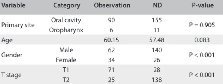
he decision between observation and neck dissection was signiicantly correlated with the T stage of the primary tumor and patient gender, but not with age or primary tumor site (Table 1). Blood vessel iniltration was found in six patients (2.42%) and lymphatic embolization in 65 patients (26.21%). Neural iniltra-tion was observed in 73 patients (29.80%). Regarding histological diferentiation, the tumors were classiied as well diferentiated in 178 patients (67.94%), moderately diferentiated in 71 patients (27.09%) and poorly diferentiated in 13 patients (4.96%). he tumor thickness measured at histological examination ranged
Variable Category Observation ND P-value
Primary site Oral cavity 90 155 P = 0.905 Oropharynx 6 11
Age 60.15 57.48 0.083
Gender Male 62 140 P < 0.001 Female 34 26
T stage T1 71 28 P < 0.001
T2 25 138
from 0.2 to 25 millimeters (mean of 5.81 and SD of 4.33 millime-ters). he number of lymph nodes recovered from the neck dis-section specimen ranged from 6 to 116 in the homolateral neck (mean of 29.51 nodes and SD of 17.59 nodes).
he number of retrieved lymph nodes ranged from 8 to 90 (mean of 44.5 nodes and SD of 17.4 nodes) in patients who underwent radical neck dissection and from 6 to 116 (mean of 58.8 nodes and SD of 13.1 nodes) in selective neck dissection patients. Among all the patients who underwent neck dissection, 120 (72.29%) had no metastatic nodes ipsilateral to the primary tumor, while 22 patients (13.25%) presented one involved node, and 24 patients (14.46%), up to eight involved nodes. In the con-tralateral neck, two patients presented involved nodes. Postoper-ative radiotherapy was used for 68 patients (25.95%).
he length of follow-up ranged from 4 to 369.6 months (mean of 70.65 and SD of 30.4 months). here were 28 cases (10.69%) of ipsilateral neck recurrence, eight cases (3.05%) of contralateral neck recurrence and three cases (1.14%) of synchronous bilat-eral recurrence. At the last follow-up, 118 patients were alive and without active disease, 53 patients had died due to disease pro-gression or recurrence, 84 patients had died from other, unrelated causes and seven patients had died following the operation.
Among the patients who underwent synchronous neck dis-section, the following factors were signiicant for the diagnosis of metastatic nodes: size of primary tumor (P = 0.047), histological diferentiation (P = 0.002), lymphatic embolization (P < 0.001), neural iniltration (P = 0.045) and tumor thickness (P = 0.018). In multivariate analysis, histological diferentiation (odds ratio, OR: 3.78; 95% conidence interval, CI: 1.62-8.78; P = 0.002) and lymphatic embolization (OR: 18.97; 95% CI: 3.98-27.51; P < 0.001) remained signiicant. Among these patients, there were eight cases of ipsilateral recurrence, eight cases of contralateral recurrence and one case of bilateral recurrence. In univariate analysis, the following factors were signiicant for neck recur-rence: lymphatic embolization (hazard ratio, HR: 1.388; 95% CI: 1.131-2.502; P < 0.001) and tumor thickness (HR: 1.170; 95% CI: 1.027-1.345; P = 0.001). In multivariate analysis, lymphatic embolization (HR: 1.042; 95% CI: 1.034-2.332; P = 0.009) and tumor thickness (HR: 1.069; 95% CI: 1.149-1.316; P = 0.002) remained statistically signiicant. Among the patients who did not undergo neck dissection, there were 20 cases of ipsilateral neck recurrence, no contralateral recurrences and two bilateral recurrences. All the ipsilateral recurrences occurred at levels I-III. In these patients, the signiicant factors for neck recurrence were: T stage (P = 0.015), perineural iniltration (P = 0.006) and tumor thickness (P = 0.022). In multivariate analysis, only tumor thickness remained signiicant (HR = 1.069; 95% CI: 1.012-1.130; P = 0.018). here was a signiicant diference in mean time that elapsed until neck recurrence between the two groups. Among the patients who underwent neck dissection, the mean time that
elapsed until recurrence was 19.75 months and in the observa-tion group, 6.49 months (P = 0.024, Table 2).
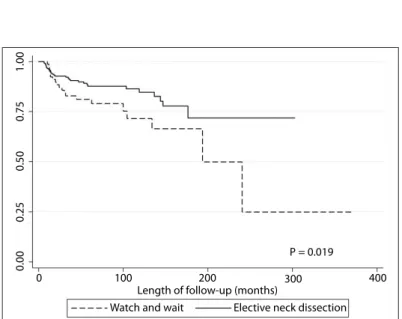
here was a statistically signiicant increase in the rate of neck recurrence risk among the patients who did not undergo elective neck dissection, in comparison with those who underwent syn-chronous neck treatment (P = 0.019, Figure 1).
In a multivariate model that included the risk factors for neck recurrence identiied in both groups (tumor thickness and lym-phatic embolization) and the type of neck treatment and adju-vant radiotherapy, only tumor thickness and synchronous neck dissection were signiicant (Table 3).
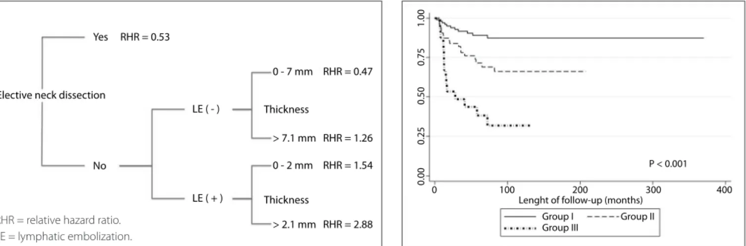
When we analyzed disease-free survival, the following fac-tors were statistically signiicant: tumor extent (P < 0.001), T stage (P < 0.001), lymphatic embolization (P < 0.001), neural iniltration (P = 0.039), tumor thickness (P = 0.021) and elective neck dissection (P = 0.023). In a multivariate analysis on survival, lymphatic embo-lization and elective neck dissection remained signiicant (Table 4). We also classiied the patients through recursive partitioning (RP). his method uses a classiication tree and its branches are deined by the variables included in the model. Terminal branches repre-sent RP-derived homogeneous categories according to a speciic outcome.
Neck recurrence and disease-speciic survival analysis showed that tumor thickness, lymphatic embolization and elective neck dis-section were the variables with the best discriminating power for drawing a classiication tree (Figure 2).
0.00
0.25
0.50
0.75
1.00
0 100 200 300
P = 0.019
Length of follow-up (months)
Watch and wait Elective neck dissection 400
Figure 1. Kaplan-Meier survival curve for patients who underwent either neck dissection or a watch-and-wait policy.
Treatment Events Mean
Observation 22 6.49
P = 0.024 Neck dissection 17 19.75
cell carcinoma.7 Occult neck metastases have a significant
impact on survival.
In a study on patients with clinically node-negative necks, the rate of occult metastases was 50% and these patients had signii-cantly worse survival (P < 0.001).8 Also, the diagnosis of node
metastases and the presence of extracapsular spread are consid-ered to be an indication for adjuvant treatment.9 On the other
hand, a neck dissection may avoid unnecessary adjuvant treat-ment and spare the use of radiotherapy.10
Surgery alone may achieve a control rate on pN0 necks of 75% and may compare favorably with radiation therapy.11 Neck
metastases have been linked to certain factors. Tumor thickness has been signiicantly linked to postoperative upstaging of the neck, and a positive correlation between tumor depth and T stag-ing has also been demonstrated.
In one study, a cutof point of 4 mm was suggested for risk stratiication, although those authors suggested that for oropha-ryngeal tumors, a lower cutof point might be required.12 his
Hazard ratio 95% conidence interval P
Neck dissection 1
Watch-and-wait 3.808 1.595-8.391 < 0.001 Tumor thickness 1.126 1.035-1.225 0.006
Table 3. Multivariate analysis of risk factors for neck recurrence in all groups
Table 4. Multivariate analysis on factors with significant impact on disease-specific survival
Variable Hazard ratio 95% conidence interval P
Neck dissection Yes
No 1.587 1.014-2.461 0.032 Lymphatic
embolization No
Yes 1.922 1.119-3.303 0.018
0.00
0.25
0.50
0.75
1.00
0 100 200 300
P = 0.007
Group I Group III
Group II
Length of follow-up (months) 400
Figure 3. Neck recurrence according to N0 stratification.
0.00
0.25
0.50
0.75
1.00
0 100 200 300
P < 0.001
Group I Group III
Group II Lenght of follow-up (months)
400
Figure 4. Disease-specific survival of N0 patients according to group stratification.
he irst division was elective neck dissection and we decided to group the patients in the observation group into three groups. Group I consisted of individuals with tumor thickness from 0 to 0.7 mm, without lymphatic embolization. his group had a simi-lar relative hazard ratio to that of patients who underwent neck dissection. Group II consisted of patients without lymphatic embolization and with tumor thickness greater than 0.7 mm or with lymphatic embolization and tumor thickness less than or equal to 2 mm. Group III consisted of individuals with tumor thickness greater than 2 mm and lymphatic embolization. here were signiicant diferences between these groups in relation to both neck recurrence rates (Figure 3) and disease-speciic sur-vival (Figure 4).
DISCUSSION
Neck staging is crucial for prognosis definition and treat-ment planning, since neck metastases are the single most important prognostic factor in head and neck squamous
Figure 2. Classification analysis diagram according to survival. Branch splits were performed at a significance level of P < 0.05.
Yes RHR = 0.53
LE ( - )
LE ( + )
0 - 7 mm
> 7.1 mm
0 - 2 mm
> 2.1 mm Thickness Thickness
RHR = 1.26
RHR = 1.54
RHR = 2.88 RHR = 0.47
No Elective neck dissection
inding had been previously demonstrated in another study that suggested that this cutof point could be used in making the deci-sion regarding elective treatment of the neck for patients with oral tongue carcinomas.5 In another report, a cutof point of 3
mm, for moderate or poor diferentiation, cases of perineural invasion and lymphovascular permeation had a signiicantly higher incidence of occult neck metastases.13
Simultaneous use of tumor thickness and histological difer-entiation has also been proposed for stage I and II tongue carci-nomas. Kurokawa et al. suggested that tumor depth > 4 mm and moderately diferentiated carcinoma should be deinitive indica-tions for neck dissection.5
Management of N0 necks may fall into three categories: elec-tive neck dissection, radiotherapy or observation. he choice between radiotherapy or neck dissection will depend essentially on the treatment for the primary tumor. An approach based on location and stage of the primary tumor was shown to be efec-tive, with 9% development of neck recurrences in early-stage oral cancers.14
Using a decision-analysis approach, Song et al. demonstrated that neck dissection was the preferred management for early-stage tongue cancer in clinical N0 necks. hese authors stated that the incidence of neck recurrences was high and that pathologi-cal analysis was more precise than imaging methods and allowed for improved deinition of postoperative chemoradiotherapy. However, if the risk of neck metastasis was lower than 0.17 and the salvage rate higher than 0.73, watchful waiting would be an appropriate choice.15
he use of irradiation, although with similar control rates when compared with neck dissection, was found to have signii-cantly higher incidence of adverse side efects.16
In patients with early-stage oral carcinoma, elective neck dissection was seen to be a signiicant factor for recurrence (8% versus 26.8%; P = 0.001) and survival rates (P < 0.01), thus sug-gesting that elective neck dissection was superior to observation alone. A signiicant beneit regarding survival and neck recur-rence rate was also observed in another series of 380 patients with early-stage oral tongue squamous cell carcinoma.17
he importance of surgical staging for treatment planning should also not be underestimated, with 40% stage migration in a series of patients with T1-T2 N0-N1 oropharyngeal cancers.18
his evidence goes against a recent report that showed that there was no survival advantage for patients who underwent neck dissection, in comparison with a watchful waiting policy.19
In a prospective, randomized clinical trial comparing elec-tive neck dissection and observation in cases of early stage oral tongue carcinoma, the ive-year disease-speciic survival was comparable, with no statistically signiicant diference between the two groups. he neck recurrence rate was higher in the obser-vation group but because of the strict follow-up schedule, salvage
was possible in all cases. hat trial supported the use of watch-and-wait and a strict observation schedule.20
his treatment choice was also supported by another report that outlined a sensitivity analysis on neck metastasis in cN0 patients.21
CONCLUSION
Our data show that clinical N0 patients with oral cancer are a heterogeneous population with diferent rates of neck recur-rence and disease-speciic survival. Our decision tree approach was able to stratify them into three distinctive groups and show the importance of neck dissection. For the patients who did not undergo neck dissection, only a deined set of individuals had comparable regional recurrence rate and survival.
his stratiication could only be performed using patholog-ical variables that became available ater the deinitive patho-logical report had been produced, thus limiting its applicability. herefore, elective neck dissection seems to be the best treatment option. Patients eligible for watch-and-wait constitute a small group, which is ideally assessed according to the postoperative pathological indings.
REFERENCES
1. Capote A, Escorial V, Muñoz-Guerra MF, et al. Elective neck dissection
in early-stage oral squamous cell carcinoma--does it influence
recurrence and survival? Head Neck. 2007;29(1):3-11.
2. Weiss MH, Harrison LB, Isaacs RS. Use of decision analysis in planning
a management strategy for the stage N0 neck. Arch Otolaryngol
Head Neck Surg. 1994;120(7):699-702.
3. Pitman KT. Rationale for elective neck dissection. Am J Otolaryngol.
2000;21(1):31-7.
4. Brennan JA, Mao L, Hruban RH, et al. Molecular assessment of
histopathological staging in squamous-cell carcinoma of the head
and neck. N Engl J Med. 1995;332(7):429-35.
5. Kurokawa H, Yamashita Y, Takeda S, et al. Risk factors for late cervical
lymph node metastases in patients with stage I or II carcinoma of the
tongue. Head Neck. 2002;24(8):731-6.
6. Greene FL, Compton CC, Fritz AG, Shah JP, Winchester DP. Head and
neck sites. In: Greene FL, Compton CC, Fritz AG, Shah JP, Winchester
DP, editors. AJCC cancer staging atlas. New York: Springer Science;
2006. p. 11-74.
7. Myers EN, Fagan JJ. Treatment of the N+ neck in squamous cell
carcinoma of the upper aerodigestive tract. Otolaryngol Clin North
Am. 1998;31(4):671-86.
8. Gourin CG, Conger BT, Porubsky ES, et al. The effect of occult nodal
metastases on survival and regional control in patients with head and
neck squamous cell carcinoma. Laryngoscope. 2008;118(7):1191-4.
9. Cooper JS, Pajak TF, Forastiere AA, et al. Postoperative concurrent
radiotherapy and chemotherapy for high-risk squamous-cell carcinoma
10. Jegoux F, Cazé A, Mohr E, Godey B, Le Clech G. Evidement cervical
dans les carcinomes de la cavité orale classes N0 [Neck dissection
for stage N0 oral cavity carcinoma]. Ann Otolaryngol Chir Cervicofac.
2006;123(5):221-6.
11. Buck G, Huguenin P, Stoeckli SJ. Efficacy of neck treatment in
patients with head and neck squamous cell carcinoma. Head Neck.
2008;30(1):50-7.
12. Alkureishi LW, Ross GL, Shoaib T, et al. Does tumor depth affect
nodal upstaging in squamous cell carcinoma of the head and neck?
Laryngoscope. 2008;118(4):629-34.
13. Chen YW, Yu EH, Wu TH, et al. Histopathological factors affecting
nodal metastasis in tongue cancer: analysis of 94 patients in Taiwan.
Int J Oral Maxillofac Surg. 2008;37(10):912-6.
14. O’Brien CJ, Traynor SJ, McNeil E, McMahon JD, Chaplin JM. The
use of clinical criteria alone in the management of the clinically
negative neck among patients with squamous cell carcinoma of
the oral cavity and oropharynx. Arch Otolaryngol Head Neck Surg.
2000;126(3):360-5.
15. Song T, Bi N, Gui L, Peng Z. Elective neck dissection or “watchful
waiting”: optimal management strategy for early stage N0 tongue
carcinoma using decision analysis techniques. Chin Med J (Engl).
2008;121(17):1646-50.
16. Chow JM, Levin BC, Krivit JS, Applebaum EL. Radiotherapy or surgery
for subclinical cervical node metastases. Arch Otolaryngol Head Neck
Surg. 1989;115(8):981-4.
17. Huang SF, Kang CJ, Lin CY, et al. Neck treatment of patients with
early stage oral tongue cancer: comparison between observation,
supraomohyoid dissection, and extended dissection. Cancer.
2008;112(5):1066-75.
18. Walvekar RR, Li RJ, Gooding WE, et al. Role of surgery in
limited (T1-2, N0-1) cancers of the oropharynx. Laryngoscope.
2008;118(12):2129-34.
19. D’Cruz AK, Siddachari RC, Walvekar RR, et al. Elective neck dissection
for the management of the N0 neck in early cancer of the oral tongue:
need for a randomized controlled trial. Head Neck. 2009;31(5):
618-24.
20. Yuen AP, Ho CM, Chow TL, et al. Prospective randomized study of
selective neck dissection versus observation for N0 neck of early
tongue carcinoma. Head Neck. 2009;31(6):765-72.
21. Kaneko S, Yoshimura T, Ikemura K, et al. Primary neck management
among patients with cancer of the oral cavity without clinical
nodal metastases: A decision and sensitivity analysis. Head Neck.
2002;24(6):582-90.
Sources of funding: None
Conflict of interest: None
Date of first submission: May 24, 2010
Last received: May 18, 2011
Accepted: May 18, 2011
Address for correspondence:
Hugo Fontan Kohler
Rua Santana, 142 — sala 42/43
Vila Marques — São Roque (SP) — Brasil
CEP 18130-555
Tel. (+55 11) 4784-4413