Rogério Lacerda dos santos(a)
Mirella de Fátima Liberato de MouRa(a)
Fabiola Galbiatti de CaRvaLho(a)
Gymenna Maria tenório Guênes(b)
Polliana Muniz aLves(c)
Matheus Melo Pithon(d)
(a) Clínica Infantil, Curso de Odontologia,
Universidade Federal de Campina Grande - UFCG, Patos, PB, Brazil.
(b) Odontologia Restauradora, Curso de
Odontologia, Universidade Federal de Campina Grande - UFCG, Patos, PB, Brazil.
(c) Patologia Bucal, Curso de Odontologia,
Universidade Estadual da Paraíba - UEPB, Campina Grande, PB, Brazil.
(d)Ortodontia, Curso de Odontologia, Universidade Estadual do Sudoeste da Bahia - UESB, Jequié, BA, Brazil.
histological analysis of biocompatibility
of ionomer cements with an acid-base
reaction
abstract: The purpose of this study was to evaluate the inlammatory and cure events of acid-based reactions using glass ionomer cement used for cementation of crowns, bridges, onlays and orthodontic bands im-planted in subcutaneous tissue, at different time intervals. A total of 48 male Wistar rats were used, distributed into 4 groups (n = 12), as follows: Group C (control, polyethylene), Group ME (Meron), Group KC (Ketac Cem) and Group PR (Precedent). The animals were sacriiced after time intervals of 7, 15 and 30 days, and their tissues were analyzed under an optical microscope for such events as inlammatory iniltrate, edema, ne-crosis, granulation tissue, multinucleated giant cells, young ibroblasts and collagen. The results was assessed using Kruskal-Wallis and Dunn’s tests (p < 0.05). In the initial period, intense inlammatory iniltrate was observed for all the materials with no signiicant difference among them (p = 0.104). Groups PR and KC showed signiicant difference in relation to Group C, at 7 days (p = 0.025) and 15 days (p = 0.006). Edema and gi-ant cells were more expressive in Group ME, differing signiicgi-antly from Groups C (p = 0.023) and KC (p = 0.039), respectively, at 7 days. Group ME showed a statistically signiicant difference in relation to Groups PR and KC for the presence of young ibroblasts (p = 0.009) and for collagen (p = 0.002), at 7 days. Within the limits of this in vivo study, Precedent and Ketac Cem glass ionomer cements showed better tissue healing with a greater number of ibroblasts and collagen, as compared to Meron.
Keywords: Biocompatible Materials; Glass Ionomer Cements; Dentistry.
introduction
There has been a growing concern about the biocompatibility of den-tal materials over the last few years, including glass ionomer cements (GICs).1,2,3 GIC powder is a calcium luoroaluminosilicate glass, frequently
associated with other components, such as lanthanum, strontium, bar-ium oxide and zinc oxide. The acid used in most GICs is an aqueous solu-tion of polyacrylic and tartaric acid.1,4 Organic acids from the polyacrylic
acid chains react with the glass powder, breaking the Al-O-Si bonds, and releasing aluminum and calcium ions in an aqueous medium.1,4
Some studies5-7 have demonstrated that the cytotoxic effects of GICs
may be attributed to the metal components released, such as aluminum and iron.8-9 Data obtained in studies of odontoblastic cell lineages report
the solubility of these experimental materials and their possible toxic declaration of interests: The authors
certify that they have no commercial or associative interest that represents a conflict of interest in connection with the manuscript.
Corresponding author:
Rogério Lacerda dos Santos Email: [email protected]
http://dx.doi.org/10.1590/S1806-83242014.50000003 Epub Jan 24, 2014
Submitted: Jan 25, 2013
effects, especially when applied in a humid environ-ment,9 as well as their potential to cause damage to
gingival tissues.10-11
Research on the biocompatibility of materials has been conducted12-14 to date; however, there are only few studies that relate the inlammatory and cure events of GICs in vivo. The proximity of the gingival tissues to prostheses and orthodontic bands cemented with GICs makes the biocompatibility of these cements an important factor that must be borne in mind when opting for their use in clinical practice. The aim of this study was to evaluate the inlammatory and cure events of GICs with an acid-base reaction, used for cementation and implanted in subcutaneous tissue, at different time intervals.
Methodology
animal model and experimental groups
A total of 48 adult male Wistar rats with a mean weight of 250 g, belonging to the vivarium of the Unidade Acadêmica de Ciências Biológicas/Universi-dade Federal de Campina Grande - UACB/UFCG, were used in this study. The animals were divided into 4 experimental groups:
• Group C (control, polyethylene tube),
• Group ME (Meron, VOCO, Cuxhaven, Germany), • Group KC (Ketac Cem, 3M ESPE, Seefeld, Germa
-ny) and
• Group PR (Precedent, Reliance Orthodontic Prod
-ucts, Inc., Itasca, USA; Table 1).
The rats were anesthetized with an intraperitoneal injection of sodium thiopental (50 mg/kg; Cristália, Campinas, Brazil). Hair removal was then performed on the dorsal region of each animal (4 × 4 cm). Animal
experimentation was approved by the Ethics Commit-tee on Animal Research, Centro de Saúde e Tecnologia Rural – CSTR/UFCG, Protocol CEP no. 0102011.
A 4% chlorhexidine gluconate solution was used for antisepsis of the operative ield.15 Two incisions
approximately 8 mm long were made in the midline, equidistant from the tail base to the head of the ani-mal, using a no. 15 scalpel blade (Embramac, Itapira, Brazil) adapted to a scalpel handle.
Using a blunt tipped scissors (Dulex, SS White Ltda., Rio de Janeiro, Brazil), the subcutaneous tis-sue was parted laterally to promote a tunnel in the lateral direction, forming two surgical recesses, each approximately 18 mm deep. Each rat received two tube implants (1.5 mm inner diameter × 5 mm long) made of polyethylene (nontoxic Scalp Vein 19G, Embramac, Itapira, Brazil). Before use, the implants were kept in 70% alcohol for 120 minutes, washed with deionized water and autoclaved at a tempera-ture of 110°C for 20 minutes, after which they were used as inoculation vehicles for the tested materials.
The experimental materials were handled according to the manufacturers’ instructions, that is, respecting the recommended ratio of powder/liquid and using paper blocks and a plastic spatula, previously auto-claved at a temperature of 110°C for 20 minutes. The GICs were introduced into the openings at the extremi-ties of the tubes, using a syringe (Centrix, Shelton, USA) supported on a glass slide at one extremity and on a small glass slide at the other, to latten the material.
After the GICs hardened, the tubes were implanted and the surgical recesses were sutured with a 4.0 suture needle and thread (Ethicon, Johnson & Johnson, São José dos Campos, Brazil). The animals then received a 0.2 mL intramuscular dose of veterinary pentabiotic
table 1. Composition of the tested glass ionomer cements (GICs).
Group GIC Composition Manufacturer Lot
ME Meron Powder: fluoroaluminosilicate glass, polyacrylic acid and pigments
Liquid: water, tartaric acid, initiators
VOCO 1123187
KC Ketac-Cem Powder: fluoroaluminosilicate glass, carbonic acid copolymers (polyacrylic and maleic) and pigments
Liquid: water, tartaric acid and benzoic acid
3M ESPE 456255
PR Precedent Powder: fluoroaluminosilicate glass, carbonic acid copolymers and pigments
Liquid: tartaric acid solution
(Wyeth Ayerst Laboratory, New York, USA), and an injection of sodium dipyrone (0.3 mL/100 g, Noval-gina, São Paulo, Brazil). All the procedures of this study were performed in accordance with the guidelines of the Canadian Council on Animals Care (1981). The ani-mals were kept in individual cages under adequate con-ditions, with appropriate rations and water ad libitum. After time intervals of 7, 15 and 30 days, the ani-mals were anesthetized to obtain excisional biopsies of the implant area, including suficient normal sur-rounding tissue. Afterwards, the rats were sacriiced using the cervical dislocation technique, after hav-ing been sedated with sodium thiopental (50 mg/kg; Cristália Ltda., Campinas, Brazil).
Biocompatibility
After the rats were sacriiced, samples were taken and submitted to ixation in 4% formaldehyde
(Mil-ony solution) for 24 h, and then embedded in paraf-in to obtaparaf-in serial and histological 6-µm thick sec-tions, and stained with hematoxylin and eosin. The inlammatory tissue reaction induced by the compos-ites was evaluated by a blind examiner using a light microscope (BX40; Olympus, Hamburg, Germany) at 100×, 200× and 400× magniication. The examiner was calibrated before data analysis (kappa = 0.7). Five representative histological sections were evaluated for each sample of the study.
The presence of inlammatory iniltrate, edema, necrosis, granulation tissue, multinuclear giant cells, young ibroblasts and collagen was assigned points according to the following scores:
1. absent, 2. scarce, 3. moderate and 4. intense.
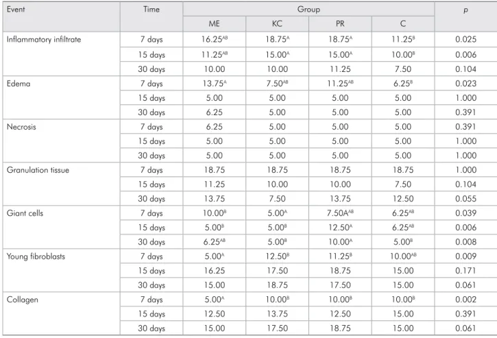
table 2. Mean scores attributed to cements and control group, after time intervals of 7, 15 and 30 days, for the 7 events evaluated.
Event Time Group p
ME KC PR C
Inflammatory infiltrate 7 days 16.25AB 18.75A 18.75A 11.25B 0.025
15 days 11.25AB 15.00A 15.00A 10.00B 0.006
30 days 10.00 10.00 11.25 7.50 0.104
Edema 7 days 13.75A 7.50AB 11.25AB 6.25B 0.023
15 days 5.00 5.00 5.00 5.00 1.000
30 days 6.25 5.00 5.00 5.00 0.391
Necrosis 7 days 6.25 5.00 5.00 5.00 0.391
15 days 5.00 5.00 5.00 5.00 1.000
30 days 5.00 5.00 5.00 5.00 1.000
Granulation tissue 7 days 18.75 18.75 18.75 18.75 1.000
15 days 11.25 10.00 10.00 7.50 0.104
30 days 13.75 7.50 13.75 12.50 0.055
Giant cells 7 days 10.00B 5.00A 7.50AAB 6.25AB 0.039
15 days 5.00B 5.00B 12.50A 6.25AB 0.006
30 days 6.25AB 5.00B 10.00A 5.00B 0.008
Young fibroblasts 7 days 5.00A 12.50B 11.25B 10.00AB 0.009
15 days 16.25 17.50 18.75 15.00 0.171
30 days 15.00 18.75 17.50 15.00 0.061
Collagen 7 days 5.00A 10.00B 10.00B 10.00B 0.002
15 days 12.50 13.75 12.50 15.00 0.391
30 days 15.00 17.50 18.75 15.00 0.061
statistical analysis
The data were tabulated and analyzed in the BioEstat statistical program version 5.0 (Instituto Mamirauá, Manaus, Brazil). The results of the cellu-lar events were submitted to the Kruskal-Wallis non-parametric test, followed by Dunn’s test to determine the differences among the groups (p < 0.05), insofar as the results did not present normal distribution.
Results
In the initial period, intense inlammatory inil-trate was observed for all the materials, with no sig-niicant statistical difference among them (p = 0.104). Groups PR (Figure 1A) and KC (Figure 1B) showed signiicant statistical difference in relation to Group C (Figure 1C), at 7 days (p = 0.025) and 15 days (p = 0.006; Table 2).
Circulatory alterations (edema) were more sig-niicant in Group ME, differing sigsig-niicantly from Group C (p = 0.023) at 7 days. In the events regard-ing tissue degeneration (necrosis) and granulation tissue around and within the cavity, the materials demonstrated similar behavior, with little or no necrosis and expressive response of tissues at the onset of the repair process, with no statistical dif-ference among the groups (p > 0.05). Multinucle-ated giant cells were more evident in Group ME on the 7th day, with a statistical difference (p = 0.039)
between Groups ME and KC, which did not per-sist at 15 and 30 days (Figure 1D). Group PR (Fig-ure 1A) presented a statistical difference in relation to Groups KC and ME at 15 days (p = 0.006), a sig-niicant difference that persisted between Group PR and KC at day 30 (p = 0.008; Table 2) in regard to the giant cells.
In relation to the tissue repair events, Group ME was less expressive and showed a statistically sig-niicant difference, in relation to Groups PR and KC, for the presence of young ibroblasts (p = 0.009) and for collagen (p = 0.002) at 7 days (Table 2).
discussion
GICs are used for cementation, especially for the ixation of crowns, bridges, onlays and orthodontic bands, because of their clinical properties, which include luoride release and bonding to tooth struc-ture.16 Nevertheless, for a cement to be adequate for
clinical use, its components must be biocompatible.15,17-21
In this context, the aim of the present experiment was to show evidence of the action of different dental ionomers on tissues, by means of histological analysis. This analysis must fundamentally be based on how live vascularized tissue is inluenced by aggression, which may be associated with cell death and necrosis, and how this aggression inluences the developmen-tal characteristics of capsule collagenization, inlam-matory iniltrate and macrophage action. Moreover, the analysis should be performed by means of quali-tative and quantiied evaluation.11-12,15
Biocompatibility studies22-23 have used polyethylene
tubes as controls,22 because they are considered
harm-less to the epithelial and conjunctive tissue. For this reason, they were used as such in this experiment.15
In this study, inlammatory iniltrate and edema events were expressive and intense. No signiicant difference among the cements was observed on the 7th day in regard to these events, and the events
became gradually less intense and similar in sub-sequent periods, as a response to the presence of metal ions,8-9,20 such as those of the aluminum
pres-ent in all the cempres-ents used in this study. These ions are released in the tissue as a result of the presence and/or concentration of the different acids compos-ing the GICs, such as polyalkenoic acids19,9 and
ben-zoic acid.7 These acids have been pointed out as being
substances capable of causing oxidative stress within the cells and interfering in cellular response.19
More-over, the low pH of these materials, especially when recently prepared, may have an influence on the potential inlammatory response,8-24 as supported by
Ribeiro et al.7 These authors demonstrated that Ketac
Cem cement powder did not induce alteration in cell DNA, whereas measurable genotoxicity was found for Ketac Cem liquid, composed of tartaric and ben-zoic acid, in all the dilutions tested in cell cultures. Similarly, Precedent and Ketac Cem cements were shown to stimulate signiicant inlammatory iniltrate in the irst 15 days, in comparison with the control, but showed no signiicant difference in relation to Meron cement. However, the process of collageniza-tion showed ascending values at 30 days, a perfor-mance similar to that observed for other materials.15,22
In addition to the inlammatory iniltrate, circu-latory alterations (edema) and multinucleated giant cells proved expressively greater with the Meron cement at 7 days, contrary to the respective results observed for the other cements. This was a propor-tional response of the body to the presence of alu-minum and/or iron ions,8-25 whereby giant cells are
released in the tissue to promote phagocytosis, a process by which the ions are surrounded and con-tained. Moreover, leachable substances from the GICs may also affect the epithelial cell rate of progression through the cellular cycle.26 There was virtually no
References
1. Wilson AD, Kent BE. The glass-ionomer cement, a new trans
-lucent dental filling material. J Appl Chem Biotechnol. 1971 Nov;21(11):313.
2. Mount GJ. Glass ionomers: a review of their current status. Oper Dent. 1999 Mar-Apr;24(2):115-24.
3. Kent BE, Lewis BG, Wilson AD. Glass ionomer cement for
-mulations: I. The preparation of novel fluoroaluminosilicate glasses high in fluorine. J Dent Res. 1979 Jun;58(6):1607-19. 4. Mickenautsch S, Yengopal V, Banerjee A. Pulp response to
resin-modified glass ionomer and calcium hydroxide cements in deep cavities: a quantitative systematic review. Dent Mater. 2010 Aug;26(8):761-70.
5. Santos RL, Pithon MM, Martins FO, Romanos MT, Ruellas AC. Evaluation of cytotoxicity and degree of conversion of glass ionomer cements reinforced with resin. Eur J Or
-thod. 2012 Jun;34(3):362-6.
6. Costa CA, Giro EM, Nascimento AB, Teixeira HM, Hebling J. Short-term evaluation of the pulpo-dentin complex response to a resin-modified glass-ionomer cement and a bonding agent applied in deep cavities. Dent Mater. 2003 Dec;19(8):739-46.
7. Ribeiro DA, Marques ME, Salvadori DM. Genotoxicity and cytotoxicity of glass ionomer cements on Chinese hamster ovary (CHO) cells. J Mater Sci Mater Med. 2006 Jun;17(6):495-500.
8. Oliva A, Salerno A, Locardi B, Riccio V, Della Ragione F, Iardino P, et al. Behaviour of human osteoblasts cultured on bioactive glass coatings. Biomaterials. 1998 Jun;19(11-12):1019-25.
9. Coimbra LR, Giro EMA, Aranha AMF, Costa CAS. Cytotox
-icity of restorative glass-ionomer cements to an odontoblast cell line. Rev Odonto Cienc. 2006 Jan;54(21):338-45.
10. Ozturk F, Yuksel S, Toy E, Kurtoglu EL, Kuçuk EB. Geno
-toxic effects of banding procedure with different orthodon
-tic cements on human oral mucosa cells. Turk J Med Sci. 2012 Jul;42(Suppl 1):1157-165.
11. Souza PP, Aranha AM, Hebling J, Giro EM, Costa CA. In vitro cytotoxicity and in vivo biocompatibility of contem
-porary resin-modified glass-ionomer cements. Dent Mater. 2006 Sep;22(9):838-44.
12. Boaventura JMC, Roberto AR, Becci ACO, Ribeiro BCI, Oliveira MRB, Andrade MF. The Importance of biocompat
-ibility of new materials: review of the glass ionomer cement. Rev Odontol Univ Cid Sao Paulo. 2012 Jan-Abr;24(1):42-50. 13. Kawai K, Takaoka T. Fluoride, hydrogen ion and HEMA release
from light-cured GIC restoratives. Am J Dent. 2002 Jun;15(3):149-52.
14. Lonnroth EC, Dahl JE. Cytotoxicity of dental glass ionomers evaluated using dimethylthiazol diphenyltetrazolium and neutral red tests. Acta Odontol Scand. 2001 Feb;59(1):34-9. days, tapering off at 15 and 30 days, demonstrating
a time-related healing process.
In comparison with Meron, Precedent and Ketac Cem cements showed more evident cellular cure-related events, signaled by the presence of young ibroblasts, and subsequent collagen ibers, as well as a reduced number of blood vessels. These histo-logical indings corroborate those of Ozturk et al.,10
who demonstrated that Meron cement presented genotoxicity, but relatively less in comparison with the resin cements27 or polycarboxylates.10
In general, a reaction of chronic development was observed throughout the experiment for all the materials. Polymorphonuclear inlammation was observed in the initial stages. However, pro-gressive collagenization became more expressive throughout the successive periods, in addition to the presence of mononuclear iniltrate and multi-nucleated giant cells.
Although a reduced number of polymorphonu-clear cells were observed in some cases, these cells
were conditioned by an active reactionary process, as seen in the time interval of 15 days for Precedent and Ketac Cem cements. Generally speaking, the expressive response in the initial events, such as the response to irritation, tended to diminish in subse-quent and inal periods.9,28-29
It can be inferred that the clinical use of materials with better biologic behavior must be encouraged, insofar as orthodontic ionomers in lowable form, used for cementation, frequently come into contact with gingival and subgingival tissues, and metal ions and intrinsic acids of the GIC composition may be released both during the setting process and in the degradation of these cements over time.13-14
Conclusion
15. Santos RL, Pithon MM, Fernandes AB, Cabral MG, Ruellas AC. Biocompatibility of orthodontic adhesives in rat sub
-cutaneous tissue. J Appl Oral Sci. 2010 Sep-Oct;18(5):503-8. 16. Paradella TC. Glass-ionomer cements in modern dentistry.
Rev Odontol UNESP. 2004 Sep-Oct;33(4):157-61.
17. Nogueira Júnior L, Araújo JEJ, Pavanelli CA, Araujo MAM. Histopatological analysis of pulp reactions, in vivo, after ce
-mentation of complete crowns with three luting agents: one phosphate and two glass ionomer cements. Rev Odontol UN
-ESP. 1997 Jul-Dec;26(2):517-31.
18. Golin C, Tavares T, Cunha AC. Biocompatibility evalua
-tion of commercial marks of glass ionomer-cement: study in mouse subcutaneous tissue. Rev Bras Odontol. 1992 Jan-Fev:49(1):35-9.
19. Costa CAS, Hebling J, Hanks CT. Effects of light-curing time on the cytotoxicity of a restorative resin composite applied to an immortalized odontoblast-cell line. Oper Dent. 2003 Jul-Aug;28(4):365-70.
20. Shelton RM, Rasmussen AC, Davies JE. Protein adsorption at the interface between charged polymer substrata and migrat
-ing osteoblasts. Biomaterials. 1988 Jan;9(1):24-9.
21. Meyer U, Szulczewski DH, Barckhaus RH, Atkinson M, Jones DB. Biological evaluation of an ionomeric bone ce
-ment by osteoblast cell culture methods. Biomaterials. 1993 Oct;14(12):917-24.
22. Grecca FS, Kopper PM, Santos RB, Fossati AC, Carrard VC, Acasigua GA, et al. Biocompatibility of RealSeal, its primer
and AH Plus implanted in subcutaneous connective tissue of rats. J Appl Oral Sci. 2011 Jan-Feb;19(1):52-6.
23. Onay EO, Ungor M, Ozdemir BH. In vivo evaluation of the biocompatibility of a new resin-based obturation system. Oral Surg Oral Med Oral Pathol Oral Radiol Endod. 2007 Sep;104(3):e60-6.
24. Pameijer CH, Segal E, Richardson J. Pulpal response to a glass-ionomer cement in primates. J Prosthet Dent. 1981 Jul;46(1):36-40.
25. Soheili Majd E, Goldberg M, Stanislawski L. In vitro effects of ascorbate and Trolox on the biocompatibility of dental restorative materials. Biomaterials. 2003 Jan;24(1):3-9. 26. Lewis J, Nix L, Schuster G, Lefebvre C, Knoernschild K,
Caughman G. Response of oral mucosal cells to glass iono
-mer cements. Biomaterials. 1996 Jun;17(11):1115-20.
27. Costa CAS, Hebling J, Garcia-Godoy F, Hanks CT. In vitro cytotoxicity of five glass-ionomer cements. Biomaterials. 2003 Sep;24(21):3853-8.
28. Silva RA, Assed S, Nelson-Filho P, Silva LA, Consolaro A. Subcutaneous tissue response of isogenic mice to calcium hydroxide-based pastes with chlorhexidine. Braz Dent J. 2009 Jan-Feb;20(2):99-106.
29. Garcia LF, Pires-de-Souza FC, Teofilo JM, Cestari A, Calefi PS, Ciuffi KJ, et al. Synthesis and biocompatibility of an ex
-perimental glass ionomer cement prepared by a non-hydro