1 . De par tame nto de B io fisic a e Radio b io lo gia da Unive r sidade Fe de r al de Pe r namb uc o , Re c ife , PE. 2 . De par tame nto de Far mác ia da Unive r sidade Fe de r al de Pe r namb uc o , Re c ife , PE. 3 . De par tame nto de Pato lo gia da Unive r sidade Fe de r al de Pe r namb uc o , Re c ife , PE. 4 . Lab o r ató r io de Imuno pato lo gia Ke izo Asami ( LIKA) da Unive r sidade Fe de r al de Pe r namb uc o , Re c ife , PE.
Financ iame nto : CAPES e FINEP
Addr e ss to: Ms. Ana Luna de Olive ir a. Unive r sity o f Alab ama at B ir mingham, 8 4 5 , 1 9 th Str e e t So uth, B e vill B io me dic al Re se ar c h B uilding ( B B RB ) – r o o m 5 3 8 , Zip 3 5 2 9 4 - 2 1 7 0 B ir mingham, AL, USA.
Te l: 1 2 0 5 9 7 5 -7 6 0 2
e - mail: lunade o live ir a@ ho tmail. c o m Re c e b ido par a pub lic aç ão e m 1 5 /1 /2 0 0 3 Ac e ito e m 2 0 /2 /2 0 0 4
Exper imental schistosomiasis in the Common
Mar moset
Callithrix jacchu s
Esquistossomose experimental no
Sagüi
Ca llithrix ja cchus
Ana Luna de Oliveir a, Elizabeth Malagueño, Adr iana Mar ia da Silva Telles,
Mar ia Helena Madr uga and José Valfr ido de Santana
ABSTRACT
In o rde r to e va lu a te Callithr ix j ac c hus a s a n a n im a l m o de l fo r m a n so n i sc histo so m ia sis, a gro u p o f 10 m a le a n im a ls we re o n c e pe rc u ta n e o u sly e xpo se d to 250 c e rc a ria e o f the Sc histo so ma manso ni SLM ( Sã o Lo u re n ç o da Ma ta ) stra in . An im a ls we re pe rio dic a lly b le d fo r m e a su rin g se ru m le ve l o f e n zym e s a n d pro te in s a n d fo r b lo o d c e ll c o u n tin g. Whe n c o m pa rin g pre - in fe c tio n to po st- in fe c tio n va lu e s, a sign ific a n t in c re a se wa s fo u n d fo r a lk a lin e pho spha ta se a t 15 to 120 da ys p.i., diffe re n tia l c o u n ts o f e o sin o phil a t 45 a n d 60 da ys, a n d to ta l pro te in a n d glo b a l e o sin o phil c o u n ts a t 120 da ys. No Sc histo so ma manso ni e ggs we re fo u n d in sto o ls. Adu lt wo rm s o f sm a ll size we re re c o ve re d fro m five a n im a ls. At da y 120, the n u m b e r o f Sc histo so ma manso ni e ggs/g o f tissu e wa s 0- 289.7 ( live r) , 0- 30.1 ( la rge in te stin e ) a n d 0- 171.4 ( sm a ll in te stin e ) . The se fin din gs le a d u s to c la ssify Callithr ix j ac c hus a s a n o n - pe rm issive ho st to the SLM stra in o f Sc histo so ma manso ni.
Ke y-words: Callithr ix j ac c hus. Sc histo so m ia sis.
RESUMO
Co m o o b je tivo de a va lia r o Callithr ix j ac c hus ( sa gü i) c o m o m o de lo e xpe rim e n ta l pa ra a e sq u isto sso m o se , u m gru po de 10 a n im a is, m a c ho s, a du lto s jo ve n s, fo ra m e xpo sto s a 250 c e rc á ria s da c e pa SLM ( Sã o Lo u re n ç o da Ma ta ) do Sc histo so ma manso ni, pe la via pe rc u tâ n e a . A in te rva lo s de 15 dia s a té 120 dia s a po s in fe c ç ã o fo ra m do sa do s o s n íve is de pro te ín a s to ta is e e n zim a s sé ric a s, a lé m de re a liza do s e xa m e s pa ra sito ló gic o e he m a to ló gic o . Au m e n to sign ific a tivo fo i o b se rva do pa ra : fo sfa ta se a lc a lin a a pa rtir do 150 dia ; c o n ta ge m glo b a l de e o sin ó filo s a o s 120 dia s, e dife re n c ia l de e o sin ó filo s a o s 45 e 60 dia s p.i. Na o se o b se rvo u o vo s de Sc histo so ma n a s fe ze s.Á pe rfu sã o c in c o a n im a is a pre se n ta ra m ve rm e s a du lto s. To do s o s ve rm e s e n c o n tra do s e ra m dim in u to s. O n ú m e ro de o vo s fo i 0- 289,7 o vo s/g de te c ido n o fíga do , 0- 30,1 n o in te stin o gro sso , 0- 171,4 n o in te stin o de lga do . Esta o b se rva ç o e s le va m - n o s a c o n c lu ir q u e o Callithr ix j ac c hus se ja n ã o -pe rm issivo a o Sc histo so ma.
Pal avr as-chave s: Callithr ix j ac c hus. Esq u isto sso m o se .
It is e stimate d that mo r e than 2 0 0 millio n pe o ple ar e
infe c te d in 7 6 diffe r e nt c o untr ie s wo r ldwide and ab o ut 6 0 0
m illio n pe o ple ar e at r isk o f b e c o m ing infe c te d with the tremato de S. m a nso ni6 2 72 9 3 1. This parasite inhabits the blo o d
veins of mammals and c auses a disease c alled sc histosomiasis.
I n B r a zil, m a n s o n i s c h is to s o m ia s is is pr e s e n t a s a n
endemic disease in so me states o f the No rtheast and So utheast
r e gio n s . Th e r e a r e a ppr o xim a te ly 1 0 m illio n B r a zilia n s i n fe c te d wi th th e d i s e a s e1. I n P e r n a m b u c o , m a n s o n i
abundant vegetation c alled Zo na da Ma ta. For instanc e, in 4
villa ge s o f Sã o Lo ur e n ç o da Ma ta , a m un ic ipa lity 2 5 k m northwest from Rec ife, approximately 9 9 .6 % of the inhabitants
were infec ted with at least one spec ies of intestinal parasites.
Prevalenc e for S. m a nso ni was 8 2 .1 % in this area1 7 1 8 4 4.
Infec tions due to Schisto so m a have been studied in many
animals inc luding mic e, rats, guinea pigs, rabbits, c ows and non-human primates. Nevertheless, doubts remain about whether or
not a spec ific animal is the c orrec t representation of the disease
in humans. Many mammals have been desc ribed as c arrying
natural infec tion with S. m a nso ni. For example, c ows have been desc ribed as having natural infec tion or as being susc eptible to S. m a nso ni infec tion2 7 1 2. Ro dentia is the Order with the largest
number of spec ies found having natural infec tions. Ne cto m ys s q u a m i p e s h a s b e e n f o u n d n a t u r a l l y i n f e c t e d wi t h S. m a n so n i in m any state s in B r azil3 1 0. I n r o de nts infe c te d
with 5 0 0 S. m a nso ni c erc ariae from any strain and by either
perc utaneous or subc utaneous routes, approximately 1 0 % of
the c erc ariae c an be rec overed as adult worms2 5. On another
study, Necto m ys ra ttus presented an infec tion rate of 7 1 %3 8.
Although monkeys are phylogenetically more closely related
to humans, their use as laboratory animals is restricted to cases in
which animals traditionally used in laboratory research can not
be used with satisfactory results2. In schistosomiasis, monkeys show
some advantages compared to the common laboratory animals
since as they maintain a semi-erect position, their portal-mesenteric
circulation dynamics may be closer to that of humans. Also they
have a higher hepatic tissue volume, enabling a better comparison to lesion in humans4. In addition, they live longer enabling their
study over a period of years, which is important when one considers
that humans usually carry their infection for many years.
A non-permissive host to S. m a nso ni is defined as an animal
in which the worm development is incomplete and/or oviposition is not sustained for a long period. As in the case of the Rhesus
monkeys, which pass S. m a nso ni eggs in their feces for only about
a year. Rats, rabbits and guinea pigs are all considered examples
of non-permissive hosts to S. m a nso ni. On the other hand, in permissive animals, oviposition is maintained for long periods.
This requires that male and female adult worms mature and migrate
together, to the lower mesenteric veins where oviposition begins.
Humans, some species of monkeys, hamsters, and mice are all
classified as permissive hosts. Parameters of infection that can be
used to identify permissive or non-permissive hosts include number,
size, morphology, and loc alization of adult worms, level and duration of oviposition, maturation of eggs, and level of pathology
observed in the host1 1 1 5.
Many pr imate spe c ie s had b e e n te ste d and ar e b e lie ve d
to b e susc e ptib le to S. m a n so n i infe c tio n. Ho we ve r, the le ve l
o f p e r m i s s i ve n e s s to Sc h i sto so m a va r i e s c o n s i de r a b l y
b e twe e n diffe r e nt spe c ie s o f mo nk e ys. Ac c o r ding to Co e lho e t a l4, Le ipe r was the fist wo r k e r to infe c t Ma n ga b y so o ty
mo nk e ys with Sc histo so m a m a n so n i in his c lassic al wo r k s
in whic h he t r i e d t o s h o w m a m m a l s ’ s u s c e p t i b i l i t y t o
t h i s p a r a s i t e . E x p e r i m e n t s o n R h e s u s m o n k e ys
c a r r yi n g a n i n i ti a l Schisto so m a m a nso ni infec tion showed
that afte r a c halle nge r infe c tio n, those animals were able to
eliminate the new infective cercariae without being able to destroy the adult worms resulting from the previous infection. This brought
up for the first time the concept of co nco m ita nt im m unity in sc histosomiasis4 1 4 3. Ce b us monkey when infec ted orally and
transc utaneously with the LE strain of Sc histosoma mansoni,
3 0 -5 0 % of the cercariae are recovered as adult worms. However, there is no evidence of concomitant immunity7 2 2. When infected
subcutaneously, tamarin monkeys met the criteria of permissiveness to S. m a nso ni: correct anatomical distribution of adult worms
and oviposition for long periods of time1 5. Baboons have been
found to be a permissive host for S. m a nso ni and a good animal
model for schistosomiasis. Besides experimentally infected with S. m a nso ni, baboons are also naturally infected with this parasite. Muller-Graf et a l3 0, studying five troops of olive baboons (Pa pio
cynocephalus anubis) in Gombe Stream National Park in Tanzania, found three troops naturally infec ted with S. m a nso ni. These
authors affirm that the epidemiology of S. m a nso ni in baboons may display characteristics that are very close to human infection.
In addition, baboons can both maintain and transmit S. m a nso ni parasite in the wild3 4. In baboons, 7 8 % of c erc ariae c an be
recovered as adult worms. This percentage is usually 2 5 to 4 0 % in mice and 1 5 to 2 0 % in rats47. However there are some disadvantages
of using baboons as a model for schistosomiasis, such as the high cost of buying and maintaining baboons in the laboratory, and the need for qualified personnel to care for the animal’s health.
Ca llithrix ja cchus, also known as common marmoset, is a small neotropical primate. It belongs to the Ca llithrichida e family and inhabits the Brazilian northeast forests2 6. Ca llithrix lives in
small groups where the majority of the members are related. In
the group there is one reproductive female and many other non-reproductive females. Its diet usually comprises of fruit, nectar,
blooms, insects, and gum from some forest trees. This marmoset usually gives birth to two or more offspring at once. Parental care,
whic h inc ludes c arrying, c aring, and sharing fo o d with the
newborn, is performed by all adults and adolescents in the group3 5.
Mar mo se ts have many advantage s as a lab o r ato r y animal.
The ir small size and high le ve l o f r e pr o duc tio n in c aptivity
ar e ve r y im po r tant fac to r s. The y ar e also e asy to handle , r e q uir e sm all am o unts o f fo o d and sho w a high le ve l o f
adaptab ility to ne w e nvir o nme nts. Ca llithrix ar e c ur r e ntly
use d in e xpe r ime nts invo lving to xic ity, vir al, par asito lo gic al
a n d b a c te r i a l d i s e a s e s , a n d i m m u n o l o gi c a l r e s e a r c h ,
c o ntr ib uting to the c o nc lusio n o f impo r tant studie s2 3 5.
The o b j e c tive o f the pr e se nt wo r k was to e valuate the
mar mo set C. ja c c hus as a po ssib le animal mo del fo r manso ni
sc histo so miasis.
MATERIAL AND METHODS
Ex pe r im e nta l infe c tio n a nd blo o d c o lle c tio n. A gr o up o f twe lve adult m ale Ca lli thri x ja c c hu s ( we ighing a ppr o xim a te ly 3 0 0 g) we r e use d in the e xpe r im e nts. Te n
c o n s titute d th e e x p e r im e n ta l gr o up a n d two s e r ve d a s
Ca lli th ri x ja cchus were anesthetized and then exposed percutaneously to a pool of 2 5 0 cercariae of S. m a nso ni, SLM strain, placed in their inguinal area for 3 0 minutes under artificial
ligh t1 5. Th is S. m a n so n i s tr a in h a d b e e n m a in ta in e d in
Bio m pha la ria gla bra ta/ Swiss mice system. As a control for the cercariae infectivity, Swiss mice were also infected with the same pool of cercariae used in the marmoset infection.
Afte r infe c tio n, at inte r vals o f two we e k s, anim als had
1 ml o f blo o d drawn fro m their Femo ral artery after anesthesia
with k e tamine . Se r um was use d fo r b io c he mic al te sts, and a
small aliq uo t o f b lo o d tr e ate d with EDTA was use d in the he mato lo gic al analysis.
Animals had the ir we ight me asur e d and sto o ls c o lle c te d
at the same time pe r io ds and in additio n any signs o f c linic al
sympto ms we r e no te d.
Biochemical tests & hematological counts. Bioc hemic al manual assays were performed using c ommerc ial kits La btest. In order to c omplete the entire battery of proposed tests with a
sm all am o unt o f b lo o d, the vo lum e o f r e age nts use d was
proportionally divided by 5 ( alanine amino-transferase, aspartate
amino -tr ansfer ase, alkaline pho sphatase, to tal pr o tein, and gamma-glutamyl transferase) or 2 ( albumin) .
Diffe r e ntial c o unts o f b lo o d c e lls we r e pe r fo r me d o n a
thic k b lo o d film staine d with Gie msa ( 1 0 0 c e lls c o unte d) . At
this time , it was po ssib le to tak e no te o f any alte r atio n in c e ll
mo r pho lo gy and staining c har ac te r istic s. Glo b al c o unts we r e pe r fo r me d in a c e ll c o unt c hamb e r.
Pa r a sito lo gica l a na lysis. Sedimentatio n2 0 and fo r malin
e th e r c o n c e n tr a ti o n3 9 te c h n i q u e s we r e u s e d to d e te c t
S. m a n so n i e ggs a n d o th e r fo r m s o f pa r a s ite s in s to o l sample s. Whe n the amo unt o f sample was no t suffic ie nt fo r pe r fo r m in g b o th m e th o ds , pr e fe r e n c e wa s give n to th e
c o nc e ntr atio n te c hnique .
Animals we r e pe r fuse d at 1 2 0 days po st infe c tio n. In the
m e th o d us e d, po r ta l h e pa tic a n d m e s e n te r ic s ys te m a r e
pe r fuse d se par ate ly. Re c o ve r e d wo r m s we r e c o unte d and so r te d b y se x unde r the ste r e o sc o py. Live r, inte stine s, and
sple e n we r e we ighte d. Par t o f live r and inte stine s was use d
b o th fo r the hydr o pr o line assays and fo r S. m a n so n i e ggs
ide ntific atio n afte r dige stio n with 5 % po tassium hydr o xide
( KOH) . Tissue dige stio n using 5 % KOH was do ne ac c o r ding
to the te c hnique de sc r ib e d b y Che e ve r e t a l 8 using 4 sample s
o f 2 g fr o m live r and inte stine s ( 2 fr o m small inte stine and 2
fr o m lar ge inte stine ) fo r e ac h animal.
Fr o m e ac h animal, six sample s o f live r, o f 0 .5 g e ac h, we r e
use d fo r the hydr o xypr o line de te r minatio n. Afte r pe r fusio n,
tissue sam ple s we r e fr o ze n until pr o c e sse d ac c o r ding to
te c hnique s de sc r ib e d b y Che e ve r e t a l, Pr o c k o p e t a l, and
B e r gman e t al4 7 3 6.
In additio n, spleen, kidneys, lungs, hear t, and par t o f liver
and inte stine s we r e k e pt in 1 0 % fo r malin b uffe r fo r po ste r io r
histo patho lo gic al analysis.
Sta tistica l a na lysis. Pair e d Stude nt’s T te st was use d whe n c o mpar ing r e sults fo r the same animal at diffe r e nt time
points (
α
= 5 % ) . Unpaired Student’s T test was used to analyzethe var iatio ns in glo b al c e ll c o unts and o r gan we ight value s (
α
= 5 % ) .RESULTS
The m e an b io c he m ic al value s fr o m day 0 to 1 2 0 days
po st infe c tio n ( dpi) we r e : alb umin 3 .9 -5 .1 g/dl, to tal proteins
6 . 7 - 7 . 6 g/dl, gamma glutamyl tr ansfe r ase 4 3 . 8 - 1 0 5 . 8 U/l,
alkaline phosphatase 2 6 .2 8 9 .2 U/l, alanine aminotransferase 1 0 3 -1 2 6 .9 U/l and aspartate amino-transferase 5 .8 -2 0 .2 U/l. When
comparing values post infection ( from 1 5 to 1 2 0 dpi) with the
values pre infection ( at 0 dpi) , there was a statistically significantly
increase in the serum levels of alkaline phosphatase from 1 5 dpi, and in the total protein levels at 1 2 0 dpi. However, the biochemical
serum values of infected animals were within the normal limits for
their species for most of the proteins measured, and considered
as a whole, did not represent the characteristic changes observed in human schistosomiasis.
The r e was no sign o f ne utr o philia, b ut e o sino philia ( 2 6 %
at 1 2 0 days p.i.) was o b se r ve d dur ing the infe c tio n pe r io d,
altho ugh o the r par asite s that c an inc r e ase e o sino phil c o unts
in b lo o d we r e also de te c te d in so me o f the animals studie d
suc h as Stro n gylo ide s. Trypa n o so m a tida e we r e fo und o n thic k blood film samples of 3 /1 0 animals and Filariidae worms
we r e iso late d fr o m the lungs o f 2 /1 0 animals at pe r fusio n.
Sc histo so m a m a n so n i e ggs c o uld no t b e fo und in the i n fe c te d a n i m a l fe c e s du r i n g a l l th e o b s e r va ti o n ti m e . Ho we ve r, fi ve a n i m a l s ( # 1 , 2 , 3 , 4 , a n d 8 ) p r e s e n te d Stro n gylo ide s lar vae in the ir fe c e s in the fir st 3 0 days o f infe c tio n. Afte r 1 0 5 dpi, thr e e o f the pr e vio us five animals
( # 2 , 3 , and 4 ) we r e po sitive again fo r Stro n gylo ide s.
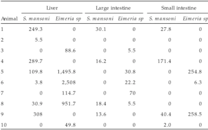
Afte r dige stio n with KOH the amo unt o f S. m a n so n i e ggs
pe r gr am o f tissue pr e se nt in live r and inte stine s o f infe c te d animals is sho wn in Tab le 1 . In the live r sample s o f infe c te d
a n im a ls , S. m a n so n i e ggs we r e fo un d in 7 /1 0 a n im a ls , Ei m e ri a sp c ys ts we r e fo und in 6 /1 0 a nim a ls , a nd b o th par asite s we r e fo und in 3 /1 0 animals. On the o the r hand, in
the inte stine sample s, S. m a n so n i e ggs we r e pr e se nt in 5 /1 0
a n im a ls , Ei m e ri a sp in 6 /1 0 a n im a ls , a n d b o th in 2 /1 0
Table 1 - Num ber of eggs of S. mansoni and cysts of Eimeria sp per g of tissue of C. jacchus infected with S. mansoni at 120 days post-infection.
Liver Large intestine Small intestine Animal S. m ansoni Eim eria sp S. m ansoni Eim eria sp S. m ansoni Eim eria sp
1 2 4 9 .3 0 3 0 .1 0 2 7 .8 0
2 5 .5 0 0 0 0 0
3 0 8 8 .6 0 5 .5 0 0
4 2 8 9 .7 0 1 6 .2 0 1 7 1 .4 0
5 1 0 9 .8 1 ,4 9 5 .8 0 3 0 .8 0 2 5 4 .8
6 3 .8 2 ,5 0 8 0 2 2 .2 0 6 .3
7 0 1 1 4 .7 0 70 0 0
8 3 0 .9 9 5 1 .7 1 8 .4 5 .5 0 0
9 3 0 8 0 1 3 .6 0 4 0 .4 2 5 8 .5
animals. S. m a n so n i tissue e gg c o unts var ie d fr o m 0 -2 8 9 .7
in the live r, 0 -1 7 1 .4 in the small inte stine , and 0 -3 0 .1 in the lar ge inte stine . Uninfe c te d animals did no t sho w any par asite
fo r m afte r KOH dige stio n.
Altho ugh S. m a n so n i e ggs we r e fo und in tissue sample s,
the mac r o sc o pic c hange s due to the ir pr e se nc e we r e no t
o b se r ve d. A statistic ally signific ant diffe r e nc e ( p < 0 .0 5 ) was fo und in the we ight o f lar ge inte stine and live r o f infe c te d
animals whe n c o mpar e d to uninfe c te d c o ntr o ls ( Tab le 2 ) .
Histopathologic examination was not one of the objectives of
the present work. However, in our pilot study 4 2, ten Ca llithrix
ja cchus were percutaneously infected with 1 5 0 cercariae of the SLM strain of Schistosom a m ansoni. Out of ten animals, 5 showed
granuloma in their livers, 2 animals had granuloma in their
intestines, and one animal had granuloma in a lung. S. m a nso ni eggs were found in the lungs of 3 animals, and in the liver of 7
animals. Out of 5 animals observed, there were no eggs in the
intestines of any animal, adult worms were found in 4 of them,
and no sign of fibrosis was found in any of the 5 animals. In another 5 animals, adult worms were found in samples of lung in one
animal, in the liver of another, and in both liver and lungs of a
third animal.
DISCUSSION
Hepatic func tion is usually well preserved in sc histosomiasis.
Patients have: 1 ) blood level of albumin bellow normal values;
2 ) levels of alkaline phosphatase and gamma glutamyl transferase
above referenc e values, 3 ) levels of aspartate amino-transferase
and alanine amino-transferase within the normal range or slightly
elevated, 4 ) normal bilirubin; and 5 ) normal or slightly higher
total protein amount in their sera1 3 1 6 3 7. In the present study,
bioc hemic al values of a set of enzymes and proteins that are
r e late d to live r func tio n and that pr o vide info r m atio n to differentiate sc histosomiasis from hepatic diseases due to other
etiologies did not c hange c harac teristic ally.
Baboons show c linic al symptoms c harac teristic of both ac ute
and c hro nic stages o f S. m a nso ni infec tio n. So me o f tho se
symptoms were diarrhea, weakness, weight loss ( ac ute stage)
and histopathologic al c hanges ( c hronic stage)1 4. On the other
hand, Cebus monkeys only presented symptomatology when
experimentally infec ted with S. m a nso ni4 7 2 2. Unlike those other
primates, C. ja cchus showed no c linic al symptoms during either
ac ute or c hronic stages of infec tion.
Leukocytosis was not present in our animal samples as has
alr eady b een desc r ib ed in humans3 2 3 7 and infec ted mic e2 3.
Eosinophilia, although present, cannot be attributed to S. m ansoni
alone, since other parasites that may also raise eosinophil counts
in the blood were found in samples of some animals. Ca llithrix
seems to bear parasite infections very well since no major clinical
symptoms or biochemical changes were seen in those animals.
The fac t that infec ted C. ja cchus do es no t pass S. m a nso ni
e ggs in the ir fe c e s is a c har ac te r istic diffe r e nt fr o m that se e n
in human and o the r animals. In ar e as o f high pr e vale nc e and
m o de r a te in te n s ity o f in fe c tio n fo r s c h is to s o m ia s is , th e
maj o r ity o f infe c te d pe o ple will pass a small numb e r o f e ggs
in the ir fe c e s, while a small numb e r o f patie nts will pass a
lar ge numb e r o f e ggs. Tanab e e t a l4 4 r e po r te d that in São
Lo ur e nç o da Mata, in Pe r namb uc o State , B r azil, 6 1 % o f the
in fe c te d p o p ula tio n p a s s e d 1 - 1 0 0 e gg/g o f fe c e s , wh ile
o n ly 6 % passed more than 4 0 1 eggs/g of fec es. Ce b us monkey,
tamar in, and Pa pio c yn o c e pha lu s all sho we d e ggs in the ir
fe c e s at ab o ut 3 5 days pi4 7 1 5 2 2. In tamar ins, this numb e r was Table 2 - Mean organ weight ( ± standard deviation) of controls and infected
C. jacchus at 120 days p.i.
Liver Large intestine Small intestine Spleen Control animals ( n= 2 ) 9 .1 6 ± 0 .3 3 5 .8 1 ± 0 .2 7 8 .7 9 ± 2 .6 4 0 .4 ± 0 .2 Infected animals ( n= 10) 1 3 .0 4 ± 1 .8 7 * 4 .4 1 ± 0 .7 7 * 7 .3 7 ± 0 .6 5 0 .5 7 ± 0 .1 3 p < 0 .0 5
As shown in Table 3 , at perfusion, the total number of adult
worms rec overed varied from 1 to 1 4 ( 0 .4 to 5 .6 % ) . The male/ female ratios were 1 0 /1 , 4 /1 , 0 /0 , 0 /1 , 1 /6 . Only one worm
pair was found. Some immature forms of worms were observed
and all worms were of small size and loc ated mainly within the
liver blood system and not in the mesenteric system.
Th e r e wa s n o s ign ific a n t in c r e a s e in th e a m o un t o f
h ydr o xypr o lin e due to S. m a n so n i in fe c tio n . Th e m e a n
amount of hydroxyproline per gram of tissue was 6 .3 3 ± 0 .7 2 mg
in the uninfe c te d animals ( n = 2 ) and 6 .4 1 ± 1 .6 5 mg in the
infe c te d animals ( n = 1 0 ) . Whe n c o mpar ing the value o f
hydr o xypr o line at 1 2 0 dpi with the value o f hydr o xypr o line
in the c o ntr o l animals, o nly in 4 /1 0 animals a hydr o xypr o line
gain o f 0 .7 7 -3 .5 5 mg was o b se r ve d. Ho we ve r, no S. m a n so n i
e gg was fo und in o ne o f tho se animals, o nly Eim e ria sp. The
me an amo unt o f hydr o xypr o line pr o duc e d b y e gg laid in the
live r was 2 6 0 hg.
Infe c te d animals did no t pr e se nt we ight lo ss o r any o the r
c linic al sympto ms dur ing the study.
Ta b le 3 - Num b e r o f S. mansoni a dult wo rm s a nd the ir lo ca liza tio n in
C. jacchus at 120 days post-infection.
2 4 7 6 9 Total
Portal- hepatic blood vessel system
male 4 2 0 0 1 7
female 1 0 0 1 6 8
immature 0 1 5 0 0 6
pair 0 0 0 0 0 0
Subtotal 5 ( 5 0 %) 3 ( 2 1 %) 5 ( 1 0 0 %) 1 ( 1 0 0 %) 7 ( 1 0 0 %) 2 1
Mesenteric blood vessel system
male 5 6 0 0 0 1 1
female 0 2 0 0 0 2
immature 0 1 0 0 0 1
pair 0 1 0 0 0 2
Subtotal 5 ( 5 0 %) 1 1 ( 7 9 %) 0 ( 0 %) 0 ( 0 %) 0 ( 0 %) 1 6
higher in animals infec ted subc utaneously. Cows infec ted with 2 5 ,0 0 0 c erc ariae first showed eggs in their fec es between 7 9 and 2 0 2 days pi2 7. The fac t that no eggs were seen in fec es of
infec ted C. ja cchus may be explained by an immunologic al fac tor c harac teristic of the spec is. For example, Karanja e t a l2 1
studying humans c o -infec ted with HIV and sc histo so miasis, observed that patients with a reduc ed number of CD4 + c ells passed less eggs in their fec es. Those authors report that some studies in humans, baboons, and mic e, suggest that CD4 + c ells are involved in the exc retion of S. m a nso ni by infec ted hosts.
Altho ugh S. m a n so n i e ggs we r e fo und in sa m ple s o f intestines and liver, their number was very small. Considering that a S. m a nso ni female adult worm lays a large number of eggs per day, and that only a small part of them passes through the feces while the majority remains in the tissue. Handerson et a l1 9,
for example, infecting inbred CB/J mice, found a range of 2 5 ,5 1 1 -4 0 ,1 -4 3 egg/liver depending on whether the animal developed m o de ra te sple no m e ga ly syndro m e o r hype rsple no m e ga ly syndro m e. In addition, in our study, macroscopic and physiologic changes due to the presence of S. m a nso ni eggs on the specified organs were not observed.
Sc h i sto so m a m a n so n i infe c tio n in hum a n a nd o the r animals is usually fo llo we d b y high le ve ls o f hydr o xypr o line , r eflec ting the level o f hepatic fib r o sis in tho se ho sts. Ho wever, hydr o xypr o line gain due to S. m a n so n i infe c tio n was lo w and unspe c ific in o ur animals.
The morphology, small size and number ( 0 .4 % to 5 .6 % rec overy) , and mainly portal hepatic loc alization of S. m a nso ni adult worms rec overed from C. ja cchus do not reflec t what has been seen so far in animals tested and c onsidered permissive to S. m a nso ni. In Pa pio a nub is and Pa pio cyno ce pha lus, the perc entage of c erc ariae rec overed as adult worms is 7 8 % and 4 2 % , r e spe c tive ly1 4. Tamar ins, if pe r c utane o usly e xpo se d,
s h o w 2 . 6 % o f c e r c ar iae r e c o ve r e d as adult wo r ms, b ut if subc utaneously exposed this value is 3 1 .3 %1 5. In mic e, this value
ranges from 2 5 to 4 0 % , while in rats from 1 5 to 2 0 %4 7. For
Ne cto m ys squa m ipe s it has been observed as being 1 1 .5 %2 5.
For c ows, this value was 2 .2 %2 7.
In baboons, maturation of cercariae often exceeds 9 0 %. Female adult worms of S. m a nso ni start laying eggs around 5 -6 weeks p.i. with a large number of eggs laid per worm pair per day. These eggs are found mostly in the intestines ( 8 0 %) while around only 1 0 % are found in the liver similar to a human infection. Infected animals present symptoms of the disease, and two clinical phases of the disease can be recognized. The first or acute phase starts at around 6 -1 2 weeks p.i., and then there is a chronic phase after which some animals show granuloma, modulation of granuloma, and development of periportal fibrosis2 8 3 4.
Lima e t a l2 4, studying dyslipo pr o te ine mia in C. ja c c hu s
( b o dy we ight 1 0 0 -3 0 0 g) infe c te d pe r c utane o usly with 1 5 0 -4 5 0 c e r c ar iae o f S. m a n so n i ( SLM str ain) , have r e po r te d th a t th e pa r a s ite in fe c tio n wa s c o n fir m e d a t a uto ps y b y h i s to l o gi c a l e x a m i n a ti o n o f th e l i ve r. Ne ve r th e l e s s , n o info r matio n was give n o n the numb e r, size and lo c alizatio n o f adult wo r ms, numb e r and viab ility o f e ggs, o r any de tail o n the histo lo gic al e xaminatio n that was pe r fo r me d.
According to Rodrigues et a l4 0,C. penicilla ta ( body weight
1 6 0 -1 7 2 g) infected by inoculation of 2 0 0 cercariae shows signs
of granuloma around S. m a nso ni eggs and presenc e of adult
worms on histopathological examination of the liver. However, the autho r s did no t pr o vide any info r matio n ab o ut the str ain o f S. m ansoni used in the infection. In addition there is no information on: number or size of granulomas, egg counts, and morphology, localization and percentage of recovery of adult worms.
Sadun et a l 4 1 also found signs of granuloma on four Ca llithrix
a urita infec ted perc utaneously with 5 0 ( two animals) and 1 0 0 0 ( two other animals) c erc ariae of S. m a nso ni Puerto Ric an strain. In this artic le, the authors investigated the perc entage of adult
worms rec overed, loc alization and morphology of worms, the length of pre-patent period, number of eggs in the fec es, number and loc alization of eggs in the tissue, egg viability, gross pathology
and histopathology of organs suc h as lungs, liver, and intestines, and for the signs of other infec tions. They tried to c lassify the 1 0 spec ies of monkeys into 3 groups ac c ording to their levels of
permissiveness to S. m a nso ni. Although they saw some signs of gr a n u l o m a i n Ca lli th ri x, b a s e d o n th i s a n d a l l o th e r c harac teristic s of this spec ies they c lassify Ca llithrix, squirrel
monkey, and the tree shrew monkey in the group they refer to as the po o rer ho sts gro up.
Similarly to Ca llithrix ja cchus in our study, owl monkeys
(Ao tus na ncym a i) when infec ted with J.L. Venezuelan strain of S. m a nso ni, passed no eggs in their fec es during a period of 1 6 weeks p.i. Although diarrhea and weight loss oc c urred, no adult
worms were found by either perfusion or examination of fresh tissue. Eggs were found in the liver and intestines of the animals, but mainly in the liver. Signs of granuloma were present in the
liver and in the intestines, but more pronounc ed in the liver. In th is s a m e s tudy o n ly o n e m o n k e y fr o m a n o th e r s p e c ie s , A. vo cife ra ns, passed S. m a nso ni eggs at 8 weeks pi3 3.
Our fin din gs a r e s im ila r to th o s e o f Wa r r e n e t a l4 6.
Wh ile wo r k in g with 9 m a r m o s e ts ( b o dy we igh t 1 3 7 - 3 4 8 g) , 3 C. ja cchus and 6 C. pe nicilla ta, 6 males and 3 females, infec ted perc utaneously with 1 0 0 or 2 0 0 c erc ariae of a B razilian S. m a n so n i str ain pe r 1 0 0 g o f we ight, the y fo und har dly any S. m a nso ni egg in the marmoset fec es, no c linic al symptoms, no weight lo ss and o n aver age the adult wo r ms r ec o ver ed
amounted to 5 .2 % of the c erc ariae inoc ulated ( 1 .9 % to 1 0 .5 % ) . Worms were smaller than those usually rec overed from infec ted mic e, and the presenc e of other parasites c o-infec ting their
animals. In animals infec ted with 1 0 0 c erc ariae, only one egg with a dead mirac idium was found after 2 0 weeks of infec tion.
We c o nc lude tha t whe n pe r c uta ne o us ly infe c te d with Sc histo so m a m a n so n i, SLM str ain, the pr imate C. ja c c hu s sho ws c har ac te r istic s o f no n-pe r missive ne ss.
ACKNOWLEDGMENTS
We wo uld lik e to thank the B r azilian age nc ie s CAPES and
FINEP fo r the ir financ ial suppo r t. We also e xte nd o ur thank s to Dr. Ro b e r to Siq ue ir a fr o m “Re fugio Ec o lo gic o Char le s Dar win”, at Igar assu-PE, B r azil, fo r the animals use d in this
study and to Mr. Hailto n and Luis Fe lipe fo r he lping to c ar e fo r the animals. We are also indebted to Jo se Felipe Go nc alves,
REFERENCES
1 . Aguiar J LA, Do mingue s ALC, Aguiar SM, Ke lne r S. Hidr o xipr o lina ur inár ia n a fa s e h e p a to e s p l ê n i c a d a E s q u i s to s s o m o s e m a n s ô n i c a . An a i s d a Fac uldade de Me dic ina da Unive r sidade Fe de r al de Pe r namb uc o 4 2 : 3 8 -4 2 , 1 9 9 7 .
2 . Andr ade MCR. A utilizaç ão de Símio s do gê ne r o Ca lli thri x c o mo mo de lo exper imental. B o letim info r mativo do Co légio B r asileir o de Exper imentaç ão Animal 0 3 - 9 7 /9 8 : 5 , 1 9 9 8 .
3 . An tun e s CM, An dr a de M, Ka tz NK, Co e lh o PM, Pe lle gr in o J . R o le o f
Ne c to m ys sq u a m i p e s sq u a m i p e s in the e pide m io lo gy o f Sc h i sto so m a m a n so n i infe c tio n. Anna ls o f Tr o pic a l Me dic ine a nd Pa r a s ito lo gy 6 7 : 6 7 - 7 3 , 1 9 7 3 .
4 . B e r gm a n I , Lo xle y R . Two im pr o ve d a n d s im plifie d m e th o ds fo r th e spe c tr o pho to me tr ic de te r minatio n o f hydr o xypr o line . Analysis Che mistr y 3 5 : 1 9 6 1 -1 9 6 5 , 1 9 6 3 .
5 . B e r gq uis t NR. Co n tr o llin g Sc h is to s o m ia s is b y Va c c in a tio n : A r e a lis tic Optio n? Par asito lo gy To day 1 1 : 1 9 1 - 1 9 4 , 1 9 9 5 .
6 . B o r d a CE , P e l l e gr i n o J , Me s c h e s s i HD. E l i m i n a c i o n d e Hu e vo s d e Sc h is to s o m a m a n s o n i p o r Ce b u s a p e lla m a c ro c e p h a lu s I n fe c ta do s Expe r ime ntalme nte . Re vista do Instituto de Me dic ina Tr o pic al de São Paulo 1 1 : 9 1 - 9 6 , 1 9 7 2 .
7 . Che e ve r AW, Duvall RH, Hallac k TA, Mink e r RG, Malle y J D, Malle y KG S p e c tr o p h o to m e tr i c As s a y m e a s u r e m e n t o f th e c o n c e n tr a ti o n o f Hydr o xypr o line in the live r s o f m ar m o se ts infe c te d with Sc h i sto so m a m a n so n i. Ame r ic an J o ur nal o f Tr o pic al Me dic ine and Hygie ne 3 7 : 4 1 5 -5 2 2 , 1 9 8 7 .
8 . Che e ve r AW, War r e n KS. He patic b lo o d flo w in mic e with ac ute he pato -sple nic sc histo so m iasis m anso ni. Tr ansac tio ns o f the Ro yal So c ie ty o f Tr o pic al Me dic ine and Hygie ne 5 : 4 0 6 - 4 0 9 , 1 9 6 4 .
9 . Ch ie ffi PP, Klo e tze l K, Siq ue ir a J GV. Ab s e n c e o f n a tur a l in fe c tio n b y
Sc hi sto so m a m a n so n i in wild r o de nts c aptur e d in e nde m ic ar e as fo r sc histo so m iasis in the state o f Alago as, B r azil. Re vista do I nstituto de Me dic ina Tr o pic al de São Paulo 3 6 : 3 7 7 - 3 7 8 , 1 9 9 4 .
1 0 . Cio li D, Kno pf PM, Se nft AW. A study o f Sc hi sto so m a m a n so n i tr ansfe r r e d into pe r missive and no n pe r missive ho sts. J o ur nal o f Par asito lo gy 7 : 2 9 3 -2 9 7 , 1 9 7 7 .
1 1 . Co e lh o B , Ma ga lh ã e s Filh o A. R e s ulta do s Pa to ló gic o s de in fe s ta ç ã o e xpe r ime ntal de Sc histo so m a m a n so n i e m mac ac o Ce b u s sp. Pub lic aç õ e s avulsas do Instituto Agge u Magalhãe s II: 6 1 - 9 8 , 1 9 5 3 .
1 2 . Co e lho PM, No gue ir a RH, Lima WS, Cunha MC. Sc hi sto so m a m a n so n i: e xpe r im e ntal b o vine sc histo so m iasis. Re vista do I nstituto de Me dic ina Tr o pic al de São Paulo 2 4 : 3 7 4 - 3 7 7 , 1 9 8 2 .
1 3 . Co sta MFFL, Katz N. Co m pa ra ti ve stu dy o f Sc hi sto so m a m a n so n i str ains is o la te d from patients with toxemic or intestinal forms of sc histosomiasis. Americ an Journal of Tropic al Medic ine and Hygiene 3 1 : 4 9 9 - 5 0 4 , 1 9 8 2 . 1 4 . Co u t i n h o AD , D o m i n g u e s ALC. I n: E s q u i s t o s s o m o s e M a n s o n i .
Ga s tr o e n te r o l o gi a Cl í n i c a 3 ª e d i to r a . E d i to r a Gu a n a b a r a Ko o ga n . Ca pítulo 1 0 8 : 1 6 9 7 - 1 7 2 8 , 1 9 9 3 .
1 5 . Damian RT, Gr e e ne ND, Me ye r KF, Che e ve r AW, Hub b ar d WJ , Hawe s ME, Cl a r k J D. Sc h i s to s o m a m a n s o n i i n Ba b o o n s I I I . T h e c o u r s e a n d c har ac te r istic s o f infe c tio n, with additio nal o b se r vatio ns o n immunity. The Ame r ic an Jo ur nal o f Tr o pic al Me dic ine and Hygie ne2 5 : 2 9 9 - 3 0 6 , 1 9 7 6 .
1 6 . De l Po r tillo HA, Damian RT. Expe r ime ntal Sc histo so m a m a n so n i infe c tio n in a s m a ll n e w wo r ld m o n k e y, th e s a ddle - b a c k ta m a r in (Sa gu i n u s
fu sc ic o llis) . Ame r ic an Jo ur nal o f Tr o pic al Me dic ine and Hygie ne , 3 5 : 5 1 5 -5 2 2 , 1 9 8 6 .
1 7 . Do mingue s ALC, Do mingue s LAW. In: Esq uisto sso mo se Mansô nic a, Edito r a Unive r sitár ia da UFPE Capítulo 5 : 9 1 - 1 0 9 , 1 9 9 4 .
1 8 . Go nc alve s JF, Co utinho A, Santana V, B ar b o sa CJ. Esq uisto sso mo se aguda de c a r á te r e pis ó dic o n a I lh a de I ta m a r a c á , Es ta do de Pe r n a m b uc o . Cade r no s de Saúde Púb lic a 7 : 4 2 4 - 4 2 5 , 1 9 9 1 .
1 9 . Go nc alve s J F, Tanab e M, Me de ir o s FPM, Go nç alve s J F, Ac a IS, Mo tta SRN, Ta te no S, Ta k e uc hi T. Pa r a s ito lo gic a l s tudie s o n a m e b ia s is a nd o the r
inte stinal par asitic infe c tio n in the r ur al se c to r ar o und Re c ife , No r the ast, B r azil. Re vista do Instituto de Me dic ina Tr o pic al de São Paulo 3 2 : 4 5 8 -4 3 5 , 1 9 9 0 .
2 0 . He nde r so n GS, Nix NA, Mo nte sano MA, Go ld D, Fr e e man J r GL, Mc Cur le y TL, Co lle y DG. Two distinc t patho lo gic al syndr o me s in male CB L/J inb r e d mic e with c hr o nic Sc hi sto so m a m a n so n i infe c tio ns. Ame r ic an J o ur nal o f Patho lo gy 1 4 2 : 7 0 3 - 7 1 4 , 1 9 9 3 .
2 1 . Ho ffman WA, Po ns JA, Jane r JL. The Se dime ntatio n – Co nc e ntr atio n Me tho d in Sc histo so miasis Manso ni. The Pue r to Ric o J o ur nal o f Pub lic He alth and Tr o pic al Me dic ine 9 : 2 8 3 - 2 9 1 , 1 9 3 4 .
2 2 . Ka r a n j a DMS, Co lle y DG, Na h le n B L, Oum a J H, Se c o r E. Studie s o n s c h is to s o m ia s is in We s te r n Ke n ya : I . Evide n c e fo r im m un e - fa c ilita te d e xc r e tio n o f Sc histo so m e e ggs fr o m patie nts with Sc hi sto so m a m a n so n i and human immuno de fic ie nc y vir us c o - infe c tio ns. Ame r ic an J o ur nal o f Tr o pic al Me dic ine and Hygie ne5 6 : 5 1 5 - 5 2 1 , 1 9 9 7 .
2 3 . Ka tz N, Pe lle gr in o J . Es tudo de Algun s As pe c to s da Es q uis to s s o m o s e Manso ni e m Mac ac o s Ce b us pe lo Me to do do Oo gr ama Quantitativo . Re vista do Instituto de Me dic ina Tr o pic al de São Paulo 1 6 : 2 4 5 - 2 5 2 , 1 9 7 4
2 4 . Lima VLM, Se na VLM, Ste war t B , Owe n J S, Do lphin PJ . An e valuatio n o f the mar mo se t Ca lli thri x ja c c hu s ( sagui) as an e xpe r ime ntal mo de l fo r the dyslipo pr o te ine m ia o f hum an Sc histo so m iasis m anso ni. B io c him ic a e t B io physic a Ac ta ( B B A) – Lipids and Lipid Metabo lism 1 3 9 3 : 2 3 5 -2 4 3 , 1 9 9 8 .
2 5 . Maldo nado J r A, Silva J RM, Silva RR, Le nzi HL, Re y L. Evaluatio n o f the r e sistanc e to Sc h i sto so m a m a n so n i infe c tio n in Ne c to m ys sq uam ipe s ( r o de ntia: c r ic e tidal) , a natur al ho st o f infe c tio n in B r azil. Re vista do Instituto de Me dic ina Tr o pic al de São Paulo 3 6 : 1 9 3 - 1 9 7 , 1 9 9 4 .
2 6 . Me ne ze s AAL, Mo r e ir a LFS, Aze ve do CVM, Co sta SF. B e havio r al r hythms in th e c a p tive c o m m o n m a r m o s e t (Ca lli th ri x ja c c h u s) un de r n a tur a l e nvir o nme ntal c o nditio ns. B r azilian J o ur nal Me dic al B io lo gic Re se ar c h 2 6 : 7 4 1 -7 4 5 , 1 9 9 3 .
2 7 . Mo de na CM, Co e lho PMZM, B ar b o sa FS, Lim a WS J r. Tr ansm issio n o f
Sc hi sto so m a m a n so n i unde r e xpe r ime ntal c o nditio ns using the b o vine
-Bi o m pha la ri a gla b ra ta b o vine mo de l. Re vista do Instituto de Me dic ina Tr o pic al São Paulo 3 5 : 1 1 - 1 6 , 1 9 9 3 .
2 8 . Mo r ale s-Mo nto r J, Ne who use E, Mo hame d F, B aghdadi, Damian RT. Alte r e d le ve ls o f Hypo thalamic -pituitar y-adr e no c o r tic al axis ho r mo ne s in b ab o o ns and mic e dur ing the c o ur se o f infe c tio n with Sc hi sto so m a m a n so n i. The J o ur nal o f Infe c tio us Dise ase s 1 8 3 : 3 1 3 - 3 2 0 , 2 0 0 1 .
2 9 . Mo stafa MH, B adawi AF, O’Co nno r PJ . B ladde r c anc e r asso c iate d with sc histo so miasis. Par asito lo gy To day 1 : 8 7 - 8 9 , 1 9 9 5 .
3 0 . Mulle r- Gr af CD M, Co llins DA, Pac k e r C, Wo o lho use MEJ . Sc histo so m a m a n so n i in fe c tio n in a n a tur a l po pula tio n o f o live b a b o o n s (Pa p i o
c yn o c e p h a lu s a n u b i s) i n Go m b e S tr e a m n a ti o n a l P a r k , Ta n za n i a . Par asito lo gy1 1 5 : 6 2 1 - 6 2 7 , 1 9 9 7 .
3 1 . Ndhlo vu P. To pic s in inte r natio nal he alth. B o o k r e vie ws. Par asito lo gy To day 5 : 3 9 2 , 1 9 9 9 .
3 2 . Ne wso me FV, Eb e igb e P. B lo o d e o sino phil de gr anulatio n and vac uo latio n in he lm inthic infe c tio n. Anna ls o f Tr o pic a l Me dic ine a nd Pa r a s ito lo gy 8 5 : 2 3 9 - 2 4 2 , 1 9 9 1 .
3 3 . No ya O, Go nzale s-Ric o S, Ro dr igue z R, Ar r e c he de r r a H, Patar r o yo ME, No ya B A. Sc h i sto so m a m a n so n i in fe c tio n in o wl m o n k e ys (Ao n tu s na nc ym a i) : Evidenc e fo r the ear ly eliminatio n o f adult wo r ms. Ac ta Tr o pic a 7 0 : 2 5 7 - 2 6 7 , 1 9 9 8 .
3 4 . Nyindo M, Far ah IO. The b ab o o n as a no n-human pr imate mo de l o f human sc histo so me infe c tio n. Par asito lo gy To day 1 5 : 4 7 8 - 4 8 2 , 1 9 9 9 .
3 5 . Po o le TB . Envir o nme ntal e nr ic hme nt fo r mar mo se ts. Animal Te c hno lo gy. 4 1 : 8 1 - 8 6 , 1 9 9 0 .
3 6 . P r o c k o p D J , Ud e n fr i e n d S . A s p e c i fi c m e th o d fo r th e a n a l ys i s o f hydroxyproline in tissues and urine. Analysis B ioc hemistry1 : 2 2 8 -2 3 9 , 1 9 6 0 .
3 7 . Re y L. Par asito lo gia 2 ª e dito r a. Edito r a Guanab ar a Ko o gan, 1 9 9 1 .
Sc histo so m a m a n so n i ( Samb o m, 1 9 0 7 ) : as a po te ntial r e se r vo ir in B r azil. Me mó r ias do Instituto Oswaldo Cr uz 9 3 : 2 9 5 - 2 9 9 , 1 9 9 8 .
3 9 . Ritc hie LS, Lin S, Mo o n AP, Fr ic k LP, Williams J E, Asak ur a S, Hishinuma Y. The po ssib le e ffe c ts o f pH and spe c ific gr avity o n the e the r- se dime ntatio n pr o c e dur e in c o nc e ntr ating e ggs and c ysts. Ame r ic an Jo ur nal o f Tr o pic al Me dic ine and Hygie ne 9 : 4 4 4 - 4 4 9 , 1 9 6 0 .
4 0 . Ro dr igue s L, Co s ta MFD, Na s c im e nto RJ M, Mir a glia J . B io q uim ic a da Esq uisto sso mo se Manso nic a. V- Atividade Mito c o ndr ial e m figado s e r ins de saguis (Ca llithrix pe n ic illa ta) infe stado s pe lo Sc histo so m a m a n so n i. Re vista de Saúde Pub lic a 1 7 : 1 3 0 - 1 3 7 , 1 9 8 3 .
4 1 . Sadun EH, von Lic htenberg F, Bruc e JI. Susc eptibility and c omparative pathology of ten spec ies of primates exposed to infec tion with Schisto so m a m a nso ni. The Americ an Journal of Tropic al Medic ine and Hygiene 1 5 : 7 0 5 -7 1 8 , 1 9 6 6 . 4 2 . Santana JV, Figueredo -Silva J, Amanc io FF, Go nc alves JF, Malagueno E, Murfin DJ , Dam ian RT. Expe r im e ntal sc histo so m iasis in Ca lli thri x ja c c hu s. A histo patho lo gic study. Memo r ias do Instituto Oswaldo Cr uz 8 4 :2 5 7 , 1 9 8 9 .
4 3 . Sm ith e r s SR, Te r r y RJ . Na tur a lly a c q uir e d r e s is ta n c e to e xpe r im e n ta l in fe c tio n s o f Sc h i sto so m a m a n so n i in th e Rh e su s m o n k e y (Ma c a c a m u lla ta) . Par asito lo gy 5 5 : 7 0 1 - 7 1 0 , 1 9 6 5 .
4 4 . Ta n a b e M , Go n ç a l ve s J F, Go n ç a l ve s F J , Ta t e n o S , Ta k e u c h i T. Oc c u r r e n c e o f a c o m m u n i ty wi th a h i gh m o r b i di ty a s s o c i a te d wi th
Sc h i s t o s o m a m a n s o n i i n f e c t i o n i n t e n s i t y i n n o r t h - e a s t B r a zi l . Tr a n s a c ti o n s o f th e R o ya l S o c i e ty o f Tr o p i c a l Me di c i n e a n d Hygi e n e 9 1 , 1 4 4 - 1 4 9 , 1 9 9 7 .
4 5 . Te r r y R J . Hum a n im m un ity to s c h is to s o m e s : Co n c o m ita n t im m un ity? Par asito lo gy To day 1 0 : 3 7 7 - 3 7 8 , 1 9 9 4 .
4 6 . War r e n KS, Simõ e s Jr J. The mar mo se t: a pr imate r e sistant to Sc histo so m a m a n so n i infe c tio n. Ame r ic an J o ur nal o f Tr o pic al Me dic ine and Hygie ne 1 5 : 1 5 3 - 1 5 5 , 1 9 6 6 .