*Study carried out at the Department of Pathology and in the Division of Pulmonology. Instituto do Coração – InCor, Heart Institute – Faculdade de Medicina da Universidade de São Paulo – FMUSP, University of São Paulo School of Medicine – São Paulo, Brazil.
1. Physician. Faculdade de Medicina da Universidade de São Paulo – FMUSP, University of São Paulo School of Medicine – São Paulo, Brazil. 2. Professor. Faculdade de Medicina da Universidade de São Paulo – FMUSP, University of São Paulo School of Medicine – São Paulo, Brazil. 3. PhD. Faculdade de Medicina da Universidade de São Paulo – FMUSP, University of São Paulo School of Medicine – São Paulo, Brazil.
4. Associate Professor in the Department of Pathology. Faculdade de Medicina da Universidade de São Paulo – FMUSP, University of São Paulo School of Medicine – São Paulo, Brazil.
Correspondence to: Vera Luiza Capelozzi or Edwin Roger Parra. Departamento de Patologia, Faculdade de Medicina da Universidade de São Paulo, Av. Dr. Arnaldo, 455, CEP 01246-903, São Paulo, SP, Brasil.
Tel 55 11 3066-7427. Fax 55 11 3064-2744. E-mail: vcapelozzi@lim05.fm.usp.br / erparra20003@yahoo.com.br Submitted: 8 November 2006. Accepted, after review: 11 April 2007.
Immunophenotyping and gene rearrangement analysis
in lymphoid/lymphoproliferative disorders of the lungs*
Imunofenotipagem e rearranjo gênico em doenças pulmonares linfocíticas e linfoproliferativas
Camila Cristina Ishikawa1, Alexandre Muxfeldt Ab’Saber2, Edwin Roger Parra2,
Chin Jia Lin2, Carmen Silvia Valente Barbas3, Vera Luiza Capelozzi4
Abstract
Objective: To determine the usefulness, in routine practice, of using polymerase chain reaction to analyze B and T lymphocyte clonality in pulmonary tissue as a tool for the diagnosis of pulmonary lymphoproliferative disorders. Methods: Immunohistochemistry and molecular gene rearrangement analysis were performed in order to assess 8 cases of lymphoid interstitial pneumonia (LIP) and 7 cases of pulmonary lymphoproliferative disorders. Results: All 8 cases of LIP presented moderate to strong immunostaining for CD3, compared with only 2 cases of lymphoma and 1 case of pseudolymphoma (p = 0.02). Gene rearrangement was detected in 4 of the 8 cases, which changed the diagnosis from LIP to lymphoma, showing the importance of gene rearrangement detection in cases of LIP. In this situation, gene rearrangement using the VH/JH and Vγ11/Jγ12 primer pairs was detected in 3 cases and 1 case, respectively, and no gene abnormalities were found using the Dβ1/Jβ2 and Vγ101/Jγ12 primer pairs in any of the cases. A significant positive association was found between the intensity of CD20 and CD68 expression and gene rearrangement using the VH/JH primer pair. Prior to the gene rearrangement, 4 patients with LIP died quickly, whereas only one patient with LIP died after the gene rearrangement. Conclusions: Detection of monoclonal B and T cells by immunophenotyping and polymerase chain reaction had an impact on the diagnosis of pulmonary lymphomas in patients previously diagnosed with LIP. Therefore, immunophenotyping and polymerase chain reaction should be used as ‘gold standard’ techniques in routine practice.
Keywords: Lymphoma; Lung diseases, interstitial; Molecular biology; Polymerase chain reaction.
Resumo
Objetivo: Determinar a utilidade, na prática rotineira, da análise da clonalidade dos linfócitos T e B nos tecidos pulmonares por reação em cadeia da polimerase no diagnóstico das doenças linfoproliferativas pulmonares. Métodos: Avaliaram-se, mediante análise imuno-histoquímica e rearranjo molecular dos genes, 8 casos de pneumonia intersticial linfocítica (PIL) e 7 casos de doenças linfoproliferativas pulmonares. Resultados: Todos os 8 casos de PIL expressaram imunocoloração moderada a forte para CD3, em contraste com apenas 2 casos de linfoma e 1 caso de pseudolinfoma. Rearranjo gênico foi detectado em 4 de 8 casos de PIL, o que mudou o diagnóstico de PIL para linfoma, indicando, assim, a importância da detecção de rearranjo gênico em casos de PIL. Nesta situação, rearranjo gênico usando-se os pares de primers VH/JH e Vγ11/Jγ12 foi detectado em 3 e 1 casos de PIL, respectivamente, e não foram detectadas anormalidades gênicas usando-se as pares Dβ1/Jβ2 e Vγ101/Jγ12. Uma associação positiva foi detectada entre a intensidade de imunoexpressão CD20 e CD68 e rearranjo gênico usando-se o par de primers VH/JH. Antes do rearranjo gênico, 4 pacientes com PIL morreram rapidamente, enquanto que, após o rearranjo gênico, apenas 1 paciente com PIL morreu. Conclusões: A detecção de células B e T monoclonais por imunofenotipagem e reação em cadeia da polimerase mostrou impacto no diagnóstico de linfomas pulmonares em pacientes previamente diagnosticados com PIL. Portanto, imunofenotipagem e reação em cadeia da polimerase devem ser incluídas como métodos de ‘padrão ouro’ na rotina diagnóstica.
Methods
This study was approved by the Ethics Committee of the University of São Paulo.
Paraffin blocks with sufficient amounts of tissue were collected from our archives in order to cut serial sections for histochemical preparations. All biopsies had been obtained during the routine clinical care of these patients. The study population consisted of 9 cases (6 females and 3 males; median age of 41 years). Experienced pulmonary pathologists eval-uated the samples and determined that 8 of the cases presented a histological pattern consistent with LIP, and that the remaining case presented a histological pattern consistent with pseudolymphoma (Table 1). The diagnosis was based on clinical and histological findings in biopsies (7 surgical lung biopsies and 2 transbronchial biopsies) obtained between 1982 and 2002 and unanimously reclassified according to the American Thoracic Society/European Respiratory Society International Multidisciplinary Consensus Classification of Idiopathic Interstitial Pneumonias.(24) Cases of LIP secondary to Sjögren’s
syndrome or acquired immunodeficiency syndrome were excluded from the study.
As a control, 6 cases of pulmonary lymphoma were included (median age, 43 years): 1 case of low-grade B-cell NHL, 1 case of low-grade B-cell lymphoma with plasmacytic differentiation, 2 cases of high-grade T-cell NHL, 1 case of lymphomatoid granulomatosis, and 1 case of Hodgkin’s disease. Tissue specimens had been obtained from autopsy (1 case) or open lung biopsy (5 cases).
Formalin-fixed, paraffin-embedded sections of 3 µm in thickness were deparaffinized with xylene and rehydrated using a graded alcohol series. Endogenous peroxidase was blocked with seven 5-min washes in 3% hydrogen peroxide. Better amplification was achieved in high temperature masking solution (10 mM citrate buffer, pH 6.0). The sections were washed in Tris-buffered saline and incubated with 2% blocking serum for 20 min. They were subsequently incubated for 12-18 h at 4 °C with primary antibodies at the appropriate dilutions and then incubated with secondary anti-bodies (LSAB kit peroxidase; Dako, Carpinteria, CA, USA) for 45 min at 37 °C. The peroxidase reaction was developed using 3,3’-diaminobenzidine tetra-chloride.(20) The sections were counterstained with
H&E.
Introduction
Lymphoid interstitial pneumonia (LIP) is a clin-icopathologic term that relates histologically to a dense interstitial infiltrate, composed primarily of T cells, plasma cells, and histiocytes, germinal centers often being identified.(1-3) This disease is
included in the spectrum of pulmonary lymphoid proliferations, ranging from follicular bronchitis/ bronchiolitis and pulmonary lymphoid hyperplasia (proliferations largely limited to the airways) to low-grade malignant lymphoma.(4,5) It appears that
LIP sometimes evolves to lymphoma. Although the frequency of such evolution is probably low, it is difficult to assess, since low-grade lymphomas can mimic LIP.(3,4)
The major differential diagnosis of LIP is low-grade lymphoma,(3) which is typically a
well-differentiated B-cell tumor that appears to arise from mucosa-associated lymphoid tissue and is the most common histological subtype of pulmonary non-Hodgkin’s lymphoma (NHL).(6,7) Drawing the
distinction between LIP and low-grade lymphoma can be difficult in routine hematoxylin and eosin (H&E)-stained sections and can require immuno-histochemical analyses including CD3, CD20, CD4, CD45RO, CD8, CD15, CD30, and CD68 antibodies,(8,9)
as well as molecular gene rearrangement studies using techniques such as polymerase chain reaction (PCR).(10,11)
The development of molecular biology tech-niques, such as Southern blotting(12) and PCR,(13-15)
has provided the ability to detect monoclonal populations of B and T lymphocytes through the detection of rearrangements of the genes that encodeB-cell immunoglobulin and T-cell receptor (TCR) proteins. This detection hasbeen applied to support the diagnosis of nodal lymphomas,(16,17) as
well as extranodal lymphomas such as gastric(18) and
pulmonary(19-23) lymphomas.
A universal VH primer and a consensus JH primer were used to detect complete IgH rearrangements. The analyses of the T-cell receptor (TCR) β- and TCR γ-chain gene rearrangement were performed using two different primer combinations: Dβ1/Jβ2 and Vγ11/Vγ101/Jγ12. These primers were obtained from Integrated DNA Technologies (Coralville, IA, USA).
Primer combinations were as follows: VH (5’-CTGTCGACACGGCCGTGTATTACTG-3’); JH (5’-AACTGCAGAGGAGACGGTGACC-3’); Dβ15’- (CAAAGCTGTAACATTG TGGGGAC-3’); Jβ2 (5’-AGCAC(T/G/C)GTGAGCC(T/G)GGTGCC-3’); Vγ115’-(TCT GG(A/G)GTCTATTACTGTGC-3’); Vγ101 (5’-CTCACACTC(C/T)CACTTC-3’); and Jγ12 (5’-CAAGTGTTGTTCCACTGCC-3’).
The reaction mixture consisted of 30 ρmol of each oligonucleotide primer; 3 µL of tissue extract; 200 µm of each deoxynucleotide triphosphate (dATP, dCTP, dGTP, dTTP); 50 mM KCl; 10 mM Tris-HCl (pH 9.0); 3 mM MgCl2; 100 µg/mL bovine serum albumin; and 0.25 µL of Taq polymerase, in a total volume of 50 µL with sterile distilled water.
Forty PCR cycles were performed, each cycle consisting of a denaturing step at 93 °C for 1 min, an annealing step for 1 min, and an elongation step at 73 °C for 1 min. In the annealing step, different temperatures were used for each primer pair: 52 °C (for VH/JH, Vγ11/Jγ12, and Vγ101/Jγ12) and 55 °C (for Dβ1/Jβ2). Following the 40 cycles, there was a Any brown cytoplasmic staining of cells
charac-terized positive expression for the following markers: CD3 (dilution 1:200; Dako USA, Santa Barbara, CA, USA); CD20 (dilution 1:800; Dako USA); CD15 (dilu-tion 1:100; Dako USA); CD30 (dilu(dilu-tion 1:50; Dako Denmark, Glostrup, Denmark); CD45 (dilution 1:25; Novocastra, Newcastle Upon Tyne, UK); CD68 (dilu-tion 1:100; Dako USA). We also graded the degree of tumor staining for CD3, CD20, CD15, CD30, CD45, and CD68. First, at low magnification, we selected the region of greatest expression. Subsequently, at a magnification of ×400, the degree of tumor staining was graded according to the sum of the intensity of staining and the proportion of cells staining, yielding a potential value of 0 to 4.
DNA was extracted from a 25-µm section of the paraffin block that best represented each case (previ-ously selected from H&E stained slides). Disposable microtome blades were used, and instruments were cleaned with xylene after each tissue section to avoid cross-contamination.
Paraffin sections were not deparaffinized. DNA was extracted by proteolysis (40 µL of 10 mg/mL of proteinase K), in the presence of 800 µL of extraction buffer consisting of 50 mM KCl; 10 mM Tris-HCl (pH 8.3); 2.5 mM MgCl2; 0.1 mg/mL gelatin; 0.45% Nonidet P40; 0.45% Tween 20. Overnight incubation at 57 °C was followed by 10 min of boiling to deactivate the proteinase K.(25,26)
Table 1 - Clinical data of the patients.
Case Age Gender Biopsy Histology Survival (months) Status
1 57 F TBB LIP 25 1
2 2 M OLB LIP 2 0
3 40 F OLB PSLY 17 1
4 17 F OLB LIP 65 1
5 52 F TBB LIP 2 0
6 20 M OLB LYG 20 0
7 38 F OLB LGBCL 46 0
8 28 M OLB LIP 30 0
9 73 F OLB LIP 48 1
10 31 M OLB LIP 69 1
11 71 F OLB LIP 24 1
12 24 F Mediast/ OLB Hodgkin’s disease 29 1
13 71 F OLB NHL (B-cell low-grade) 17 1
14 81 F Lymph node NHL (T-cell high-grade) NE 0
15 67 M Lymph node NHL (T-cell high-grade) NE 0
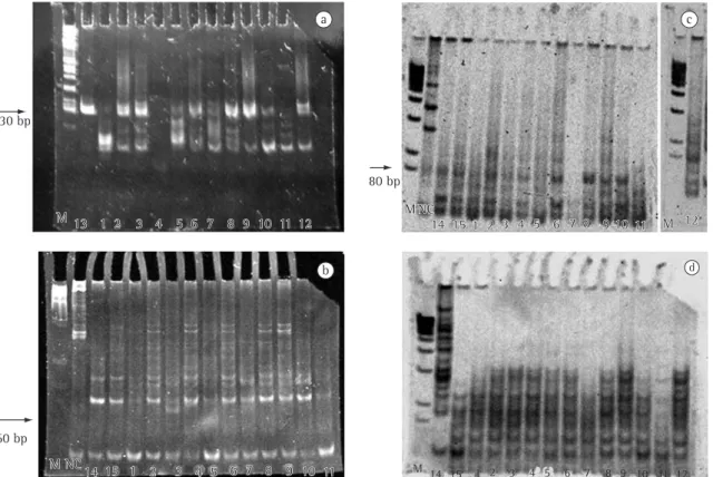
of 130 bp (predicted size range, 100-150 bp) indicating the presence of a predominant clonal lymphocyte population in high-grade T lymphoma (Case 13); LIP (Cases 2, 8, and 9); pseudolymphoma (Case 3); lymphomatoid granulomatosis (Case 6); and Hodgkin’s disease (Case 12). The cases exhib-iting more than one evident band were considered polyclonal, whereas bands lower than 75 bp were assumed to be nonspecific (Figure 2a).
TCR β-chain gene rearrangement analysis using the Dβ1/Jβ2 primer pair showed a band of 60 bp (predicted size range, 55-100 bp) in pseudolym-phoma (Case 3) (Figure 2b), probably indicating a monoclonal amplification.
A band of 80 bp (predicted size range, 70-110 bp) was considered as evidence of amplification of a monoclonal population for the Vγ11/Jγ12 primer pair in high-grade T lymphoma (Cases 14 and 15); LIP (Cases 2, 8, and 10); lymphomatoid granulo-matosis (Case 6); and Hodgkin’s disease (Case 12) (Figure 2c). In contrast, no bands, and therefore no monoclonal amplification, were visualized using the Vγ101/Jγ12 primer pair (Figure 2d).
Table 1 depicts clinical data. The median age of the patients was 40 years (range, 2-81 years). There were 10 females and 5 males. Patients younger than 40 years presented greater CD15 expression in lymphomas (p = 0.04). All male patients presented VY11 expression, whereas 8 of the 10 female patients did not express VY11 (p = 0.007). No differences were detected in terms of immunophenotyping, gene rearrangement, gender, or age.
Table 2 depicts the results regarding tumor staining intensity for CD3, CD20, CD15, CD30, CD45, and CD68 immunophenotyping stratified by diagnosis (LIP or lymphoma). All 8 cases of LIP presented moderate to strong immunostaining for CD3, whereas only 2 cases of lymphomas and 1 case of pseudolymphoma presented similar immunostaining, and this difference was statis-tically significant (p = 0.02). All 8 cases of LIP also presented CD20 expression, as did 6 cases of lymphoma and 1 case of pseudolymphoma, with no differences in terms of the staining intensity. Similarly, no differences were observed for CD15, CD30, CD45, or CD68.
Table 2 depicts the results regarding gene rear-rangement detection using the VH/JH, Dβ1/Jβ2, Vγ11/Jγ12, and Vγ101/Jγ12 primer pairs stratified by diagnosis (LIP or lymphoma). Gene rearrange-5-min period at 73 °C to ensure complete extension
and annealing of the PCR products.
Aliquots of 30 µL were then analyzed by electro-phoresis on a 10% polyacrylamide gel stained with ethidium bromide and photographed under ultra-violet light. Bands of relevant size were identified by comparison with a molecular weight marker (1 kb DNA ladder; Invitrogen Carlsbad, CA, USA).(10)
All necessary precautions were taken in order to prevent contamination.
In order to identify any correlations among immunohistochemical findings, PCR results, and clinicaldata, the chi-square test, Fisher’s exact test, and the Student’s t-test were used. Survival curves were created using theKaplan-Meier method, and the statistical significance of differenceswas calcu-lated using the log-rank test, with p < 0.05 indicating a significant difference, and the Statistical Package for Social Science (version 10.0 for Windows; SPSS Inc., Chicago, IL, USA).
Results
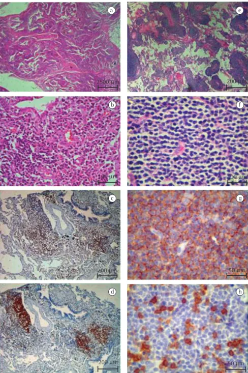
Figure 1 shows the lymphocytic infiltrates in different histological patterns of lymphoproliferative pulmonary disorders seen after H&E and immuno-histochemical staining. In the cases of LIP (Figures 1a to 1d), there was marked lymphocytic infiltra-tion with extensive involvement of the alveolar septa (Figures 1a and 1b). The lymphocytic infiltrate consisted mostly of lymphocytes (B and T cells) with varying numbers of plasma cells. The T lymphocytes (CD3 cells) were seen predominantly in the alveolar septal interstitium (Figure 1c), whereas B lymphocytes (CD20 cells) were primarily found in aggregates within lymphoid follicles and focally in thickening alveolar walls (Figure 1d). In contrast, the cases of low-grade lymphoma (Figures 1e to 1h) presented diffuse, dense, and monomorphous infiltration of small lymphoid cells with irregular nuclear contours (Figures 1e and 1f) remodeling the lung architecture. The neoplastic cells were CD20-positive monoclonal B cells (Figure 1g), with a background population of variable reactive CD3-positive T-cells (Figure 1h).
The molecular biology results are listed in Table 2, which includes the total number of cases and controls, as well as the presence of mono-clonality for the genes studied.
2500 µm 200 µm
100 µm 100 µm
200 µm 50 µm
200 µm 50 µm
a e
b f
c g
d h
A significant positive association was found between the intensity of CD20 and CD68 expression and VH rearrangement (p = 0.01 and p = 0.002, respectively).
The relationships among clinical data, lymphoid/ lymphoproliferative disorders, and overall survival were examined using the Kaplan-Meier method and the log-rank test prior to and after the gene rear-rangement. No age- or gender-related differences were found. Prior to the gene rearrangement, the mean survival was 48 months for the 8 patients with ment was detected in 4 of the 8 cases of LIP, which
changed the diagnosis from LIP to lymphoma, thus showing the importance of gene rearrangement detection in cases of LIP. In this situation, gene rearrangement using the VH/JH and Vγ11/Jγ12 primer pairs was detected in 3 and 1 cases of LIP, respectively, and no gene abnormalities were found using the Dβ1/Jβ2 and Vγ101/Jγ12 primer pairs in any of the cases.
Table 3 depicts the associations between immunophenotyping and gene rearrangement.
b
M NC14 15 1 2 3
4 5 6 7 8 9 10 11
M NC14 15 1 2 3
4 5 6 7 8 9 10 11 130 bp
a
M 13 1 2 3 4 5 6 7 8 9 10 11 12
M 13 1 2 3 4 5 6 7 8 9 10 11 12
60 bp
M NC
14 15 1 2 3 4 5 6 7 8 9 10 11 M 12
M M NC
14 15 1 2 3 4 5 6 7 8 9 10 11 M 12 c
d
M 14 15 1 2 3 4 5 6 7 8 9 10 11 12 80 bp
tration of small lymphoid cells in lymphomas. All 8 cases of LIP presented significant moderate-to-strong CD3 immunostaining, whereas only 2 cases of lymphoma and 1 case of pseudolymphoma had similar immunostaining. In contrast, CD20, CD15, CD30, CD45, and CD68 immunostaining was seen in nearly all cases. Although CD20, CD45, and CD68 antibodies are routinely employed to determine clonality in different types of lymphoproliferative disorders, our findings demonstrated that only CD3 was specifically related to cases of LIP. This finding suggests that the differential diagnosis between LIP and lymphomas is difficult due to the (not always detectable in routine practice) presence of clonal B-cell or T-cell populations in lymphocytes. There have been a few reports in the literature of clonal B-lymphocyte or T-lymphocyte populations in the pulmonary tissue of patients with B-cell or T-cell pulmonary lymphoma.(11,22,24,29) In this context, the
PCR technique is promising.
In fact, by employing the PCR technique to detect complete IgH rearrangements and analyzing TCR β- and TCR γ-chain using two different primer combinations such as Dβ1/Jβ2 and Vγ11/ Vγ101/Jγ12, we found that the VH/JH primer pair indicated the presence of a predominant clonal lymphocyte population in high-grade T lymphoma LIP and 46 months for the 7 patients with lymphoma
and pseudolymphoma. After the gene rearrange-ment, although not statistically significant, the mean survival for the patients with LIP was 58 months, compared with 30 months for patients with lymphoma (log-rank = 2.64; p = 0.10). Prior to the gene rearrangement, 4 patients with LIP died quickly, whereas only one patient with LIP died after the gene rearrangement. This difference did not achieve statis-tical significance (log-rank = 0.20; p = 0.65).
Discussion
The position of LIP within classification systems has changed with advances in the understanding of the nature of pulmonary lymphocytic infil-trates, and many groups prefer to classify LIP under the heading of pulmonary lymphoproliferative disorders.(27) A diagnosis of pulmonary
lymphopro-liferative disorders can be evoked in many clinical conditions, such as LIP, pseudolymphoma, primary lymphoma, and pulmonary localization of an other-wise extended NHL.
In our study, we found that LIP was characterized by marked lymphocytic infiltration with extensive involvement of the alveolar septa, similar to that described by other authors in 1969,(28) contrasting
with the diffuse, dense and monomorphous
infil-Table 2 - Lymphocyte immunophenotyping and gene rearrangement by tumor staining degree.
Diagnosis Imunohistochemistry Primers
Initial/Final CD3 CD15 CD20 CD30 CD45 CD68 VH/JH Dβ1/Jβ2 Vγ11/Jγ12 Vγ101/Jγ12
LIP/LIP 2 NP 1 NP NP NP 0 0 0 0
LIP/LGB 3 0 2 0 2 2 1 0 1 0
PSLY/LGB 4 0 2 0 4 3 1 1 0 0
LIP/LIP 2 3 1 1 2 1 0 0 0 0
LIP/LIP 3 NP 2 NP NP NP 0 0 0 0
LYG/LGB 1 2 2 0 3 3 1 0 1 0
LGBCL/LGBCL 1 0 1 0 2 2 0 0 0 0
LIP/LGB 3 0 2 0 3 1 1 0 1 0
LIP/LGB 3 0 2 0 3 1 1 0 0 0
LIP/LGT 2 1 1 1 0 2 0 0 1 0
LIP/LIP 2 0 1 0 0 0 0 0 0 0
HD/HD 0 2 0 3 2 1 1 0 1 0
LGB/LGB 1 0 4 0 4 1 1 0 0 0
HGT/HGT 3 NP 0 1 NP NP 0 0 1 0
HGT/HGT 4 0 0 1 0 0 0 0 1 0
contributing to better identification/classification of lymphoma cases. In fact, we found a significant positive association between immunohistochem-istry and the PCR technique (Table 3). In addition, our study showed that immunohistochemistry and molecular analysis can separate reactive and neoplastic infiltrates, and that 50% of the cases of LIP were actually found to be malignant transfor-mations, including Hodgkin’s disease (a variant of lymphocytic predominance). We also confirmed that pseudolymphoma actually represents a low-grade lymphoma. These findings call for modifications in the therapeutic protocol employed for this subgroup of patients. However, this might not occur in all cases of LIP, suggesting that some cases of LIP meet the criteria for benign status and should be treated only with steroids, whereas cases of LIP with gene rear-rangement detection should be seen as lymphoma cases and treated with chemotherapy.
In this study, we emphasized the diagnosis of LIP using immunohistochemistry and PCR analysis of the IgH gene rearrangement. This study is very important for oncologists, pulmonologists, and pulmonary pathologists because it allows the estab-lishment of the correct diagnosis of lymphoma and the introduction of the appropriate treatment for (Case 13), LIP (Cases 2, 8 and 9), pseudolymphoma
(Case 3), lymphomatoid granulomatosis (Case 6), and Hodgkin’s disease (Case 12). The analysis of TCR β-chain gene rearrangement using the Dβ1/ Jβ2 primer pair identified monoclonal amplification in pseudolymphoma (Case 3). Amplification of a monoclonal population using the Vγ11/Jγ12 primer pair was found in high-grade T lymphomas (cases 14 and 15); LIP (Cases 2, 8, and 10); lymphoma-toid granulomatosis (Case 6); and Hodgkin’s disease (Case 12). These results suggest that VH/JH and Vγ11/Jγ12 are the best combinations for detecting gene rearrangement in cases of lymphoma that present a histological pattern consistent with LIP.
Our findings underscore the suggestion that, since many cases of LIP evolve to lymphoma, LIP should be considered preneoplastic or seen as a true lymphoma if any gene rearrangement is detected. In fact, our study showed that 4 of the 8 cases of LIP were cases of low-grade B-cell or T-cell lymphoma and cases of Hodgkin’s disease from the outset.
We also found that it is difficult to draw the distinctions among idiopathic LIP, lymphoma, and Hodgkin’s disease based solely on the routine anal-ysis of histological sections,(30) which should not be
undervalued but rather should be complemented with immunophenotyping and PCR analysis, thus
Table 3 - Correlations between immunophenotyping and gene rearrangement. Lymphocyte
immunophenotyping
Gene rearrangement
VH/JH Dβ1/Jβ2 Vγ11/Jγ12 Vγ101/Jγ12
CD3 Correlation
coefficient
−0.048 0.415 0.064 0.0
(two-tailed) 0.865 0.124 0.821 0.0
CD15 Correlation
coefficient
−0.117 −0.208 0.259 0.0
(two-tailed) 0.718 0.516 0.416 0.0
CD20 Correlation
coefficient
0.605 0.229 −0.311 0.0
(two-tailed) 0.017a 0.412 0.260 0.0
CD30 Correlation
coefficient
−0.431 −0.224 0.431 0.0
(two-tailed) 0.142 0.462 0.142 0.0
CD45 Correlation
coefficient
0.786 0.452 −0.300 0.0
(two-tailed) 0.002b 0.140 0.344 0.0
CD68 Correlation
coefficient
0.334 0.458 0.101 0.0
(two-tailed) 0.289 0.134 0.754 0.0
the Laboratory of Immunohistochemistry and Histotechnology, for their technical assistance.
References
1. Nicholson AG. Lymphocytic interstitial pneumonia and other lymphoproliferative disorders in the lung. Semin Respir Crit Care Med. 2001;22(4):409-22.
2. Nicholson AG, Wotherspoon AC, Diss TC, Hansell DM, Du Bois R, Sheppard MN, et al. Reactive pulmonary lymphoid disorders. Histopathology. 1995;26(5):405-12.
3. Fishback N, Koss M. Update on lymphoid interstitial pneumonitis. Curr Opin Pulm Med. 1996;2(5):429-33. 4. Swigris JJ, Berry GJ, Raffin TA, Kuschner WG. Lymphoid
interstitial pneumonia: a narrative review. Chest. 2002;122(6):2150-64.
5. Habermann TM, Ryu JH, Inwards DJ, Kurtin PJ. Primary pulmonary lymphoma. Semin Oncol. 1999;26(3):307-15. 6. Koss MN, Hochholzer L, Nichols PW, Wehunt WD,
Lazarus AA. Primary non-Hodgkin’s lymphoma and pseudolymphoma of lung: a study of 161 patients. Hum Pathol. 1983;14(12):1024-38.
7. Teruya-Feldstein J, Temeck BK, Sloas MM, Kingma DW, Raffeld M, Pass HI, et al. Pulmonary lymphoma of mucosa-associated lymphoid tissue (MALT) arising in a pediatric HIV positive patient. Am J Surg Pathol. 1995;19(3):357-63. 8. Mazur G, Halo A, Wróbel T, Kuliczkowski K. Macrophage/
histiocytic antigen CD68 expression in neoplastic and reactive lymph nodes. Rocz Akad Med Bialymst. 2004;49 Suppl 1:73-5.
9. Carbone A, Gloghini A, Volpe R. Immunohistochemistry of Hodgkin and non-Hodgkin lymphomas with emphasis on the diagnostic significance of the BNH9 antibody reactivity with anaplastic large cell (CD30 positive) lymphomas. Cancer. 1992;70(11):2691-8.
10. Lobo A, Okhravi N, Adamson P, Clark BJ, Lightman S. Protocol for the use of polymerase chain reaction in the detection of intraocular large B-cell lymphoma in ocular samples. J Mol Diagn. 2007;9(1):113-21.
11. Betsuyaku T, Munakata M, Yamaguchi E, Ohe S, Hizawa N, Sukoh N, et al. Establishing diagnosis of pulmonary malignant lymphoma by gene rearrangement analysis of lymphocytes in bronchoalveolar lavage fluid. Am J Respir Crit Care Med. 1994;149(2 Pt 1):526-9.
12. Cleary ML, Chao J, Warnke R, Sklar J. Immunoglobulin gene rearrangement as a diagnostic criterion of B-cell lymphoma. Proc Natl Acad Sci U S A. 1984;81(2):593-7.
13. Derksen PW, Langerak AW, Kerkhof E, Wolvers-Tettero IL, Boor PP, Mulder AH, et al. Comparison of different polymerase chain reaction-based approaches for clonality assessment of immunoglobulin heavy-chain gene rearrangements in B-cell neoplasia. Mod Pathol. 1999;12(8):794-805.
14. McCarthy KP, Sloane JP, Kabarowski JH, Matutes E, Wiedemann LM. A simplified method of detection of clonal rearrangements of the T-cell receptor-gamma chain gene. Diagn Mol Pathol. 1992;1(3):173-9.
15. Slack DN, McCarthy KP, Wiedemann LM, Sloane JP. Evaluation of sensitivity, specificity, and reproducibility of an optimized method for detecting clonal rearrangements of immunoglobulin and T-cell receptor genes in formalin-fixed, paraffin-embedded sections. Diagn Mol Pathol. 1993;2(4):223-32.
the disease, as well as, perhaps, a better chance of survival for these patients.
Since LIP and primary lymphomas of the lung are rare, accounting for less than 1% of all lung pathologies in most studies, the small size of our study sample was to be expected. However, the clinical impact of our findings was investigated. No differences were detected in terms of immunophe-notyping, gene rearrangement, gender, or age. We also examined survival curves prior to and after the gene rearrangement. The differences did not achieve statistical significance, probably due to the limited number of patients and to the fact that the clinicians were unaware of the fact that some of the cases of LIP were lymphomas. The diagnosis of lymphoma in some of the cases of LIP was made only after the gene rearrangement. The analysis of the survival curves prior to and after the gene rearrangement revealed that 4 LIP patients died quickly prior, whereas only 1 LIP patient died after, confirming that the more aggressive stage of the disease occurs prior to the gene rearrangement.
Regardless of the lymphocytic/lymphopro-liferative pathogenetic mechanism, detection of monoclonal B and T cells by immunophenotyping and PCR had an impact on the diagnosis of pulmo-nary lymphomas in patients previously diagnosed with LIP. Therefore, immunophenotyping and PCR should be used as the ‘gold standard’ techniques in routine practice. Interpretable PCR results were obtained in the majority of the cases analyzed, demonstrating that our PCR analysis could become a routine procedure. The detection of gene rear-rangement in lung biopsies, especially in cases diagnosed as LIP, is very important for the estab-lishment of an accurate diagnosis of lymphoma. In order to determine whether or not these cases represent malignant transformation from LIP, rand-omized, prospective trials involving large patient samples are needed. In addition, we believe that further studies of lymphoid/lymphoproliferative disorders are warranted in order to validate the results of our immunophenotyping and gene rear-rangement analysis.
Acknowledgments
24. American Thoracic Society; European Respiratory Society. American Thoracic Society/European Respiratory Society International Multidisciplinary Consensus Classification of the Idiopathic Interstitial Pneumonias. This joint statement of the American Thoracic Society (ATS), and the European Respiratory Society (ERS) was adopted by the ATS board of directors, June 2001 and by the ERS Executive Committee, June 2001. Am J Respir Crit Care Med. 2002;165(2):277-304.
25. Bagg A, Braziel RM, Arber DA, Bijwaard KE, Chu AY. Immunoglobulin heavy chain gene analysis in lymphomas: a multi-center study demonstrating the heterogeneity of performance of polymerase chain reaction assays. J Mol Diagn. 2002;4(2):81-9.
26. Inghirami G, Szabolcs MJ, Yee HT, Corradini P, Cesarman E, Knowles DM. Detection of immunoglobulin gene rearrangement of B cell non-Hodgkin’s lymphomas and leukemias in fresh, unfixed and formalin-fixed, paraffin-embedded tissue by polymerase chain reaction. Lab Invest. 1993;68(6):746-57.
27. Epstein DM, Glickstein MF. Pulmonary lymphoproliferative disorders. Radiol Clin North Am. 1989;27(6):1077-84. 28. Liebow AA, Carrington CB. The interstitial pneumonias. In:
Simon M, Potchen EJ, Le May MJ, Fleischner FG, editors. Frontiers of Pulmonary Radiology. New York: Grune and Stratton Inc, 1969. p. 102-41.
29. Elenitoba-Johnson K, Medeiros LJ, Khorsand J, King TC. Lymphoma of the mucosa-associated lymphoid tissue of the lung. A multifocal case of common clonal origin. Am J Clin Pathol. 1995;103(3):341-5. Erratum in: Am J Clin Pathol. 1995;103(6):773.
30. Szalay F, Szathmári M, Pálóczi K, Földi J, Demeter J. Immunologic and molecular biologic characterization of pleural involvement in a case of T-chronic lymphocytic leukemia. Chest. 1994;106(4):1283-5.
16. Aubin J, Davi F, Nguyen-Salomon F, Leboeuf D, Debert C, Taher M, et al. Description of a novel FR1 IgH PCR strategy and its comparison with three other strategies for the detection of clonality in B cell malignancies. Leukemia. 1995;9(3):471-9.
17. Trainor KJ, Brisco MJ, Story CJ, Morley AA. Monoclonality in B-lymphoproliferative disorders detected at the DNA level. Blood. 1990;75(11):2220-2.
18. Savio A, Franzin G, Wotherspoon AC, Zamboni G, Negrini R, Buffoli F, et al. Diagnosis and posttreatment follow-up of Helicobacter pylori-positive gastric lymphoma of mucosa-associated lymphoid tissue: histology, polymerase chain reaction, or both? Blood. 1996;87(4):1255-60.
19. Kurosu K, Yumoto N, Mikata A, Taniguchi M, Kuriyama T. Monoclonality of B-cell lineage in primary pulmonary lymphoma demonstrated by immunoglobulin heavy chain gene sequence analysis of histologically non-definitive transbronchial biopsy specimens. J Pathol. 1996;178(3):316-22.
20. Nicholson AG, Wotherspoon AC, Diss TC, Butcher DN, Sheppard MN, Isaacson PG, et al. Pulmonary B-cell non-Hodgkin’s lymphomas. The value of immunohistochemistry and gene analysis in diagnosis. Histopathology. 1995;26(5):395-403. 21. Philippe B, Delfau-Larue MH, Epardeau B, Autran B, Clauvel
JP, Farcet JP, et al. B-cell pulmonary lymphoma: gene rearrangement analysis of bronchoalveolar lymphocytes by polymerase chain reaction. Chest. 1999;115(5):1242-7. 22. Subramanian D, Albrecht S, Gonzalez JM, Cagle PT. Primary
pulmonary lymphoma. Diagnosis by immunoglobulin gene rearrangement study using a novel polymerase chain reaction technique. Am Rev Respir Dis. 1993;148(1):222-6.