* Study carried out in the Department of Thoracic Surgery of the São Lucas Hospital. Pontifícia Universidade Católica do Rio Grande do Sul – PUCRS, Pontifical Catholic University of Rio Grande do Sul – Porto Alegre (RS) Brazil.
1. Resident in the Department of Thoracic Surgery of the São Lucas Hospital. Pontifícia Universidade Católica do Rio Grande do Sul – PUCRS, Pontifical Catholic University of Rio Grande do Sul – Porto Alegre (RS) Brazil.
2. Thoracic Surgeon at the São Lucas Hospital. Pontifícia Universidade Católica do Rio Grande do Sul – PUCRS, Pontifical Catholic University of Rio Grande do Sul – Porto Alegre (RS) Brazil.
3. Fellow in the Department of Thoracic Surgery. University of Toronto, Toronto, Ontario, Canada.
4. Adjunct Professor in the Department of Thoracic Surgery. Pontifícia Universidade Católica do Rio Grande do Sul – PUCRS, Pontifical Catholic University of Rio Grande do Sul – School of Medicine, Porto Alegre (RS) Brazil.
5. Full Professor in the Department of Thoracic Surgery. Pontifícia Universidade Católica do Rio Grande do Sul – PUCRS, Pontifical Catholic University of Rio Grande do Sul – School of Medicine, Porto Alegre (RS) Brazil.
Correspondence to: Pedro Augusto Reck dos Santos. Rua Sofia Veloso, 104, apto 903, Cidade Baixa, CEP 90040-150, Porto Alegre, RS, Brasil. Tel 55 51 3407-4120. E-mail: [email protected]
Submitted: 1 December 2006. Accepted, after review: 3 April 2007.
Concordance between cl
i
n
i
cal and patholog
i
cal stag
i
ng
i
n pat
i
ents w
i
th stages
I
or
II
non-small cell lung
cancer subjected to surg
i
cal treatment*
Pedro Augusto Reck dos Santos1, Rodrigo Sponchiado da Rocha1,
Maurício Pipkin1, Marner Lopes da Silveira2, Marcelo Cypel3,
Jayme Oliveira Rios4, José Antonio Lopes de Figueiredo Pinto5
Abstract
Objective: To compare clinical and pathological staging in patients with non-small cell lung cancer submitted to surgical treatment, as well
as to identify the causes of discordance. Methods: Data related to patients treated at the Department of Thoracic Surgery of the Pontifical
Catholic University of Rio Grande do Sul São Lucas Hospital were analyzed retrospectively. Sensitivity, specificity, positive predictive value,
negative predictive value, and accuracy were calculated for clinical stages IA, IB, and IIB. The kappa index was used to determine the
concordance between clinical and pathological staging. Results: Of the 92 patients studied, 33.7% were classified as clinical stage IA, 50%
as IB, and 16.3% as IIB. The concordance between clinical and pathological staging was 67.5% for stage IA, 54.3% for IB, and 66.6% for IIB. The accuracy of the clinical staging was greater for stage IA, and a kappa of 0.74, in this case, confirmed a substantial association with
pathological staging. The difficulty in evaluating nodal metastatic disease is responsible for the low concordance in patients with clinical
stage IB. Conclusions: The concordance between clinical and pathological staging is low, and patients are frequently understaged (in the
present study, only one case was overstaged). Strategies are necessary to improve clinical staging and, consequently, the treatment and
prognosis of patients with non-small cell lung cancer.
neoplastic disease and for whom surgery is not indicated.
The objective of this study was to determine
the concordance between CS and PS, in addition to identifying the causal factors that lead to di
scord-ance between them.
Methods
Between January of 2002 and January of 2006, 125 patients, all of whom were diagnosed with
NSCLC via fiberoptic bronchoscopy, percutaneous
puncture, thoracoscopy, or intraoperative biopsy,
underwent surgical treatment in the Department
of Thoracic Surgery of the Pontifícia Universidade
Católica do Rio Grande do Sul (PUCRS, Pontifical
Catholic University of Rio Grande do Sul) São
Lucas Hospital. Those patients were clinically
staged by computed tomography (CT) of the chest and abdomen, CT of the skull (in the presence of
neurological symptoms), or bone scintigraphy (in
the presence of related symptoms). Cervical med
i-astinoscopy was the method used to evaluate the
paratracheal and subcarinal lymph nodes. The
patients with tumors located in the left upper lobe
underwent parasternal mediastinotomy for the
evaluation of the subaortic and aortopulmonary
window stations. Thoracoscopy was used for the investigation of pleural effusion.
All patients underwent thoracotomy with
standard resection for NSCLC, according to resp
i-ratory function, with systematic lymph node
dissection (paratracheal, subcarinal, and hilar lymph
nodes on the right side; and subaortic,
aortopul-monary window, subcarinal, and hilar lymph nodes
on the left side). The postoperative
anatomopatho-logical analysis revealed a particular PS, which was
then compared to the CS in order to identify where
there was discordance.
There were 92 patients with CS I or II, data
collection being performed retrospectively. Patients
with incomplete CS or PS, those with histology other
than NSCLC, those with CS III or IV submitted to
surgical treatment, and those submitted to
neoadju-vant therapy were excluded. This study was approved
by the Ethics in Human Research Committee of the
PUCRS São Lucas Hospital.
The data related to the patients were analyzed
using Microsoft Office Excel 2003, and the stati
s-tical analysis was performed using the R for
Introduct
i
on
Lung cancer is the most common cause of death
from malignant neoplasia, in males and females
alike. In addition, it accounts for 28% of all cancer
deaths, and only breast cancer has a higher inc
i-dence. There are approximately 180,000 new cases
of pulmonary neoplasia in the United States each
year.(1)
The prognosis of patients with pulmonary
neoplasia depends on early diagnosis and
accu-rate oncologic staging. The type of treatment used
differs according to the time (stage) at which the
neoplasia is diagnosed. Therefore, it is important
to identify subgroups of patients with the same
prognosis and define treatment strategies for each
subgroup.
The current staging system for non-small cell
lung cancer (NSCLC) is tumor-node-metastasis
(TNM) staging,(2) which provides only a definition
of the anatomic extent of the neoplasia: size,
loca-tion, and extent of the primary lesion; presence and
location of the locoregional lymph node
metas-tasis; and systemic metastatic disease. This system is recognized as having significant limitations, such
as that of not considering clinical aspects (including
the general status of the patient, weight loss,
func-tional capacity, and the histologic subtype of the
tumor) or the influence of prognostic factors that
are primarily biological (such as the mutation in the
p53 gene).
From this results a special problem,
under-staging, which is caused by the limitations in the
sensitivity of the various diagnostic methods used in the preoperative staging. The staging obtained
after all tests, invasive or not, that are performed
prior to pulmonary resection is designated clinical
staging (CS). For patients undergoing surgery, CS is
confirmed or reviewed according to the operative
findings. Pathological staging (PS) is determined by
the analysis of the surgical sample, and this result
plays an important role in the subsequent
thera-peutic decisions.
Disparities between CS and PS occur frequently,
thus compromising the design of clinical studies
and, more importantly, the individual treatment
of patients, since inadequate staging can result in losing the benefit of the neoadjuvant therapy
left upper lobe in 32.6% of the patients and in the
right upper lobe in 27.5% of the patients.
With regard to CS, 33.7% of the patients were
classified as IA, 50% as IB, and 16.3% as IIB
(Table 2). All patients classified as IIB were so
clas-sified because tomographic findings led to the
suspicion of thoracic wall or parietal pleura i
nva-sion, since there were no patients with CS IIB
- T2N1M0.
In relation to CS IA, the concordance between
CS and PS was 67.5%. There were 4 patients who
migrated to PS IB due to visceral pleura invasion,
1 who migrated to IIA due to the presence of a
metastatic hilar lymph node, 1 who migrated to IIB
due to parietal pleura invasion, 2 who migrated to IIIA due to the presence of metastatic disease in ipsilateral mediastinal lymph node, 1 who migrated
to IIIB because there was another lesion in the same
lobe, and 1 who migrated to IV due to the presence
of a lesion in another lobe. In the group of patients
classified as CS IB, in which most of the changes
occurred, the concordance was 54.3%, and there were 12 patients who migrated to PS IIB, 9 due to
the presence of pathological N1 lymph nodes and the
remaining 3 due to previously undetected parietal
pleura invasion. However, there were 9 patients who
migrated to PS III, 8 to stage IIIA because there was
metastasis in N2 ganglia and 1 to IIIB due to the
presence of another lesion in the same lobe. In the
group of patients classified as CS IIB, the
concord-ance was 66.6%, and there was 1 patient who
migrated to PS IB since no parietal pleura invasion
was found. Of the remaining patients, 2 migrated
to IIIA (neoplasia with costal invasion and presence
of a metastatic N1 lymph node), 1 migrated to IIIB,
again due to the presence of another lesion in the
lobe resected, and 1 migrated to IV, also due to the
presence of another lesion, albeit in another lobe
(Table 2 and Figure 1).
A classification of CS IA had a sensitivity of
100%, specificity of 85%, positive predictive value
of 67%, negative predictive value of 100%, and a
quite high accuracy (89%). The power of
concord-ance, calculated using the kappa index, was 0.74.
Sensitivity for stage IB was 98%, with a low
specificity of 35%, positive predictive value of 66%,
and negative predictive value of 93%. Accuracy was
70%, and the kappa index was 0.33.
Finally, the patients with CS IIB presented
low sensitivity (43%) and high specificity (92%).
Windows software.(3) Sensitivity, specificity, pos
i-tive predictive value, negative predictive value, and
accuracy were calculated for CS IA (T1N0M0), CS IB (T2N0M0), and CS IIB (T3N0M0). None of the
patients with CS IIB were found to present T2N1M0
preoperative staging. Cohen’s kappa index was used
to determine the power of concordance between CS
and PS in the stages studied.(4)
Results
Of the 92 patients studied, 65 (70.6%) were
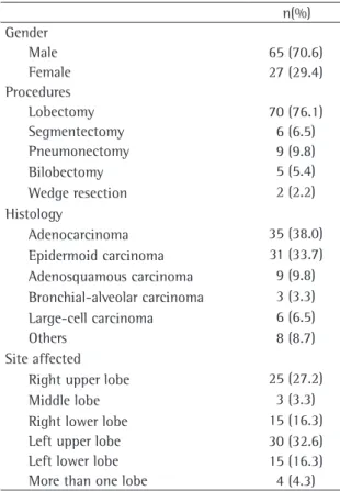
male and 27 (29.4%) were female. The mean age was 63.2 years (median, 67 years). Table 1 shows
that pulmonary lobectomy (76.1%) was the surgical
procedure most commonly used, followed by
pneumonectomy, conservative segmentectomy,
bilobectomy, and wedge resection. The predom
i-nant histological type was adenocarcinoma (in 38%
of the cases), followed by epidermoid carcinoma (in
33.7%). Most of the neoplasms were located in the
Table 1 - Characteristics of the patients.
n(%) Gender
Male 65 (70.6)
Female 27 (29.4)
Procedures
Lobectomy 70 (76.1)
Segmentectomy 6 (6.5)
Pneumonectomy 9 (9.8)
Bilobectomy 5 (5.4)
Wedge resection 2 (2.2)
Histology
Adenocarcinoma 35 (38.0)
Epidermoid carcinoma 31 (33.7)
Adenosquamous carcinoma 9 (9.8)
Bronchial-alveolar carcinoma 3 (3.3)
Large-cell carcinoma 6 (6.5)
Others 8 (8.7)
Site affected
Right upper lobe 25 (27.2)
Middle lobe 3 (3.3)
Right lower lobe 15 (16.3)
Left upper lobe 30 (32.6)
Left lower lobe 15 (16.3)
to mediastinal lymph node sampling or dissection
during surgery, and this compromised the PS. Most
patients received surgical treatment only, and it is important to emphasize that, in stages II, III, and IV, no adjuvant treatment was used in 61, 38, and
37%, respectively. It becomes evident that there is
lack of consensus on this point.
In our study, 32.2% of the patients with CS IA
were reclassified as being in more advanced stages
when the PS was analyzed. It is important to hi
gh-light that 6.4% of the patients were reclassified
as PS II, and that, despite having undergone the
standard treatment for the stage, those patients
clearly have a worse prognosis, as has been
demon-strated by other researchers.(10) However, 12.9% of
the patients were found to be in PS III or IV and, in this context, lost the benefits of the multimodal
treatment or had no indication for surgery at any
time during treatment. Patients with small (T1)
tumors are hardly ever understaged, and, when this
occurs, the tumors are usually reclassified as T2 due
to undiagnosed visceral pleura invasion in the CS.
With regard to stage IB, the concordance
between CS and PS was slightly lower (approximately
54%). In this group, 26% of the patients migrated
to PS IIB, the main reason for this being the
pres-ence of hilar lymph nodes affected by the disease,
whereas 19.5% of the patients migrated to stages in
which surgery was not the first therapeutic option.
Approximately 43% of the T2 patients were
reclas-sified as being in more advanced stages because
they presented lymph node metastasis in N1 or N2
stations. We observe, therefore, a deficiency in the
evaluation of nodal metastatic disease during CS
using the methods currently available.
The reason why 16.3% of the patients
evalu-ated were classified as CS IIB was the suspicion of
thoracic wall or parietal pleura invasion, and the
Positive predictive value was 66%, and negative
predictive value was 83%, with an accuracy of
80%. Concordance, using the kappa index, was 0.41
(Figure 2).
D
i
scuss
i
on
The PS is the most important factor in the
prog-nostic evaluation of patients with NSCLC submitted
to surgical treatment. As some studies have shown,
the benefits of the neoadjuvant treatment, especially in relation to the increased survival of patients with
stage IIIA, the preoperative identification of this
subgroup of patients has acquired greater signif
i-cance.(5,6)
The importance of the preoperative identifi
ca-tion of those patients who, in the PS, proved to
belong to stage IV should also be stressed, since
those patients did not benefit, at any time, from
the surgical procedure they were submitted to.(7) In
this sense, studies that determine the concordance
between CS and PS are of utmost importance.
However, a clinical pathological comparison is
only valid for T and N, since thoracotomy, with the
subsequent analysis of the surgical sample, does
not typically alter the M factor. In general, what is
found is that this concordance deteriorates as T and
N increase. Even with an appropriate CS, the rate
of exploratory thoracotomy or incomplete resection
ultimately ranges from 8 to 10%.(8)
This objective has been given considerable
weight, as have and the refinement of CS and PS.
However, one group of authors,(9) in a series of
11,668 patients submitted to surgical treatment for
NSCLC, found that only 27% had been submitted
to cervical mediastinoscopy and that, of this group,
only 46% had undergone lymph node biopsy. In add
i-tion, only 60% of the patients had been submitted
Table 2 - Concordance between clinical and pathological staging.
Clinical staging Pathological staging % of total Probability CS = PS
IA IB IIA IIB IIIA IIIB IV Total
IA 21 4 1 1 2 1 1 31 33.7 67.54
IB 0 25 0 12 8 1 0 46 50.0 54.34
IIB 0 1 0 10 2 1 1 15 16.3 66.6
Total 21 30 1 23 12 3 2 92 100.0 62.82
IA, 51.6% for IB, 48.2% for IIB, and 33.3% for IIIA. In the analysis of the mediastinal lymph nodes,
26% of the patients were classified as presenting
clinical N0, when, in fact, they were patients with
pathological N2. Nevertheless, in the subgroup of
patients with pathological N2, 46.3% were found
to have been correctly staged, and, of those, only 3 had been submitted to preoperative cervical
mediastinoscopy.
Other researchers,(13) by means of a prospective
analysis of their patients using chest CT, found that
the concordance between CS and PS was 66% for stage I, 82% for stage II, and 69% for stage III.
The benefits of highly accurate staging in
defining prognosis should always be emphasized,
since the most appropriate therapy for the patient is thereby determined. In this sense, the evaluation
of the nodal extent of the NSCLC plays an i
mpor-tant role. The removal of all mediastinal lymph
nodes allows the extent of the disease to be
evalu-ated with high accuracy, as well as being associated
with lower recurrence and better survival, since it
provides the appropriate PS.(14)
In the study of nodal metastatic disease, its
presence in the hilar groups has been highlighted as
another factor with prognostic importance, showing
that the group of N1 patients is heterogeneous. The involvement of the hilar groups is associated with
a 39% five-year survival, being significantly lower if compared to the involvement of the interlobar,
lobar, and segmental groups.(15) Some authors(16)
have also demonstrated that the prognosis is better
for patients in whom the lobar N1 was resected
than for those in whom the hilar N1 was resected.
As currently performed, CS has its problems. In
general, the limitations of chest CT and those of
cervical mediastinoscopy ultimately contribute to
decreasing the accuracy of CS. The concordance
between CS and PS in the evaluation of metastatic
disease in mediastinal lymph nodes using chest CT is low, with a false positive rate of 44% and a false
negative rate of 17%.(17) Cervical mediatinoscopy,
despite having advantages over chest CT, since it
determines the presence of N1 or N3 and provides
histological diagnosis, has its range restricted to
the upper lymph node groups and does not fully approach the subcarinal station,(18) as well as having
risks and accompanying complications. Therefore,
new procedures have helped to optimize CS.
concordance between CS and PS was 66.6%, which
reflects, in our study, the precision of chest CT in identifying this impairment. Here, the loss of the
benefits of the neoadjuvant therapy or even the
absence of surgical indication occurred in 26.6% of
the cases due to the migration of patients to stages IIIA, IIIB, or IVB. There was only one patient who
was downstaged, being reclassified as IB. The kappa index shows that the concordance for CS IA (0.74)
was much better than that found for CS IB (0.33).
Typically, concordance decreases as CS increases. In
our study, the kappa for stage IIB was greater than
that for stage IB, probably because the former only
evaluates the T factor, since there were no patients
presenting CS IIB - T2N1M0.
The relatively low positive predictive values
and the relatively high negative predictive values
found corroborate the results of other studies and
demonstrate that CS typically understages patients,
rarely overstaging them. On group of authors,(11)in
a study of 2994 patients with pulmonary neoplasia
submitted to surgical treatment, found a
concord-ance of 75% for stages IA and IB and of 23.5%
for stage IIB, although, in that study, the patients
were submitted to cervical mediastinoscopy only in
the presence of lymph nodes greater than 1 cm in
diameter.
Another group of authors,(12) by means of a
retrospective analysis of 180 patients with NSCLC
submitted to surgical treatment between 1994
and 2000, in whom cervical mediastinoscopy was
performed only in the presence of lymph nodes
greater than 1 cm in diameter, found that the
concordance between CS and PS was 77% for stage
0 5 10 15 20 25 30 35 40 45 50
IA IB IIA IIB IIIA IIIB IV
EC EP
Figure 1 - Results of the comparison between clinical
the presence of mediastinal lymph node metastasis in 42% of the patients in whom chest CT findings
were normal.
Endoscopic ultrasonography, a procedure that
has been introduced in the evaluation of patients
with pulmonary neoplasia, has contributed to better
CS evaluation. It can visualize the layers of the
trachea and bronchiin detail, thereby distinguishing
neoplastic invasion from extrinsic compression, as
well as making it possible to evaluate the
rela-tionship between extraluminal pulmonary lesions
and adjacent vessels. It can also facilitate biopsy
of mediastinal and hilar lymph nodes as well as of
parenchymal lesions.(22)
Thoracoscopy has been used in order to increase
CS accuracy. One group of authors,(23) analyzing
patients with negative cervical mediastinoscopy
results whose T and N were evaluated with the aid
of thoracoscopy, through pleural lavage cytology, in
addition to biopsy of paraesophageal lymph node
groups and of the pulmonary ligament in tumors
located in lower lobes (when possible, biopsy of the interlobar or intersegmental groups was performed,
and the subcarinal and paratracheal lymph nodes
were only sampled if they had a pathological aspect),
found that thoracoscopy presented no compli
ca-tions, being considered more accurate than chest CT
since it staged 88% of the cases correctly, compared
with 42% for CT. Its greatest benefit was staging
the factor T correctly in 96% of the cases.
The factor N was staged correctly in 74% of the
cases, revealing metastatic disease in paraesophageal
and pulmonary ligament lymph nodes in patients
with negative cervical mediastinoscopy results.
Despite the limitation of thoracoscopy in relation
to the evaluation of the aortopulmonary window in patients with large lesions in the left upper lobe, it was found that it can identify malignant pleural
effusion that is too small for visualization on chest
CT, evaluate thoracic wall invasion, in addition to
sampling posterior subcarinal lymph nodes, as well
as pulmonary ligament and paraesophageal lymph
nodes, which is very important in patients with
negative cervical mediastinoscopy results.
Considering the mean difference of 35%
between CS and PS, it is very difficult to compare
a study that uses CS to one that uses PS. It is
always important to remember that each invasive
staging method has the potential to better evaluate
different lymph node stations. It is essential that
Positron emission tomography (PET) scan,
due to its capacity of detecting areas with greater
metabolic activity in the human body, in add
i-tion to helping differentiate between malignant
and benign pulmonary nodules, has also allowed
better selection of patients for surgical treatment, improving CS, since it has higher accuracy to
eval-uate mediastinal lymph nodes than does chest CT,
especially in relation to specific lymph node groups
such as those of the subaortic, aortopulmonary
window, and subcarinal stations.(19) The PET scan
does not replace mediastinal lymph node biopsy,
and surgical treatment should not be denied to any
patient based exclusively on this test. However, its
significance lies in selecting areas to be sampled,
and this often leads to the identification of lymph
node disease that was previously unsuspected.
Another testing modality that has participated in this sense is endoscopic endosonography with
fine-needle aspiration. Through the esophagus,
an echoendoscope is used to access the posterior
mediastinum and adjacent lymph nodes
(aortop-ulmonary window, subcarinal, paraesophageal,
and pulmonary ligament lymph nodes), sampling
these stations. It is a safe method of evaluating
the stations cited above, with an accuracy of 97%,
avoiding exploratory procedures in approximately
57% of the patients,(20) a percentage that is greater
than that achieved through chest CT or PET scan.
Usingh this test, one group of researchers(21) found
0 0,1 0,2 0,3 0,4 0,5 0,6 0,7 0,8 0,9 1
Sens itivi
ty
Specifi city
Positi ve pre
dicti ve v
alue
Nega tive
predi ctive
value Accu
racy Kappa
CS IA CS IB CS IIB
Figure 2 - Statistical evaluation of the clinical staging
6. Albain KS, Rusch VW, Crowley JJ, Rice TW, Turrisi AT 3rd, Weick JK, et al. Concurrent cisplatin/etoposide plus chest radiotherapy followed by surgery for stages IIIA (N2) and IIIB non-small-cell lung cancer: mature results of Southwest Oncology Group phase II study 8805. J Clin Oncol. 1995;13(8):1880-92.
7. Spira A, Ettinger DS. Multidisciplinary management of lung cancer.N Engl J Med. 2004;350(4):379-92.
8. Deslauriers J, Gregoire J. Clinical and surgical staging of non-small cell lung cancer. Chest. 2000;117(4 Suppl 1):S96-S103.
9. Little AG, Rusch VW, Bonner JA, Gaspar LE, Green MR, Webb WR, et al. Patterns of surgical care of lung cancer patients. Ann Thorac Surg. 2005;80(6):2051-6; discussion 2056. 10. Naruke T, Tsuchiya R, Kondo H, Asamura H. Prognosis and
survival after resection for bronchogenic carcinoma based on the 1997 TNM-staging classification: the Japanese experience. Ann Thorac Surg. 2001;71(6):1759-64. 11. López-Encuentra A, García-Luján R, Rivas JJ,
Rodríguez-Rodríguez J, Torres-Lanza J, Varela-Simo G, et al. Comparison between clinical and pathologic staging in 2,994 cases of lung cancer. Ann Thorac Surg. 2005;79(3):974-9; discussion 979.
12. Cetinkaya E, Turna A, Yildiz P, Dodurgali R, Bedirhan MA, Gürses A, et al. Comparison of clinical and surgical-pathologic staging of the patients with non-small cell lung carcinoma. Eur J Cardiothorac Surg. 2002;22(6):1000-5. 13. Cerfolio RJ, Bryant AS, Ojha B, Eloubeidi M. Improving the
inaccuracies of clinical staging of patients with NSCLC: a prospective trial. Ann Thorac Surg. 2005;80(4):1207-13; discussion 1213-4.
14. Moskovitz AH, Rusch VW. Resection and mediastinal lymph node dissection. Op Tech Thorac Cardiovasc Surg. 2005;10(2):166-77.
15. Tanaka F, Yanagihara K, Otake Y, Yamada T, Shoji T, Miyahara R, et al. Prognostic factors in patients with resected pathologic (p-) T1-2N1M0 non-small cell lung cancer (NSCLC). Eur J Cardiothorac Surg. 2001;19(5):555-61. 16. Yano T, Yokoyama H, Inoue T, Asoh H, Tayama K, Ichinose
Y. Surgical results and prognostic factors of pathologic N1 disease in non-small-cell carcinoma of the lung. Significance of N1 level: lobar or hilar nodes. J Thorac Cardiovasc Surg. 1994;107(6):1398-402.
17. Toloza EM, Harpole L, McCrory DC. Noninvasive staging of non-small cell lung cancer: a review of the current evidence. Chest. 2003;123(1 Suppl):S137-S46.
18. Hoffmann H. Invasive staging of lung cancer by mediastinoscopy and video-assisted thoracoscopy. Lung Cancer. 2001;34(Suppl 3):S3-S5.
19. Cerfolio RJ, Ojha B, Bryant AS, Bass CS, Bartalucci AA, Mountz JM. The role of FDG-PET scan in staging patients with nonsmall cell carcinoma. Ann Thorac Surg. 2003;76(3):861-6.
20. Eloubeidi MA, Cerfolio RJ, Chen VK, Desmond R, Syed S, Ojha B. Endoscopic ultrasound-guided fine needle aspiration of mediastinal lymph node in patients with suspected lung cancer after positron emission tomography and computed tomography scans. Ann Thorac Surg. 2005;79(1):263-8. 21. Wallace MB, Silvestri GA, Sahai AV, Hawes RH, Hoffman BJ,
Durkalski V, et al. Endoscopic ultrasound-guided fine needle aspiration for staging patients with carcinoma of the lung. Ann Thorac Surg. 2001;72(6):1861-7.
these methods be used in conjunction in order to
achieve greater concordance between CS and PS.
For ideal intrathoracic staging, we suggest the
following measures:
1) a better definition of tomographic criteria for
the presence of pathological N1 lymph nodes
and visceral pleura invasion;
2) performance of cervical mediastinoscopy in
all patients, even in those whose
preopera-tive tests show no mediastinal adenopathy,
as well as systematic evaluation of the
parat-racheal and subcarinal stations; and
3) performance of thoracoscopy also in all
patients for the evaluation of the par
i-etal and visceral pleurae, as well as for the
evaluation of the subcarinal lymph nodes,
especially when they are not accessible
through cervical mediastinoscopy, in add
i-tion to the paraesophageal group and the
pulmonary ligament.
In the present study, the concordance between CS and PS was substantial for stage IA, decreasing
for IB and IIB. The patients were understaged,
except in one case, in which the patient was
over-staged. The main source of discordance was the
difficulty in the preoperative evaluation of
meta-static lymph node involvement. It is necessary that
tomographic criteria for N1 be defined and that
staging using surgical methods be systematically indicated in order to increase CS yield. Therefore,
we expect to optimize the treatment and improve
the prognosis of patients with NSCLC.
Referênc
i
as
1. Schneider A, Schwartzmann G. Tratamento cirúrgico do carcinoma brônquico. In: Pinto Filho DR, Cardoso PFG, Figueiredo Pinto JAL, Schneider A, editors. Manual de cirurgia torácica. Rio de Janeiro: Editora Revinter, 2001. p. 245-6.
2. Mountain CF. Revisions in the International System for Staging Lung Cancer. Chest. 1997;111(6):1710-7.
3. R-project [homepage on Internet]. Vienna: The R Project for Statistical Computing, c2006. [cited 2006 Nov 09]. Available from: http://www.r-project.org
4. Fleiss LJ, Levin B, Paik MC, editors. Statistical methods for rates and proportions. 2nd ed. New York, NY: John Wiley and Sons, Inc.; 1981. p 218.
23. Roberts JR, Blum MG, Arildsen R, Drinkwater DC Jr, Christian KR, Powers TA, et al. Prospective comparison of radiologic, thoracoscopic, and pathologic staging in patients with early non-small cell lung cancer. Ann Thorac Surg. 1999;68(4):1154-8.