Haplotypes
β
s-globin and its clinical-haematological
correlation in patients with sickle-cell anemia in
Triângulo Mineiro, Minas Gerais, Brazil
Haplótipos da
β
s-globina e sua correlação clínica-hematológica em portadores
de anemia falciforme no Triângulo Mineiro, Minas Gerais, Brasil
Alexandra S. Leal1; Paulo Roberto J. Martins1, 2; Marly Aparecida S. Balarin1;Gilberto A. Pereira1; Gláucia Aparecida D. Resende1
1. Universidade Federal do Triângulo Mineiro (UFTM), Minas Gerais, Brasil. 2. Fundação Hemominas, Minas Gerais, Brasil.
First submission on 19/08/15; last submission on 20/08/15; accepted for publication on 18/09/15; published on 20/02/16
ABSTRACT
Introduction: Sickle-cell anemia (SCA) is the most severe form of sickle-cell disease, and is characterized by homozygous hemoglobin
S (α2βS2). Objective: Determine the haplotypes frequency in patients with SCA and their correlation with clinical and hematological proile. Method: We performed a retrospective descriptive study by reading the charts and a cross-sectional study for molecular analysis to determine the haplotypes of the gene pS globin in 61 patients with sickle-cell anemia (SS) by polymerase chain reaction-restriction fragment length polymorphism (PCR-RFLP), using restriction endonucleases Xmn I, Hind III, Hinf I and Hinc II for analysis of six polymorphic sites in the beta cluster. Result: The genotypes were Central African Republic (CAR)/CAR (50.8%), CAR/Benin type (BEN) (13.1%), CAR/ Cameroon type (CAM) (1.6%), CAR/atypical (ATP) (13.1%), BEN/BEN (13.1%), BEN/ATP (4.9%) and ATP/ATP (3.3%). Among the analyzed chromosomes, 64.8% were CAR type, 22.1% were BEN, 12.3% ATP and 0.8% CAM. Levels of fetal hemoglobin (HbF) were signiicantly lower in CAR/CAR than in ATP/ATP, BEN/ATP and CAR/BEN. No association was observed between the different genotypes and clinical manifestations. Conclusion: Despite the lack of association between genotypes and clinical proiles, higher frequency of clinical events was observed in patients with at least one type of CAR chromosome. A signiicant association was also observed between lower average levels of HbF and CAR/CAR genotype compared to other genotypes.
Key words: sickle-cell anemia; haplotypes; fetal hemoglobin.
INTRODUCTION
Sickle-cell anemia (SCA) is caused by homozygous
hemoglobin S (α2βS2) that derives from a point mutation in
the β-globin (βS) chain gene, located on chromosome 11, which
leads to the replacement of adenine (A) for thymine (T) at codon 6
(GAG → GTG), resulting in the switch from glutamic acid amino
acid to valine in the sixth position of the βS(1).
It is clinically characterized by chronic hemolytic anemia and repeated vaso-occlusive episodes(2). As it is extremely variable, the
severity of clinical and hematological manifestations may range from a serious illness to an almost asymptomatic condition only accidentally detected. Its hematological features, as well as clinical severity, are affected by variations in the levels of fetal hemoglobin,
simultaneous presence of α-thalassemia, deiciency of the glucose-6-phosphate dehydrogenase enzyme, haplotypes related
to the β-globin gene cluster, and endothelial dysfunction(3-5).
The set of polymorphic regions of a chromosome is referred to as haplotype(6). Haplotypes have been useful markers for
anthropological studies and to deine the low of βS allele in
human populations. They have different ethnic and geographical origins: the Central African Republic (CAR), or Bantu kind, in South-Central and Eastern Africa; the Benin type (BEN), originated in the African Midwest; the Senegal type (SEN), characteristic of Atlantic Africa; the Cameroon type (CAM), found within the geographical boundaries of that country and at a small part of the west coast of Africa; and the Arabian-Indian or Asian, present in the Arabian Peninsula and India. Those that do not correspond
to the ive standard types commonly associated with βS gene are
called atypical(7).
The different haplotypes of SCA are related to variable levels of fetal hemoglobin (HbF) and, consequently, also to varied clinical features: the SEN and Arab-Indian haplotypes are associated with high levels of HbF (> 15%) and a milder course of disease; BEN and CAM, to median HbF levels (5% to 15%) and an intermediate clinical course; the Bantu or CAR shows decreased levels of HbF (< 5%) and more severe clinical signs(8).
The incidence of SCA and the absence of a study on the frequency of haplotypes in our region justify the conduction of this work, aiming to determine the haplotypes of patients with SCA treated at the Hematology Service of the university hospital, and to establish a possible association of these haplotypes with clinical and hematologic manifestations.
METHODS
The target population of this study consisted of SCA patients treated at the clinical hospital of Universidade Federal do Triângulo Mineiro (UFTM) and at Hemocentro Regional de Uberaba, state of Minas Gerais, in the period 2008-2012. This is a quantitative cross-sectional analytical study of institutional base, carried out in two steps.
The irst step was performed retrospectively. The records of the research participants in the studied period were read considering variables such as age, gender, origin, use of hydroxyurea (HU), diagnosis by neonatal screening, occurrence of major clinical events (vaso-occlusive crisis, leg ulcers, stroke, gallstones, acute chest syndrome, splenic sequestration, acute insuficiency, history of transfusion and hospitalization), and hematological data (erythrocytes, hemoglobin, hematocrit, platelets and HbF), provided that the patients lie off the period of painful crisis.
The second stage of the study was performed transversely. From each of the 61 patients with SCA, homozygous SS, 10 ml of peripheral blood were collected by venipuncture into sterile
collection vacuum tubes containing ethylenediaminetetraacetic acid (EDTA), to determine haplotype of βS globin gene. DNA
extraction was performed by the phenol-chloroform method. For the identiication of haplotypes βS mutation, we used the technique of polymerase chain reaction-restriction fragment length polymorphism (PCR-RFLP) and analyzed six polymorphic restriction sites following the method of Sutton et al. (1989)(9): 1)
5’γG XmnI; 2) HindIII γG; 3) Hind III γA; 4) Hinc IIψβ; 5) Hinc II
3’δ; and 6) Hinf I 5’β.
For statistical analysis, the categorical variables were examined by a descriptive analysis based on absolute and percentage frequencies; and the numeric variables, by the descriptive measures based on centrality and dispersion. Hematologic parameters were compared between groups of genotypes (CAR/CAR; CAR/BEN; CAR/atypical (ATP); BEN/BEN; BEN/ATP and ATP/ATP), using the analysis of variance (ANOVA). The association of clinical manifestations with groups of haplotypes was checked by the chi-square test. The level of statistical signiicance for all tests was 5%
(p < 0.05). The normality of data was checked by the Kolmogorov-Smirnov test; and homogeneity of variances, by the Bartlett test.
The research project was approved by the ethics committee of UFTM, as protocol 1949/2011; and by Fundação Hemominas, under registration number 324/2011. All study subjects signed a free informed consent term.
RESULTS
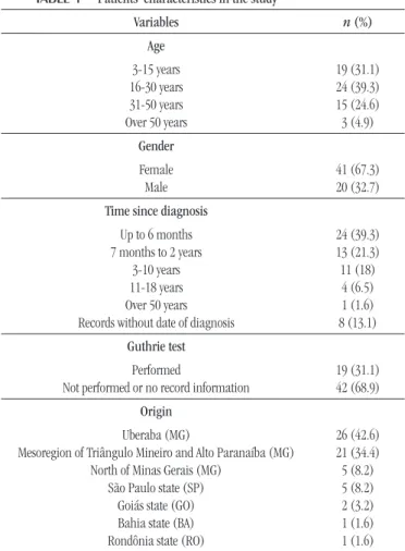
The epidemiological proiles of the patients participating in the study are shown in Table 1.
TABLE 1 − Patients’ characteristics in the study
Variables n (%)
Age
3-15 years 16-30 years 31-50 years Over 50 years
19 (31.1) 24 (39.3) 15 (24.6) 3 (4.9)
Gender
Female Male
41 (67.3) 20 (32.7)
Time since diagnosis
Up to 6 months 7 months to 2 years
3-10 years 11-18 years Over 50 years Records without date of diagnosis
24 (39.3) 13 (21.3) 11 (18) 4 (6.5) 1 (1.6) 8 (13.1)
Guthrie test
Performed
Not performed or no record information
19 (31.1) 42 (68.9)
Origin
Uberaba (MG)
Mesoregion of Triângulo Mineiro and Alto Paranaíba (MG) North of Minas Gerais (MG)
São Paulo state (SP) Goiás state (GO)
Bahia state (BA)
Rondônia state (RO)
TABLE 4 − Clinical manifestations in relation to genotype (n = 61 patients)
Clinical manifestations CAR/CAR
(n = 31)
CAR/BEN
(n = 13)
CAR/CAM
(n = 1)
CAR/ATP
(n = 8)
BEN/BEN
(n = 8)
BEN/ATP
(n = 3)
ATP/ATP
(n = 2) Total
Gallstones (p = 0.79)
Leg ulcers (p = 0.32) Acute chest syndrome (p = 0.3)
Stroke (p = 0.76)
HF (p = 0.83) HT (p = 0.36)
HH (p = 0.43)
13 (46.4) 5 (45.4) 10 (66.7) 5 (62.5) 8 (47) 26 (51) 25 (49) 3 (10.7) 1 (9.1) 1 (6.7) -3 (17.6) 5 (9.8) 6 (11.8) -1 (6.7) -1 (2) 1 (2) 4 (14.3) 1 (9.1) 1 (6.7) 1 (12.5) 3 (17.6) 7 (13.7) 6 (11.8) 5 (17.8) 1 (9.1) 1 (6.7) 2 (25) 2 (11.7) 8 (15.7) 8 (15.7) 2 (7.1) 2 (18.2) 1 (6.7) -1 (5.9) 2 (3.9) 3 (5.9) 1 (3.6) 1 (9.1) -2 (3.9) 2 (3.9) 28 (100) 11 (100) 15 (100) 8 (100) 17 (100) 51 (100) 51 (100)
Analysis using chi-square test, p < 0.05. Values presented as n (%).
CAR: Central African Republic; BEN: Benin; CAM: Cameron; ATP: atypical; HF: heart failure; HT: history of transfusion; HH: history hospitalization.
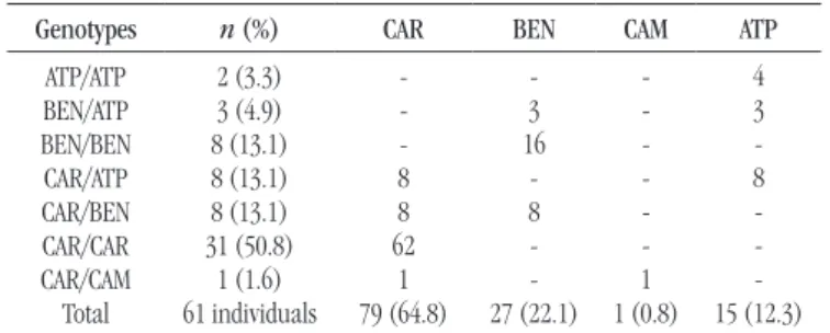
TABLE 2 −Distribution of genotypes and haplotypes
Genotypes n (%) CAR BEN CAM ATP
ATP/ATP BEN/ATP BEN/BEN CAR/ATP CAR/BEN CAR/CAR CAR/CAM Total 2 (3.3) 3 (4.9) 8 (13.1) 8 (13.1) 8 (13.1) 31 (50.8) 1 (1.6) 61 individuals -8 8 62 1 79 (64.8) -3 16 -8 -27 (22.1) -1 1 (0.8) 4 3 -8 -15 (12.3)
Haplotypes – chromosomes 122 (100%).
CAR: Central African Republic; BEN: Benin; CAM: Cameroon; ATP: atypical.
TABLE 3 −Hematologic characteristics in association with genotype
Genotypes Erythrocytes (× 109/l) Hemoglobin (g/dl) Hematocrit (%) Platelets (× 109/l) HbF (%)
CAR/CAR CAR/BEN CAR/CAM CAR/ATP BEN/BEN BEN/ATP ATP/ATP
p value (ANOVA)
2.6 ± 0.1 2.8 ± 0.2 3.4 ± 0.5 2.5 ± 0.2 2.4 ± 0.5 2.7 ± 0.5 2.9 ± 0.36
0.44
8.1 ± 1.1 8.2 ± 1.1 7.3 ± 1 8.1 ± 0.8 8.2 ± 1.1 8.8 ± 1.1
8.9 0.77
23.3 ± 3.7 23.3 ± 2.9
21.2 23.6 ± 2.6
23.3 ± 3 20.1 ± 2.5
26.6 0.64
439,878 ± 156,378 336,315 ± 120,972
160,966 446,694 ± 242,737
347,895 ± 92,743 300,111 ± 89,881
238,600 0.98
4.6 ± 0.4 7.7 ± 0.8
7.2 4.9 ± 0.8 5.3 ± 0.8 9.7 ± 1.3 8.2 ± 1.6 0.008*
The results were reported in average values ± standard deviation. ANOVA test was used for multiple comparison of means between haplotypes, *p < 0.05. HbF: fetal hemoglobin; CAR: Central African Republic; BEN: Benin; CAM: Cameron; ATP: atypical; ANOVA: analysis of variance.
The occurrence of the haplotypes and combinations thereof are shown in Table 2.
Twenty-seven patients (44.3%) had a history of HU use sometime in the studied period; however, attendance and adherence to treatment were neither monitored nor evaluated. Among these patients, 13 (48.1%) were identiied as clearly predominant CAR/CAR genotype carriers. In the multiple comparison of the hematologic proile with seven groups of genotypes, only HbF showed a statistically signiicant difference (Table 3 and Figure).
When HbF levels between the different genotypes were compared, we observed that CAR/CAR presented the lowest value and was statistically signiicant when compared with ATP/ATP
(p = 0.03), BEN/ATP (p = 0.0006) and CAR/BEN (p = 0.0001). We also emphasize that BEN/ATP had the highest concentration of mean HbF levels among the genotypes and was statistically signiicant when compared with BEN/BEN (p = 0.007), CAR/ATP
(p = 0.004) and CAR/CAR (p = 0.0006).
The associations between genotypes and the presence of clinical complications showed no statistically signiicant difference, but individuals with the homozygous CAR haplotype showed higher frequency of clinical events. Consolidating these indings, we also found that the group of patients with a CAR chromosome had also increased frequency and severity of clinical events in relation to the groups of patients BEN/BEN, BEN/ATP and ATP/ATP, as shown in Table 4.
FIGURE − Representation of average HbF level in genotypes
F: fetal; HbF-M: mean fetal hemoglobin; ATP: atypical; BEN: Benin; CAR: Central African Republic; p < 0.05.
ATP/ATP BEN/ATP BEN/BEN CAR/ATP CAR/BEN CAR/CAR
HbF-M 14 13 12 11 10 9 8 7 6 5 4 3 2
*p = 0.03
*p = 0.0006
*p = 0.0001 Haplotype; LS Means
DISCUSSION
The predominant age group in this study was 3-30 years, corresponding to 70.4% of patients. Only three (4.9%) were older than 50 years. These indings are similar to those described in the literature, which report a shortened life expectancy in this population(10). A study by Martins et al. (2010)(11), in our region,
revealed predominant age of 0-29 years (82.5%) and a small contingent over 40 years (8.7%), suggesting that sickle-cell patients die early in Brazil.
We had a patient diagnosed with SCA later, after 52 years old, what characterizes the phenotypic heterogeneity of this disease(4).
As to identiication of haplotypes in relation to chromosomes, there was a clear predominance of the CAR haplotype (64.8%) compared to BEN (22.1%), ATP (12.3%) and CAM (0.8%). Findings are similar to those described in different regions of Brazil. A study conducted in Ribeirão Preto (SP) revealed a predominance of the CAR haplotype (66.2%) compared to BEN (23%) in individuals with SCA(12). The predominance of CAR, followed by BEN, was also
observed in two other studies, revealing frequencies of 62.2% and 73.1% for the CAR versus 23% and 25.4% for BEN, respectively(13, 14).
In Rio de Janeiro, CAR (54%) was more frequent than BEN (44.6%)
(15), as observed in Porto Alegre(16), where the identiied frequency
was 79.6% for CAR and 18.4% for BEN. In Pernambuco, the CAR frequency was 79.1%(17); in Rio Grande do Norte, 75.5%(18); and
in Fortaleza, 66.2%(19). Nevertheless in Bahia, in the Northeast
Region, we found the prevalence of BEN (48.8%)(20).
The CAM haplotype in this study showed a low rate (0.8%), similar to those frequencies described in Salvador (BA), Belém (PA), and Pernambuco (PE): 1.2%, 1.3% and 0.8%, respectively(17, 21, 22).
The presence of 12.3% of atypical chromosomes was similar to the 11.8% occurrence observed by Silva et al. (2009)(19).
These indings probably relect the various genetic mechanisms of association with the sickle gene, i.e. the atypical differ from the ive common haplotypes observed in the world, conirming the hypothesis that these different structures are generated by recombination, speciic replacement and/or non-reciprocal transfers between common preexisting haplotypes instead of new
mutations in the β-globin genes(23). We emphasize that a patient
diagnosed at age 52 in our study showed the ATP/ATP genotype, suggesting, once again, the heterogeneity of the disease.
The highest frequency of the BEN haplotype documented in
Bahia(20), different from that found in other regions of Brazil, was
similar to those in other countries, such as Venezuela (51.1%) and Cuba (51.0%)(3). In Jamaica and the United States there are reports
of the apparent predominance of this haplotype, with frequencies of 72% and 62%, respectively(12).
Regarding the combinations of haplotypes, the CAR/CAR genotype predominated (50.8%), followed by CAR/ATP, CAR/BEN and BEN/BEN with the same frequencies (13.1%). These indings are consistent with those by Cabral et al. (2010)(18), 58.5% for
CAR/CAR and 16.9% for CAR/BEN. By contrast, our results were not similar to those described by Belisário et al. (2010)(24), in
which 39.4% were CAR/CAR, 33.2% CAR/BEN, 23.6% BEN/BEN, 0.9% CAR/ATP, 0.9% BEN/ATP and 0.9% BEN/Arab-Indian. This difference might be explained by the size of the state of Minas Gerais and its territorial boundary with several other states, enabling considerations of interest on the origin and internal migration of Africans in Brazil and in our state.
Nine of the 61 patients (14.7%) came from other states: ive from São Paulo (two CAR/CAR, two CAR/BEN and one ATP/ ATP), two from Goiás (CAR/CAR, CAR/ATP), one from Rondônia (BEN/BEN) and one from Bahia (BEN/ATP). We believe that the frequency of these genotypes did not impact on our results, because the CAR haplotype also showed a higher frequency in this subgroup. Just as expected, in the state of Bahia, where there is a higher incidence of the BEN haplotype, one patient had the combination BEN/ATP.
Regarding the hematological proile, just HbF showed average values with signiicant differences among the seven groups of genotypes. The inding of the signiicant decrease in the average levels of HbF in the CAR/CAR genotype relative to other genotypes of our study is similar to those described in the literature, which are also associated with greater clinical severity(25). In a study by
Fleury(15), as shown in our results, there was signiicant difference
only for HbF among different genotypes.
The heritage of at least one CAR chromosome is associated with more serious pictures than those related to the presence of other haplotypes. Therefore, the genotypes that have the CAR haplotype
in common are assumed to be related to similar laboratory and clinical courses(26). In our study, we observed a higher frequency
of clinical complications in patients with at least one CAR chromosome, what is consistent with Cançado(27), who reported a
twofold increase in the relative risk of stroke and a ivefold risk for kidney complication when compared to CAR-negative patients.
Thus, the CAR haplotype has the worst prognosis, with greater severity of clinical complications, more hospitalizations and increased
morbidity and mortality(28). Although we found no signiicant
association between the CAR/CAR genotype and clinical events, indings similar to those described by Silva Filho et al. (2012)(29), we
using HU, what may be indicative of clinical severity. The absence of this association can be attributed to a small sample, heterogeneity of age, the small group of patients with certain genotypes, and the quality of clinical information, which is controlled, because this is a retrospective study of medical records.
CONCLUSION
In the present study, as in most national reports, the CAR haplotype was predominant, followed by BEN. On associations
between clinical/hematological proile and the different genotypes, the only signiicant inding was a decrease in the average level of HbF in the CAR/CAR genotype compared with other genotypes. However, higher frequency of clinical events in patients having one or two CAR chromosomes was observed. This was the irst work in the Triângulo Mineiro region, and it revealed a small variation in the frequency of haplotypes in relation to that found in Belo Horizonte. It may contribute to determining the low of βS-globin in the state of Minas Gerais. In addition, this study can serve as a possible tool for anthropological studies of African origin in our state and country.
REFERENCES
1. Steinberg MH. Modulation of phenotypic diversity of sickle cell anemia. Hemoglobin. 1996; 20(1): 1-19.
2. Ashley-Koch A, Yang Q, Olney RS. Sickle hemoglobin (HbS) allele and sickle cell anemia: a huge review. Am J Epidemiology. 2000; 151(9): 839-45.
3. Moreno N, Martinez JA, Blanco Z, Osorio L, Hackshaw P. Beta-globin gene cluster haplotypes in Venezuelan sickle cell patients from the State of Aragua.Genet Mol Biol. 2002; 25: 21-4.
4. Gonçalves MS, Bomim GC, Maciel E, et al.Beta S haplotypes in sickle cell anemia patients from Salvador, Bahia, Northeastern Brazil. Braz J Med Biol Res. 2003 Oct; 36(10): 1283-8.
5. Rusanova I, Escames G, Cossio G, et al. Oxidative stress status, clinical outcome, and β-globin gene cluster haplotypes in pediatric patients with sickle cell disease. European J Haematol. 2010; 85(6): 529-37.
RESUMO
Introdução: A anemia falciforme (AF) é a forma mais grave da doença falciforme, sendo caracterizada por homozigotos de hemoglobina S (α2βS2). Objetivo: Determinar a frequência dos haplótipos de pacientes com AF e sua correlação com o perfil clínico e hematológico. Método: Realizado estudo descritivo e retrospectivo, por meio da leitura dos prontuários, e estudo transversal para análise molecular a fim de determinar os haplótipos da globina do gene pS de 61 pacientes com AF (SS) por reação em cadeia da polimerase-polimorfismo de fragmentos de restrição (PCR-RFLP), utilizando endonucleases de restrição Xmn I, Hind III, Hinf I e Hinc II para análise de seis locais polimórficos no cluster beta. Resultado: Os genótipos foram Central African Republic (CAR)/CAR (50,8%), CAR/Benin type (BEN) (13,1%), CAR/Cameroon type (CAM) (1,6%), CAR/atypical (ATP) (13,1%), BEN/BEN (13,1%), BEN/ ATP (4,9%) e ATP/ATP (3,3%). Dos cromossomos analisados, 64,8% eram do tipo CAR; 22,1%, BEN; 12,3%, ATP; e 0,8%, CAM. Os níveis de hemoglobina fetal (HbF) foram significativamente menores no CAR/CAR em relação a ATP/ATP, BEN/ATP e CAR/BEN. Não houve associação entre os diferentes genótipos e as manifestações clínicas. Conclusão: Apesar da falta de associação entre genótipos e perfis clínicos, foi observada maior frequência de eventos clínicos em pacientes com pelo menos um tipo de cromossomo CAR. Observou-se também associação significativa dos níveis médios mais baixos de HbF em genótipo CAR/CAR, em comparação com outros genótipos.
Unitermos: anemia falciforme; haplótipos; hemoglobina fetal.
6. Antonarakis SE, Kazazian Jr HH, Orkin SH. DNA polymorphism and molecular pathology of the human globin gene clusters. Hum Genet. 1985; 69: 1-14.
7. Embury SH, Hebbel RP, Mohandas N, Steinberg MH. Sickle cell disease: basic principles and clinical practice. New York: Raven Press; 1994. 8. Hirokawa K, Ohene-Frempong K, Horiushi K. Determination of HbF level, maturation and morphology of individual sickle cell image cytometry. Blood. 1995; 86(10): 139.
9. Sutton M, Bouhassira EE, Nagel RL. Polymerase chain reaction ampliication applied to the determination of beta-like globin gene cluster haplotypes.American J Hematol. 1989; 32(1): 66-9.
10. Pereira SAS, Cardoso CS, Brener S, Proietti AFC.Doença falciforme e qualidade de vida: um estudo da percepção subjetiva dos pacientes da Fundação Hemominas, Minas Gerais, Brasil.Rev Bras Hematol Hemoter. 2008; 30(5): 411-6.
11.Martins PRJ, Moraes-Souza H, Silveira TB. Morbimortalidade em
12. Zago MA, Figueiredo MS, Ogo SH.BantuβS cluster haplotype predominates
among Brazilian Blacks.Am J Phys Anthropol. 1992; 88(3): 295-8. 13. Gonçalves MS, Bomim GC, Maciel E, et al.βS-haplotypes in sickle
cell anemia patients from Salvador, Bahia, Northeastern Brazil. Braz J Med Biol Res. 2003; 36: 1283-8.
14. Figueiredo MS, Kerbauy J, Gonçalves MS, et al. Effect of alpha-thalassemia and beta-globin gene cluster haplotypes on the hematological and clinical features of sickle-cell anemia in Brazil.Am J Hematol. 1996; 53(2): 72-6.
15. Fleury MK. Haplótipos do cluster da globina beta em pacientes com anemia falciforme no Rio de Janeiro: aspectos clínicos e laboratoriais. Rev Bras Anal Clin. 2007; 39(2): 89-93.
16. Wagner S, Fredrish J, Job F, Hutz M. Caracterização molecular de pacientes com anemia falciforme de Porto Alegre. Rev Bras Genética. 1996; 19: 244.
17. Bezerra MA, Santos MN, Araújo AS, Gomes YM, Abath FG, Bandeira FM. Molecular variations linked to the grouping of beta- and alpha-globin genes in neonatal patients with sickle cell disease in the State of Pernambuco, Brazil.Hemoglobin. 2007; 31(1): 83-8.
18. Cabral CHK, Seraim ESS, Medeiros WRDB, et al. Determinação de haplótipos do geneβS em pacientes com anemia falciforme no estado
do Rio Grande do Norte. Rev Bras Hematol Hemoter. 2010; 32(4): 205-6. 19. Silva LB, Gonçalves RP, Robenhorst SHB. Análise dos haplótipos da anemia falciforme em Fortaleza revela as origens étnicas da população cearense. J Bras Patol Med Lab. 2009; 45(2): 115-8.
20. Adorno EV, Zanette A, Lyra I, et al. The beta-globin gene cluster haplotypes in sickle cell anemia patients from Northeast Brazil: a clinical and molecular view.Hemoglobin. 2004; 28(3): 267-71.
21. Adorno EV, Zabette A, Lyra I, Seixas MO, Reis MG, Gonçalves MS. Clinical and molecular characteristics of sickle cell anemia in the northeast of Brazil. Genet Mol Biol. 2008; 31(3): 621-5.
22. Cardoso GL, Guerreiro JF. Molecular characteristics of sickle cell anemia in the Northern Brazilian State of Pará. Am J Hum Biol. 2010; 22(5): 573-7.
23. Zago MA, Silva WA Jr, Dalle B, et al. Atypical βS haplotypes are generated by diverse genetic mechanisms. Am J Hematol. 2000; 63: 79-84.
24. Belisário AR, Martins ML, Brito AM, Rodrigues CV, Silva CM, Viana MB.
β-globin gene cluster haplotypes in a cohort of 221 children with sickle
cell anemia or Sβ-thalassemia and their association with clinical and
hematological features. Acta Hematol. 2010; 124(3): 162-70.
25. Silva LB, Gonçalves RP. Características fenotípicas dos pacientes com anemia falciforme de acordo com os haplótipos do gene daβS-globina
em Fortaleza, Ceará. Rev Bras Hematol Hemoter. 2010; 32(1): 40-4. 26. Rodriguez R,Sáenz RG, Chaves VMA. Haplótipos de la hemoglobina S: importancia epidemiológica, antropológica y clínica. Rev Pan Sal Public. 1998; 3(1): 1-8.
27. Cançado RD. Doenças falciformes. Prat Hosp. 2007; 50: 61-4. 28. Costa PJMS, Vilela RQB, Cipolotti R, Figueiredo MS. Diversidade clínica e laboratorial no haplótipo Bantu da anemia falciforme. Rev Bras Hematol Hemoter. 2006; 28(1): 40-4.
29. Silva Filho IL, Ribeiro GS, Moura PG, Vechi ML, Cavalcante AC, Andrada-Serpa MJ. Manifestações clínicas agudas na primeira e segunda infâncias e características moleculares da doença falciforme em um grupo de crianças do Rio de Janeiro. Rev Bras Hematol Hemoter. 2012; 34(3): 196-201.
CORRESPONDING AUTHOR
Alexandra Silva Leal