31
CASE REPORTAggressive papillary tumor of endolymphatic sac:
case report of a rare neoplasia
Tumor papilar agressivo do saco endolinfático: relato de caso de uma rara neoplasia
Eduardo Cambruzzi1; Karla Lais Pêgas2; Leandro P. Almeida3; Gerson Evandro Perondi3;Leandro I. Dini3
1. Instituto de Cardiologia – Fundação Universitária de Cardiologia (ICFUC), Rio Grande do Sul, Brasil. 2. Santa Casa de Porto Alegre, Rio Grande do Sul, Brasil. 3. Hospital Cristo Redentor, Rio Grande do Sul, Brasil.
First submission on 10/09/15; last submission on 28/11/15; accepted for publication on 28/11/15; published on 20/02/16
ABSTRACT
Aggressive papillary endolymphatic sac tumor (ELST) is a rare neoplasm, occasionally related to von Hippel-Lindau disease, characterized by locally aggressive growth with temporal bone destruction. The authors report a case of ELST in a female patient exhibiting ifth through eighth cranial nerve paralysis. Computed tomography (CT) revealed a large lytic process involving the right temporal bone. The patient underwent surgical resection. At microscopy, a neoplastic process was identiied exhibiting monomorphic columnar cells with mild atypias, arranged in a papillary pattern. The lesion exhibited positivity for A31/AE3, epithelial membrane antigen (EMA), and vimentin; and negativity for synaptophysin, glial ibrillary acidic protein (GFAP), neuron-speciic enolase (NSE), thyroglobulin, transthyretin, chromogranin, thyroid transcription factor 1 (TTF-1), trans-acting T-cell speciic transcription factor GATA-3, and intestinal transcription factor CDX-2. The diagnosis of ELST was then established. Six years after surgical resection, lesion recurrence was observed.
Key words: endolymphatic sac; temporal bone; pathology; immunohistochemistry; prognosis.
J Bras Patol Med Lab, v. 52, n. 1, p. 31-34, February 2016
INTRODUCTION
Aggressive papillary tumor of the endolymphatic sac is an uncommon process arising in temporal bone/endolymphatic sac that is characterized by an aggressive local behavior without metastasizing potential(1-3). It has also been reported as an
aggressive papillary middle-ear tumor, endolymphatic sac tumor (ELST), aggressive papillary tumor of temporal bone, low-grade adenocarcinoma of endolymphatic sac origin, among others(1-5).
ELST occurs in both genders with a similar distribution, affecting patients with a median age of 42 years. A similar number of ELST involves the right and left side, and bilateral tumors are extremely rare lesions. The most frequent clinical complaints are unilateral
hearing loss and vertigo(1-5).
Convincing anatomic, morphological and immunohistochemical data support the origin of ELST in the epithelium of the inner ear. Most lesions arise in the petrous portion of temporal bone, but the tumor is also described in the areas of the jugular bulb, mastoid, posterior cranial fossa, and middle ear,
without unequivocal radiologic evidence of endolymphatic sac
involvement(2-7). Herein, the authors report an example of large
recurrent ELST arising in the temporal bone, and discuss clinical and pathologic aspects of this rare tumor.
CASE REPORT
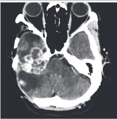
A 38-year-old female patient was taken to the neurology service with clinical complaint of right facial paralysis in the last two months. On physical/neurological examination, right ifth through eighth cranial nerve paralysis was identiied. Ophthalmic examination exhibited a normal optic nerve papilla. The other organs and systems showed no clinical alterations, as there was no previous history of relevant disease. Computed tomography (CT) imaging revealed a large, hypodense, expansive lytic lesion involving right temporal bone and middle
ear (Figure 1). CT/magnetic resonance (RM) imaging of the
chest and abdomen did not identify signiicant alterations. The patient underwent resection of the lesion. The pathologic
32
specimen was composed of some irregular gray fragments of tissue, the largest measuring 1.5 cm in the largest dimension. At microscopy, a neoplastic process was identiied exhibiting monomorphic epithelial columnar cells with clear/eosinophilic cytoplasm, round to oval nuclei, and mild atypias, arranged in a papillary pattern (Figure 2). No mitotic igures or necrosis were noted. The lesion exhibited positive immunoexpression for pancytokeratins, epithelial membrane antigen, and vimentin; and negative stains for synaptophysin, glial ibrillary acid protein, enolase neuron-speciic, thyroglobulin, transthyretin, chromogranin, thyroid transcription factor 1 (TTF-1), trans-acting T-cell speciic transcription factor GATA-3, intestinal transcription factor CDX-2, villin, napsin-A, and desmin. The diagnosis of ELST was then established. Six years after initial therapy, CT identiied a large mass on the right temporal bone, compatible with lesion recurrence (Figure 3).DISCUSSION
ELST is a rare invasive tumor of the temporal bone/ middle ear that is described in patients aged 4 years to 85
years(1, 5, 6, 8-10). In general, the affected patients complain of
audiological symptoms, vertigo, cerebellar disorders, and facial nerve paralysis. Rare cases are related to a tumor mass that ills the tympanic cavity. A long-standing symptom history and indolent clinical course are typical of ELST(1, 3, 5, 6, 8-10). Bilateral
ELST are very rare, and these cases are commonly associated with von Hippel-Lindau syndrome (VHLS). This syndrome is a rare, genetically based, autosomal dominant disorder that affects around 1:39,000 people, and until 20% of ELST cases are related to VHLS. The gene associated with VHLS is mapped on chromosome 3p25-p26, and pheochromocytomas, paragangliomas, pancreatic neuroendocrine tumors, pancreatic islet tumors, clear cell renal cell carcinoma, and central nervous system hemangioblastomas, are important manifestations of this
disease(2, 3, 7, 11-13). In the inner ear, the membranous labyrinth,
which is a closed system with a sac, the endolymphatic sac, is lined by a simple epithelium except in the endolymphatic sac, where the cells are columnar and probably associated to FIGURE 1 − Aggressive papillary tumor of endolymphatic sac: CT showing an expansile
lytic lesion arising in the right temporal bone (A and B)
CT: computed tomography.
FIGURE 3 − Aggressive papillary tumor of endolymphatic sac. Six years after surgical resection, CT scans revealed tumor recurrence
CT: computed tomography.
FIGURE 2 − Aggressive papillary tumor of endolymphatic sac. A) a neoplastic process exhibiting papillary structures lined by a single layer of epithelial cells, 100×; B) monomorphic columnar cells with clear cytoplasm, ovoid central nuclei, and mild atypias covering papillary structures with numerous capillaries, 200×
33
endolymph absorption. The endolymphatic sac, lying in the subdural space of the underlying brain, and the choroid plexushave neuroectodermal origin(6, 7, 10, 14, 15).
ELST are usually reddish brown gritty tumors, exhibiting soft tissue density on radiological scans. The characteristic invasive aspect of the tumor can be identiied on computed tomography, and the petrous temporal bone, mastoid process, or even the cerebellum, are frequently compromised(1-3, 6, 8, 9, 14). Advanced
tumors can extend into cranial fossa or to skull base near jugular foramen. Reported tumor sizes in the international literature ranged from 7.5 cm to 0.5 cm(1-3, 6, 8, 9, 14). At microscopy, the process
shows a complex interdigitating papillary pattern with variable-sized ibrovascular septa, which are covered by a simple layer of low, cuboidal to columnar epithelial cells. In some cases, the neoplastic cells are disposed in cords and/or trabeculae(2-4, 7, 9, 12, 16). An acinar
pattern or microcysts are rarely found. Fibrotic response varies from minimal to desmoplastic reactions. Extensive iniltration of bone and soft tissue, cholesterol clefts, and hemorrhagic areas are common indings(2-4, 7, 9, 12, 16). Tumoral cells can demonstrate an
eosinophilic, clear, or vacuolated cytoplasm, with indistinct cell borders and centrally placed oval nuclei with granular chromatin. A low mitotic index is the rule(6, 8, 9, 12, 14, 17). ELST shows positive
TABLE − Similar cases of aggressive papillary tumor of endolymphatic sac reported in the literature
Authors Age/gender Clinical findings Topography Treatment modality Linked to von
Hippel-Lindau syndrome Outcome
Patel et al.(8) 63/F Facial right paralysis portion of the temporal Mastoid and petrous
bone
Total surgical resection No No signs of recurrence after 24 months
Horiguchi et al.(7) 20/F Asymptomatic Left temporal bone Partial surgical
resection Yes
Large mass after 19 months
Kupferman et al.(10) 4/M Otitis media and facial
paralysis Right temporal bone Surgical resection No
No signs of recurrence after a six-month
follow-up
Reijneveld et al.(9) 63/F
Fifth through eleventh cranial nerve paralysis, ataxia, pyramidal/
pseudobulbar syndrome
Pars squamous and petrosa of temporal bone
Partial surgical
resection No Unavailable
Schick et al.(1) 32/M Hearing loss
and vertigo Temporal bone Surgical resection No
No signs of recurrence after 22 months
Sun et al.(5) 31/M Hearing loss Temporal bone Partial surgical
resection and radiotherapy
No Unavailable
Ferri et al.(18) 37/F Hearing loss Mastoid process of temporal
bone and middle ear Surgical resection No
No signs of recurrence after 36 months
Present case 38/F Fifth through eight
cranial nerve paralysis Right temporal bone Surgical resection No
Large mass after six years F: female; M: male.
immunoexpression for cytokeratin, vimentin, and epithelial membrane antigen; and negative expression for thyroglobulin, transthyretin, synaptophysin, chromogranin, desmin, and actin. Some tumors can show positive expression for glial ibrillary acid protein (GFAP), S100, and neuron-speciic enolase (NSE). Cytokeratin 7 (CK7), cytokeratin 20 (CK20), napsin-a, CDX-2, villin, GATA-3, and mammaglobin antibodies can be used to exclude metastatic lesions from colon, lung and breast(2, 3, 7, 10, 13, 18).
The Table shows some cases of ELST found in the international literature that are comparable to the reported case.
Differential diagnosis includes middle-ear adenoma, a typically non-invasive benign tumor; metastatic papillary carcinoma, especially papillary carcinoma of the thyroid gland; choroid plexus papillomas/carcinomas, which show positive expression for transthyretin; papillary meningiomas, which exhibit positive expression for epithelial membrane antigen and progesterone receptors; papillary ependymomas; and paraganglioma(1-4, 6, 7, 10, 14). Surgical treatment is the only
effective treatment, and the prognosis is good in patients who undergo total tumor resection. Adjuvant radiotherapy has been
used in some cases(6, 11, 15, 18).
34
RESUMO
O tumor papilar agressivo do saco endolinfático (TPASE) é uma neoplasia rara, ocasionalmente relacionada com a doença de von Hippel-Lindau, que se caracteriza pelo crescimento agressivo local com destruição do osso temporal. Os autores relatam um caso de TPASE em paciente do sexo feminino, exibindo paralisia do quinto ao oitavo par craniano. A tomografia computadorizada (TC) revelou grande processo lítico comprometendo o osso temporal direito. A paciente foi submetida a ressecção cirúrgica. À microscopia, identificou-se processo neoplásico que exibiu células cilíndricas monomórficas com atipias leves, dispostas em padrão papilar. A lesão apresentou positividade para AE1/ AE3, antígeno da membrana epitelial (EMA) e vimentina; e negatividade para sinaptofisina, proteína ácida fibrilar glial (GFAP), enolase específica do neurônio (NSE), tireoglobulina, transtirretina, cromogranina, fator de transcrição
da tireoide 1 (TTF-1), fator de transcrição de ação trans específico de células GATA-3 e fator de transcrição intestinal CDX-2.
O diagnóstico de TPASE foi então estabelecido. Após seis anos da ressecção cirúrgica, foi identificada recorrência da lesão.
Unitermos: saco endolinfático; osso temporal; patologia; imuno-histoquímica; prognóstico.
REFERENCES
1. Schick B, Kronsbein H, Kahle G, et al. Papillary tumor of the temporal bone. Skull Base. 2001; 11(1): 25-33.
2. Skalova A, Sima R, Bohus P, et al. Endolymphatic sac tumor (aggressive papillary tumor of middle ear and temporal bone): report of two cases with analysis of the VHL gene. Pathol Res Pract. 2008; 204(8): 599-606. 3. Bell D, Gidley P, Levine N, et al. Endolymphatic sac tumor (aggressive papillary tumor of the middle ear and temporal bone): sine qua non radiology-pathology and the University of Texas MD Anderson Cancer Center experience. Ann Diagn Pathol. 2011; 15(2): 117-23.
4. Diaz RC, Amjad EH, Sargent EW, et al. Tumors and pseudotumors of the endolymphatic sac. Skull Base. 2007; 17: 379-93.
5. Sun YH, Wen W, Wu JH, et al. Endolymphatic sac tumor: case report and review of the literature. Diagnostic Pathology. 2012; 7: 36.
6. Heffner DK. Low-grade adenocarcinoma of probable endolymphatic sac origin: a clinicopathologic study of 20 cases. Cancer. 1989; 64: 2292-2302. 7. Horiguchi H, Sano T, Toi H, et al. Endolymphatic sac tumor associated with a von Hippel-Lindau disease patient: an immunohistochemical study. Mod Pathol. 2001; 14(7): 727-32.
8. Patel PC, Pellitteri DO, Reams CL. Aggressive papillary tumor of the temporal bone: delayed extensive recurrence following radiation therapy. Skull Base Surg. 1997; 7(1): 45-8.
9. Reijneveld J, Hanlo P, Groenewoud G, et al. Endolymphatic sac tumor: a case report and review of the literature. Surg Pathol. 1997; 48(4): 368-73.
10. Kupferman ME, Bigelow DC, Carpentieri DF, et al. Endolymphatic sac tumor in a 4-year-old boy. Otol Neurotol. 2004; 25(5): 782-6.
11. Nevoux J, Nowak C, Vellin JF, et al. Management of endolymphatic sac tumors: sporadic cases and von Hippel-Lindau disease. Otol Neurotol. 2014; 35(5): 899-904.
12. Megerian CA, McKenna MJ, Nuss RC, et al. Endolymphatic sac tumors: histopathologic conirmation, clinical characterization, and implication in von Hippel-Lindau disease. Laryngoscope. 1995; 105: 801-8. 13. Bausch B, Wellner U, Peyre M, et al. Characterization of endolymphatic sac tumors and von Hippel-Lindau disease in the International Endolymphatic Sac Tumor Registry. Head Neck. 2015. doi: 10.1002/ hed.24067.
14. Cazals-Hatem D, Henin D, Bouccara D, et al. Endolymphatic sac tumor: a rare tumor of internal ear. Report of two cases. Ann Pathol. 2000; 20(4): 349-52.
15. Dadas B, Alkan S, Turgut S, et al. Primary papillary adenocarcinoma conined to the middle ear and mastoid. Eur Arch Othorhinolaryngol. 2001; 258: 93-5.
16. Batsakis JG, El Naggar AK. Papillary neoplasms (Heffner’s tumors) of the endolymphatic sac. Ann Otol Rhinol Laryngol. 1993; 102: 648-51. 17. Mukherji SK, Albernaz VS, Lo WW, et al. Papillary endolymphatic sac tumors: CT, MR imaging, and angiographic indings in 20 patients. Radiology. 1997; 202: 801-8.
18. Ferri E, Amadori M, Armato E, Pavon I. A rare case of endolymphatic sac tumour: clinicopathologic study and surgical management. Case Rep Otolaryngol. 2014; 2014: 376761. doi: 10.1155/2014/376761.
CORRESPONDING AUTHOR
Eduardo Cambruzzi
Universidade Luterana do Brasil (Ulbra); Av. Farroupilha, 8001; São José; CEP: 92425-900; Canoas-RS, Brasil; Phone: +55 (51) 9979-6026; e-mail: [email protected].