71 1. MSc. Doctoral student, Univ. do Estado do Rio de Janeiro (UERJ).
Instituto Nacional de Cardiologia Laranjeiras, Rio de Janeiro, RJ, Brazil. 2. Professor, Universidade do Estado do Rio de Janeiro (UERJ), Rio de
Janeiro, RJ, Brazil.
3. MSc. Instituto Nacional de Cardiologia Laranjeiras, Rio de Janeiro, RJ, Brazil.
4. PhD. Universidade do Estado do Rio de Janeiro (UERJ), Rio de Janeiro, RJ, Brazil.
5. MSC. Instituto Nacional de Cardiologia Laranjeiras, Rio de Janeiro, RJ, Brazil.
Manuscript received Jul 04 2003, accepted for publication Oct 15 2003. Abstract
Objective: To analyze the prognostic value of cardiomegaly, pulmonary congestion and cardiothoracic ratio as indicators of death and survival in children with idiopathic dilated cardiomyopathy (IDCM).
Methods: We carried out a retrospective review of 152 patients with idiopathic dilated cardiomyopathy diagnosed between September 1979 and March 2003. In the first 72 months, 722 exams were performed (100 in the first 15 days). Statistical analysis: chi-square, Students t test, ANOVA and Kaplan-Meier curves. Alpha = 0.05; beta = 0.80.
Results: The mean age at presentation was 2.2±3.2 years. Idiopathic dilated cardiomyopathy incidence was higher in children younger than 2 years (76.3% 95% CI = 68.7% to 82.8%) (p < 0.0001). Sex (p = 0.07) and color (p = 0.11) were not significant and mortality was not influenced by age (p = 0.73), sex (p = 0.78) or color (p = 0.20). Most patients were severely ill (84.2% 95% CI = 77.4% to 89.6%; functional class III and IV; p < 0.0001). All 43 deaths occurred in this group (p = 0.0008). Cardiomegaly at presentation was observed in 94.1% (95% CI = 89.1% to 97.2%) (p < 0.0001), and pulmonary congestion in 75.6% (95% CI = 68.0% to 82.2%) (p < 0.0001). Pulmonary congestion and cardiomegaly were more frequent in functional class III/IV patients (RC = 8.03 95% CI = 2.85% to 23.1%) (p < 0.0001). Pulmonary congestion was a marker of death (RC = 3.16 95% CI = 1.06% to 10.07) (p = 0.0222), but not cardiomegaly (p = 0.1185). Survival was influenced by both cardiomegaly (p = 0.0189) and pulmonary congestion (p = 0.0050). Mean and maximum cardiothoracic ratio were higher in the death group (0.749±0.053 vs. 0.662±0.080) (p < 0.0001) and (0.716±0.059 vs. 0.620±0.085) (p < 0.0001). ANOVA revealed a progressive decrease in cardiothoracic ratio in the survival group (p < 0.0001).
Conclusion: In children with idiopathic dilated cardiomyopathy, the presence of pulmonary congestion at presentation and increased cardiothoracic ratio are associated with poor survival.
J Pediatr (Rio J). 2004;80(1):71-6: Idiopathic dilated cardiomyopathy, child, radiology.
Prognostic value of chest roentgenograms
in children with idiopathic dilated cardiomyopathy
Vitor M.P. Azevedo1, Francisco M. Albanesi Filho2, Marco A. Santos3,
Márcia B. Castier4, Bernardo R. Tura5
Copyright © 2004 by Sociedade Brasileira de Pediatria
O
RIGINALA
RTICLEIntroduction
Heart failure (HF) may be defined as a clinical syndrome in which the heart pump function is impaired, not supplying the oxygenated blood necessary for normal tissue metabolism, growth and development.1
Ventricular dysfunction may be caused by an increase in afterload (severe aortic stenosis or chronic hypertension), by an increase in preload (mitral regurgitation or left-right shunt) or by intrinsic lesion to the heart muscle (myocarditis or dilated cardiomyopathy).2
According to the World Health Organization, dilated cardiomyopathy is characterized by dilatation and impaired contraction of the left ventricle or both ventricles.3 It may be idiopathic, familial/genetic,
damage. Histology is nonspecific. Presentation is usually with heart failure, which is often progressive. Arrhythmias, thromboembolism, and sudden death are common and may occur at any stage.4-6
Idiopathic dilated cardiomyopathy (IDCM) in pediatric patients is responsible for a large number of emergency treatments and hospitalizations due to heart failure not associated with congenital heart diseases, accounting for up to 29% of emergency treatments of children aged less than two years.7 It has a high mortality rate, with
reported rates of 16%8 at 10 years, up to 49%
,966%10
and even 80%,11 at five years. In patients who do not
show a good response to clinical treatment, heart transplant is the treatment of choice; world experience presents an actuarial survival curve of 75 to 80% at one year and of 60 to 75% at 5 years.12-14
IDCM is characterized by HF associated with cardiomegaly and pulmonary congestion on chest X-ray and/or left ventricle dilatation with reduced contractility on echocardiogram.
A radiological examination of the study population was made in order to determine the importance of chest X-ray as a prognostic factor for death in IDCM. The presence of cardiomegaly and pulmonary congestion was assessed, and the cardiothoracic ratio was used as prognostic factor and as survival marker.
Patients and methods
A retrospective study based on the data from Instituto
Nacional de Cardiologia Laranjeiras (National Institute
of Cardiology Laranjeiras) was carried out for later analysis of the medical charts of 165 consecutive patients with IDCM diagnosed between September 1979 and March 2003, with an age range from 1 to 15.6 years. Chest X-ray of 152 (92.1%) out of 165 patients were retrieved and analyzed. The inclusion criteria were presence of HF (dyspnea, dry cough, râles, tachycardia, S3 rhythm, sweating and hepatomegaly), associated with the presence of cardiomegaly on chest X-ray and/ or left ventricle dilatation with reduced contractility on echocardiogram. Patients with clinical diagnosis of myocarditis were not excluded from the study. The clinical criteria suggestive of myocarditis were: fever; chest pain; electrocardiogram (ECG) with low QRS voltages or conduction and heart rhythm defects and elevated creatinine phosphokinase (CK) and its myocardial fraction (CKmb).7 Patients with the following
conditions were excluded from the study: congenital heart diseases, anomalous coronary arteries, Kawasaki disease, ventricular arrhythmogenic cardiomyopathy, ischemic damage due to neonatal asphyxia or after cardiorespiratory resuscitation, use of cancer drugs, primary arrhythmias, rheumatic valvular disease, neur o mus c ul ar di s eas es , ar t er i al hy per t ens i o n, septicemia, HIV infection, Chagas disease and diphtheria.
The following variables were analyzed:
Age at presentation and range (before the age of two years vs after the age of two years), gender and color. The patients were classified at presentation according to the
New York Heart Association (NYHA) functional classes
(FC): FC I without restrictions on age-appropriate activities; FC II comfortable at rest, however age-appropriate activities result in symptoms of HF; FC III comfortable at rest, however mild physical activity results in symptoms of HF, and FC IV symptoms of HF even at rest, any physical activity increases discomfort.15
Initial and follow-up chest X-ray : cardiothoracic ratio (CTR) and presence or absence of cardiomegaly and pulmonary congestion were assessed on first chest X-ray. Cardiomegaly was considered to be present when CTR was greater than 0.55 in the first year of life and greater than 0.50 after the first year of life.16 Initial
chest X-ray was carried out in the first 15 days of follow-up on 100/67 patients (x = 1.49). The highest CTR values (maximum CTR) presented by each patient were r e c o r d e d . A t o t a l o f 7 7 3 c h e s t X - r a y s , e i t h e r posteroanterior (PA) or anteroposterior (AP), depending on patient age (x = 5.09/patient), were performed, corresponding to 172 months of radiological follow-up, 722 (x = 4.75/patient) patients in the first 72 months of follow-up. The chest X-ray was assessed by two researchers on different occasions, and the mean CTR was calculated for each X-ray. The concordance between CTR values was 92.6%. The presence or absence of pulmonary congestion was also analyzed by two researchers on different occasions, and if they disagreed about the results, it was reassessed by a third researcher.
Classic drug therapy for heart failure, consisting of oral digoxin, furosemide, spironolactone, captopril and acetylsalicylic acid (ASA) was utilized for the prevention of thromboembolic events. The analysis of the effect of treatment on patient outcome was not within the scope of this study.
The statistical analysis was made using Epi-Info 6.04 of CDC (Centers for Disease Control & Prevention) and Statistica 6.0 (Statsoft Inc.).
Dichotomous data were assessed by the chi-square test (χ²), and when appropriate, the odds ratio (OR) and a 95%CI were used.
Descriptive data were expressed as mean ± standard deviation (SD) being assessed by Students t test.
Continuous dependent time variables were assessed by repeated measure analysis of variance for unbalanced data, being grouped according to the type of outcome (survivor vs nonsurvivor) and according to time to outcome.
Survival analysis was made using Kaplan-Meier method.
Ethical aspects
The study was approved by the Research Ethics Committee of Instituto Nacional de Cardiologia Laranjeiras and of Universidade do Estado do Rio de Janeiro.
Results
General aspects
Mean age at presentation was 26.4±39.5 months (median = 8.40 months 0 to 188 months) or 2.2±3.2 years (median = 0.69 year 0 to 15.4 years). Mean follow-up period was 3.56 years (median = 2.23 years 0 to 15.94 years). At the end of the study, 72 (47.3%) patients were followed up as outpatients, 10 (6.6%) were discharged from hospital, 43 (28.3%) died and 27 (17.8%) were lost to follow-up. The mean follow-up period in the latter group was 2.47 (0.1 to 7.89) years.
The onset of the disease occurred preferably in children less than two years old (116 76.3% - 95%CI = 68.7% to 82.8%; x = 0.67±0.50 years), comparatively to the group aged two years or older (36 23.7% -95%CI = 17.2% to 31.2%; x = 6.98±3.64 years) (p < 0.0001). No difference was observed as to incidence in terms of gender: females 84 (55.3% - 95%CI =
47.0% to 63.3%) vs male 68 (44.7% - 95%CI =
36.7% to 53.0%) (p = 0.07) and skin color white: 69 (45.4% - 95%CI = 37.3% to 53.7%) vs nonwhite: 83 (54.6% - 95%CI = 46.3% to 62.7%) (p = 0.11). No difference in mortality was found at presentation between age groups (p = 0.73), between genders (p = 0.78), and between ethnic groups (p = 0.20).
NYHA functional classes were used to assess the severity of the clinical picture, with 10 (6.6%) patients in FC I, 14 (9.2%) in FC II, 32 (21.0%) in FC III and 96 (63.2%) in FC IV, therefore most patients (128 84.2% 95%CI = 77.4% to 89.6%) were severely ill (FC III and IV) (p < 0.0001). All deaths corresponded to patients classified as FC III/IV (p= 0.0008).
Chest X-ray
Cardiomegaly and pulmonary congestion on initial chest X-ray
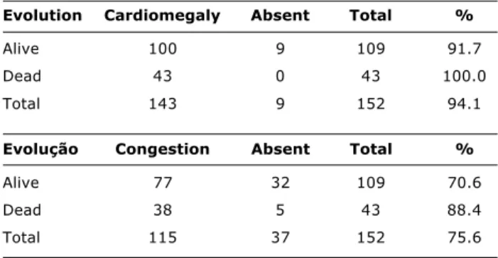
Cardiomegaly was found in 94.1% (143) of the cases (p < 0.0001) (95%CI = 89.1% to 97.2%). Pulmonary congestion was diagnosed in 75.6% (115) of the patients (p < 0.0001) (95%CI = 68% to 82.2%), being more frequent in FC III and IV (82.8%) than in FC I and II (37.5%) (p < 0.0001) (OR = 8.03 95% CI = 2.85% to 23.1%).
With regard to cardiomegaly, no difference was noted between its incidence in the survivor and nonsurvivor groups (p = 0.1185). Pulmonary congestion was a prognostic factor for death (p = 0.0222) (OR = 3.16 95% CI = 1.06% to 10.07) (Table 1).
Table 1 - Incidence of cardiomegaly and pulmonary congestion in the chest X-ray regarding the evolution
Evolution Cardiomegaly Absent Total %
Alive 100 9 109 91.7
Dead 43 0 43 100.0
Total 143 9 152 94.1
Evolução Congestion Absent Total %
Alive 77 32 109 70.6
Dead 38 5 43 88.4
Total 115 37 152 75.6
Survival curve in the presence of cardiomegaly and pulmonary congestion at presentation
As to the finding of cardiomegaly on chest X-ray, a clear difference in survival rates was observed (p = 0.0189), indicating a worse prognosis in the presence of this finding (Figure 1). The same occurred in the presence of pulmonary congestion (p = 0.0050) (Figure 2).
Figure 1 - Survival curve (Kaplan-Meier) in the presence or absence of cardiomegaly in the chest X-ray and 95% confidence interval.
Figure 2 - Survival curve (Kaplan-Meier) in the presence or absence of pulmonary congestion in the chest X-ray and 95% confidence interval.
1.0
0.9
0.8
0.7
0.6
0.5
0.4
0.3
0.2
0.1
0.0
0 4 6 8 10 12 14 16 18
Time (years)
P
ro
p
o
rt
io
n
a
l
o
v
e
ra
ll
s
u
rv
iv
a
l
Death Alive
p = 0.0189
2
Absent
Cardiomegaly
1.0
0.9
0.8
0.7
0.6
0.5
0.4
0.3
0.2
0.1
0.0
0 2 4 6 8 10 12 14 16 18
Time (years)
P
ro
p
o
rt
io
n
a
l
o
v
e
ra
ll
s
u
rv
iv
a
l
p = 0.0050 Absent
Pulmonary congestion
Cardiothoracic ratio (CTR)
Maximum CTR observed throughout the follow-up period showed significant difference between the groups. The mean in the nonsurvivor group was 0.749±0.053 while in the survivor group it was 0.662±0.080 (p < 0.0001) (Figure 3).
may be inferred that the preference of the disease for this age group (less than two years old) is due to the higher incidence of viral infections in these patients.
Age at diagnosis did not influence mortality,17 similarly
to what was found in the literature;25-27 however Arola et
al.9 observed a higher mortality rate among infants
younger than one year old who suffer from endocardial fibroelastosis and among male adolescents in the Finnish population, but other authors found a higher mortality in patients older than two years.18,23,28
No difference was observed as to the incidence of the disease with regard to gender,17 which is in agreement
with the available literature.7-11,23,27,29-31 Gender did not
show to have any influence on mortality in this study,17
which is in line with the findings of other authors.11,19,27
Figure 3 - Box & Whisker Plot of the maximum cardiothoracic index regarding deaths (p < 0.0001).
0.90
0.85
0.80
0.75
0.70
0.65
0.60
0.55
0.50
0.45
M
a
x
im
u
m
C
T
R
Death Alive
± 2 SD ± 1 SD Mean
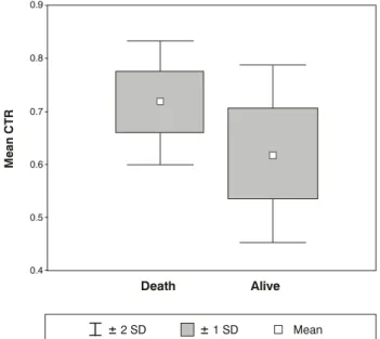
CTR was evaluated at diagnosis and at 1, 3, 6, 12, 18, 24, 36, 48, 60 and 72 months. Mean CTR was 0.620±0.085 in the survivor group and 0.716±0.059 in the nonsurvivor group, showing statistical significance (p< 0.0001) (Figure 4). Follow-up CTR was different between the survivor and nonsurvivor groups with a gradual decrease in the survivor group (p < 0.0001). Figure 5 shows the follow-up of CTR in the first 72 months between both groups and the respective 95% confidence intervals, with a clear distinction between the groups after three months.
Discussion
The sample described in the present study is the most comprehensive one ever reported in the literature, with the longest observation of childhood idiopathic dilated cardiomyopathy, which reduces the possibility of sampling biases.
Mean age at presentation was 2.2 years,17 similar to
most cases reported in the literature;9,10,18-20 however,
some authors found higher means.8,11, 21,22 The onset of
the disease was found to preferably occur before the age of two years (76.3%), in comparison with the group > 2 years.17 Literature data are controversial.18,19,23,24 It
Figure 4 - Box & Whisker Plot mean cardiothoracic index regarding deaths (p < 0.0001).
0.9
0.8
0.7
0.6
0.5
0.4
M
e
a
n
C
T
R
Death
± 2 SD ± 1 SD Mean
Alive
Figure 5 - Evolution of mean cardiothoracic index and 95% confidence interval between the survivor and nonsurvivor groups (p < 0.0001).
0.80
0.75
0.70
0.65
0.60
0.55
0.50
0.45
0 1 3 6 12 18 24 36 48 60 72
Time (months)
Death
Alive
C
T
R
Despite extensive search, we have not found other studies on the incidence and prognosis of the disease with regard to ethnicity.
At initial diagnosis, most patients (84.2%) were considered to have the severe form of the disease (FC III and IV), 43 deaths occurring in this group of patients. In the study conducted in Poland,22 no difference was noted
as to the distribution of disease severity and as to FC between the survivor and nonsurvivor groups. Silva et al.29 showed predominance of less severe cases (FC I and
II), but they do not mention anything about the influence of FC on mortality. Patients whose disease is classified as FC III and IV right at the beginning and who do not respond to clinical treatment are more likely to die.
The methodology used for analyzing the chest X-ray may result in an inherent interpretation bias, but we believe that this bias could have been reduced by having two researchers analyze the exams on different occasions, and by reaching a concordance of 92.6%.
Initial chest X-ray revealed that cardiomegaly was present in most patients, thus showing that this condition is a characteristic of the disease; however, pulmonary congestion was characteristic of more severe cases (FC III and IV). Death could not be predicted by the presence of cardiomegaly, but the presence of pulmonary congestion on initial chest X-ray was a prognostic factor for death (p = 0.0222) (OR = 3.16). The analysis of the survival curve showed that the presence of cardiomegaly influenced survival time, but the presence of pulmonary congestion is detrimental to survival, and in its absence, after one year of follow-up, no patient died.
In the literature, the incidence of cardiomegaly has
been reported to be between 75.0% and 100%,29,32
depending on the severity of the studied patients; no association between cardiomegaly and death was reported. Pulmonary congestion was described by Arola et al.10 in 78.0% of patients, being a predictor of poorer outcome (p = 0.06). Pulmonary congestion reveals left ventricle diastolic dysfunction in which the final diastolic pressure of the left ventricle increases, being aggravated by mitral regurgitation often found in IDCM, both of which elevate the pressure inside the left atrium and pulmonary veins, causing pulmonary edema into the alveoli, and different degrees of dyspnea.
Maximum CTR obtained during follow-up was significantly lower in the survivor group than in the nonsurvivor group; however, values overlapped, making the distinction between these groups difficult. An identical finding was reported for mean CTR, which was lower in the survivor group in comparison to the nonsurvivor group, with overlapping values between groups, which did not allow us to determine the thresholds for each type of outcome.
The literature shows a distinct approach to the measurement of CTR, taking into account the initial and follow-up CTR and its influence on death. Initial CTR was
described to be 0.600±0.080 and 0.660±0.058,19,23
with no difference as to death; however, only one study revealed some difference between groups (survivor =
0.571±0.061 v s nonsurvivor 0.651±0.068, p <
0.0001).11 Follow-up CTR was a prognostic factor for
death in only one report (survivor= 0.573±0.068 vs nonsurvivor = 0.697±0.058, p < 0.0001).23
In the analysis of variance of CTR in relation to death, a clear distinction between groups (p < 0.0001) was noted after three months, with a 95%CI, being lower or equal to 0.68 in the survivor group and greater than 0.68 in the nonsurvivor group. The reviewed literature does not analyze CTR changes over time in relation to death during patient follow-up.
Chest X-ray was one of the first ancillary exams used in cardiology and still is considered useful nowadays because of its ease of use and low cost. The assessment of the cardiothoracic ratio is extremely simple and useful for the longitudinal follow-up of pediatric patients with idiopathic dilated cardiomyopathy, serving as an indicative sign of unfavorable outcome and probable necessity for cardiac transplantation.
References
1. Talner NS. Heart failure. In: Moss AJ, Adams FH, Emmanouilides GC, editors. Moss and Adams Heart Disease in Infants, Children, and Adolescents: Including the Fetus and Young Adult (2 Volume Set). 5th ed. Baltimore: Lippincott, Williams & Wilkins; 1995. p. 1746-73.
2. Lewis AB. The failing myocardium. In: Chang AC, Hanley FL, Wernovsky G, Wessel DL. Pediatric Cardiac Intensive Care. 1st ed. Baltimore: Lippincott, Williams & Wilkins; 1998. p. 483-96. 3. Richardson P. Report of the 1995 World Health Organization/ International Society and Federation of Cardiology Task Force on the Definition and Classification of Cardiomyopathies. Circulation. 1996;93:841-2.
4. Günthard J. Dilated cardiomyopathy in children and thrombo-embolism. Eur J Pediatr. 1997;156:3-6.
5. Chang YC. Left ventricular thrombi in children with dilated cardiomyopathy. J Formos Med Assoc. 1995;94:469-73. 6. Berger S. Sudden cardiac death in infants, children and
adolescents. Pediatr Clin North Am. 1999;46:221-34. 7. Matitiau A, Perez-Atayde A, Sanders SP, Sluysmans T, Parness
IA, Spevak PJ, et al. Infantile dilated cardiomyopathy relation of outcome to left ventricular mechanics, hemodynamics and h i s t o l o g y a t t h e t i m e o f p r e s e n t a t i o n . C i r c u l a t i o n . 1994;90:1310-8.
8. Friedman RA, Moak JP, Garson A. Clinical course of idiopathic dilated cardiomyopathy in children. J Am Coll Cardiol. 1991;18:152-6.
9. Arola A, Tuominen J, Ruuskanen O, Jokinen E. Idiopathic dilated cardiomyopathy in children: prognostic indicators and outcome. Pediatrics. 1998;101:369-76.
10. Taliercio CP, Seward JB, Driscoll DJ, Fisher LD, Gersh BJ, Tajik AJ. Idiopathic dilated cardiomyopathy in the young: clinical profile and natural history. J Am Coll Cardiol. 1985;6:1126-31. 11. Akagi T, Benson LN, Lightfoot N, Chin K, Wilson G, Freedom RM. Natural history of dilated cardiomyopathy in children. Am Heart J. 1991;121:1502-6.
12. Wong PC, Starnes VA. Pediatric heart and lung transplantation. In: Chang AC, Hanley FL, Wernovsky G, Wessel DL. Pediatric Cardiac Intensive Care. 1st ed. Baltimore: Lippincott, Williams & Wilkins; 1998. p. 327-43.
14. Azeka E, Barbero-Marcial M, Jatene M, Camargo PR, Auler JOC, Atik E, et al. Transplante cardíaco no neonato e na infância. resultados a médio prazo. Arq Bras Cardiol. 2000;74:197-202. 15. The Criteria Committee of the New York Heart Association: Nomenclature and Criteria for Diagnosis. 9th ed. Boston: Little Brown; 1994.
16. Paul LW, Juhl JH. Princípio de Interpretação Radiológica. 6ª ed. Rio de Janeiro: Guanabara; 1980. 883p.
17. Azevedo VMP, Santos MA, Albanesi Filho FM. Características epidemiológicas da cardiomiopatia dilatada idiopática na infância. Rev Socerj. 2000;13 Supl A:56.
18. Burch M, Siddiqi SA, Celermajer DS, Scott C, Bull C, Deanfield JE. Dilated cardiomyopathy in children: determinants of outcome. Br Heart J. 1994;72:246-50.
19. Torres F, Anguita M, Gimenez TD, Franco M, Zayas R, Gallardo A, et al. Miocarditis aguda con disfunción cardíaca severa en la población pediátrica. Evolución y características diferenciales con respecto a la miocarditis del adulto. Rev Esp Cardiol. 1995;48:660-5.
20. Müller G, Ulmer HE, Hagel KJ, Wolf D. Cardiac dysrhythmias in children with idiopathic dilated or hypertrophic cardiomyopathy. Pediatr Cardiol. 1995;16:56-60.
21. Lewis AB. Prognostic value of echocardiography in children with idiopathic dilated cardiomyopathy. Am Heart J. 1994;128:133-6. 22. Ciszewski A, Bilinska ZT, Lubiszewska B, Ksiezycka E, Poplawska W, Michalak E, et al. Dilated cardiomyopathy in children: clinical course and prognosis. Pediatr Cardiol. 1994;15:121-6. 23. Griffin ML, Hernandez A, Martin TC, Goldring D, Bolman M, Spray
TL, et al. Dilated cardiomyopathy in infants and children. J Am Coll Cardiol. 1988;11:139-44.
24. Silva MAD, Silva RP, Morais SC, Fragata Filho AA, Correia EB. Aspectos clínicos e evolutivos da miocardiopatia dilatada nos lactentes e na infância. Arq Bras Cardiol. 1991;56:213-8.
25. Chen SC, Nouri S, Balfour I, Appleton RS. Clinical profile of congestive cardiomyopathy in children. J Am Coll Cardiol. 1990;15:189-93.
26. Lewis AB. Outcome of infants and children with dilated cardiomyopathy. Am J Cardiol. 1991;68:365-9.
27. Nogueira G. Miocardiopatia dilatada idiopática na criança: perfil clínico e determinantes do prognóstico. Rev Port Cardiol. 2000;19:191-200.
28. Di Filippo S. Les myocardiopathies dilatees idiopathiques de lenfant. Evolution et facteurs pronostiques. Arch Mal Coeur Vaiss. 1991;84:721-6.
29. Venugopalan P. Improved prognosis of heart failure due to idiopathic dilated cardiomyopathy in children. Int J Cardiol. 1998;65:125-8.
30. Cabrera A. Dilated myocardiopathy in children. Rev Esp Cardiol. 1990;43:246-50.
31. Herdy GVH, Menezes DMF, Lopes VGS, Azevedo SM, Odeh CSA, Mendonça C, et al. Miocardite por citomegalovírus em lactentes. Arq Bras Cardiol. 1988;50:397-400.