359
PRE-PUBERTAL TESTICULAR DERMOID CYST Case Report
International Braz J Urol
Official Journal of the Brazilian Society of Urology
Vol. 31 (4): 359-361, July - August, 2005
PRE-PUBERTAL TESTICULAR DERMOID CYST TREATED WITH
CONSERVATIVE SURGERY
LISIEUX E. JESUS, CLAUDIA R.R. PENA, ANA P.S. LEAO
Departments of Pediatric Surgery and Radiology, Hospital Municipal Jesus, Rio de Janeiro, RJ, Brazil
ABSTRACT
We present a case of testicular dermoid cyst that was treated with transinguinal excision of the testicular cyst with preservation of the healthy testicular parenchyma. We have reviewed the literature for clinical features and therapeutic approach in benign cystic tumors in the pre-pubertal testis.
Key words: testis; benign neoplasms; dermoid cyst; infant Int Braz J Urol. 2005; 31: 359-61
INTRODUCTION
The most common testicular tumors in chil-dren are teratomas with a predominance of benign lesions. Simple cysts and dermoid cysts are rare but uniformly benign, thus enabling surgery with gonadic preservation. It is important to recognize their clini-cal and radiologiclini-cal features so that the selected therapy implies minimal sequelae. We present one case of testicular dermoid tumor in an infant, which was surgically treated and with preservation of the healthy ipsilateral testicular parenchyma.
MATERIALS AND METHODS
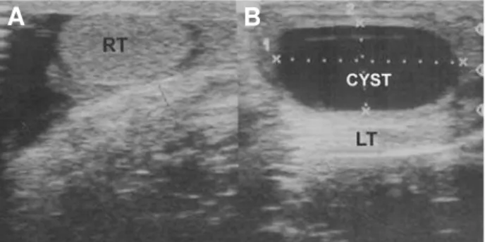
A 5-month old patient was brought to our service for evaluating an increase in scrotal volume that had been perceived some months earlier. He pre-sented normal male genitalia with increased volume of the left gland, which was painless and regular with no transillumination. Ultrasound revealed a left tes-ticular cyst measuring 23 mm at its maximal diam-eter and a well-defined and regular wall with no cal-cification, suggesting it was benign (Figure-1). The patient returned when he was 10 months old, main-taining the same clinical picture, with no increase in
the lesion. A new ultrasound demonstrated a left tes-ticular cyst measuring 17 mm at its maximal diam-eter that was causing parenchymal compression and atrophy. The affected testis was then explored by in-guinal access. We verified an increased volume of the gland resulting from an ovoid cystic lesion, which was entirely intratesticular (Figure-2). The lesion was regular and featured thickened walls and smooth in-ternal and exin-ternal contours, and it measured 20 mm at its maximal diameter, and was enucleated through compression of the spermatic cord using a longitudi-nal anterior testicular incision with preservation of the surrounding testicular parenchyma (Figure-2). The histological examination described a cystic structure
Figure 1 – Preoperative ultrasound assessment. A) Normal right testis (RT). B) Cyst and compressed left testis (LT).
360
PRE-PUBERTAL TESTICULAR DERMOID CYST
with typical squamous epithelium and fibrous connec-tive external wall amidst normal testicular parenchyma, compatible with a dermoid cyst (Figure-2). After an 18-month follow-up, the patient remains asymptom-atic, the operated testis is topical and normal on palpa-tion, with similar size to the contralateral testis and normal parenchyma on ultrasound (maximum diam-eter: 16 mm in right testis and 20 mm in left testis).
COMMENTS
Testicular tumors are uncommon in pediat-rics (1 case in 100,000 individuals per year), with incidence peaks in infants and teenagers, and less than 1% of cases are benign cysts (1). In infants, teratomas predominate. The literature mentions ap-proximately 300 cases of testicular dermoid cysts occurring mostly in young adults (only 23 cases in children) (2). Recently, Metcalfe et al. (3) have ques-tioned these data, presenting 10% dermoid cysts among testicular tumors in children aged up to 16 years in an institutional sample of 51 lesions over 18 years.
Simple and dermoid testicular tumors clini-cally manifest as an increase in scrotal volume and painless scrotal mass. Ultrasound treatments of tes-ticular dermoid cysts reveal typical properties, such as regular cystic lesions whose content has variable
sonographic features. The lesion shows echogenic thickened walls, which can be viewed as internal ech-oes with no acoustic enhancement, or as “onion-skin” patterns caused by the accumulation of multiple lay-ers of keratin debris, which are avascular on Doppler image. Possibly the presentation is less typical in younger patients due to the shorter progression time, with less marked internal tumoral desquamation which, as seen in this case, makes differential diag-nosis with simple cysts difficult since the latter are characterized by thin walls and anechoic regular con-tent. Teratomas, which are typically solid-cystic, can be exclusively cystic, and complex lesions with mul-tiple septations strongly suggest teratoma. Surgery is indicated in cases where there is diagnostic uncer-tainty involving malignant tumors and for resolving or preventing atrophy due to secondary compression by the cyst.
The traditional surgery for testicular tumors is transinguinal orchiectomy. However, since the ‘90s, the predominance of benign features among cystic testicular tumors in children and among pre-pubertal testicular teratomas, as well as the demonstration of cases with successful gonadic preservation and ab-sence of malignancy in peritumor biopsies from tes-ticular dermoid cysts, have led several authors to in-dicate simple inguinal enucleation of the tumor with compression of the spermatic cord until its benign Figure 2 – A) Aspect of gonad following reconstruction. B) Histological examination showing epithelium and support connective tissue of the cyst amidst the testicular parenchyma, compatible with dermoid cyst.
D
A
B
361
PRE-PUBERTAL TESTICULAR DERMOID CYST
features have been confirmed (3). Histopathological criteria include entirely intraparenchymal lesion with keratin debris, fibrous external wall with squamous epithelial cells on the inside, no evidence of meso-dermal or endomeso-dermal tissue, and no abnormalities in the remaining testicular parenchyma. The albug-inea layer is sutured and the gonad is returned to the scrotum. Some authors suggest that biopsies should be made on adjacent parenchymal areas in order to exclude teratomatous elements or “in situ” testicu-lar carcinoma. When considering the uniformly fa-vorable results of simple enucleation, the use of the inguinal access has been questioned since it is es-thetically inferior, and transcrotal surgery has been preferred. The limited experience, the possibility of diagnostic error and the difficulty of analyzing the alpha-fetoprotein values in children during the first semester of life have, for safety and legal reasons, maintained the transinguinal access as the rule so far.
The authors acknowledge Dr. Leonardo Rizzo and the pathology services of Jesus Municipal Hospital and Antonio Pedro Hospital.
REFERENCES
1. Ross JH, Rybicki L, Kay R: Clinical behavior and a contemporary management algorithm for pre-puber-tal testis tumors: a summary of the Pre-puberpre-puber-tal Testis Tumor Registry. J Urol 2002; 168: 1675-8; discussion 1678-9.
2. Neumann DP, Abrams GS, Hight DW: Testicular epi-dermoid cysts in pre-pubertal children: case report and review of the world literature. J Pediatr Surg. 1997; 32: 1786-9.
3. Metcalfe PD, Farivar-Mohseni H, Farhat W, McLorie G, Khoury A, Bagli DJ: Pediatric testicular tumors: contemporary incidence and efficacy of testicular pre-serving surgery. J Urol. 2003; 170: 2412-5; discussion 2415-6.
Received: January 26, 2005 Accepted after revision: May 26, 2005
Correspondence address: Dr. Lisieux E. de Jesus