r
A
a
l
þ
a
t
n
ý
i
r
j
m
ir
a
O
O
h
r
c
i
r
g
a
in
e
a
s
e
R
l
Seray Hazer¹, Koray Aydogdu², Leyla Nesrin Ustun Acar², Pınar Bıcakcıoglu², Selim Sakir Erkmen Gulhan², Sadi Kaya², Gokturk Fındık² 1Thoracic Surgery Department, Bingol State Hospital, Bingöl, 2Thoracic Surgery Department, Ataturk Chest Diseases and Thoracic Surgery Training and Research Hospital, Ankara, Türkiye Benign Mediastinal Tümörlerin Cerrahi Tedavi Sonuçları / Surgical Treatment Results of Benign Mediastinal Tumors
Surgical Treatment Results of Benign Mediastinal
Tumors; The Largest Single Institution Experience
Benign Mediastinal Tümörlerin Cerrahi
Tedavi Sonuçları, Tek Merkezli En Geniş Seri
DOI: 10.4328/JCAM.3627 Received: 25.05.2015 Accepted: 26.06.2015 Printed: 01.12.2015 J Clin Anal Med 2015;6(suppl 6): 774-8
Corresponding Author: Seray Hazer, İnalı Mahallesi, Dicle Sokak, Kenan Aybek Sitesi, Emre-1 Apt. 10/9 P.K. 12000, Bingöl, Türkiye. GSM: +905052942974 E-Mail: [email protected]
Özet
Amaç: Benign mediastinal tümörler klinikte karşılaşılan nadir tümörlerdendir ve diğer lezyonlara kıyasla bu konuda çalışmalar daha az yapılmıştır. Bu ça-lışmada, benign mediastinal kitlelerin klinik özellikleri ve cerrahi tedavi so-nuçları değerlendirilip, literatür bilgileri ile karşılaştırılmıştır. Gereç ve Yön-tem: Ocak 1999 ile Aralık 2009 tarihleri arasında, tek bir göğüs cerrahisi kli-niğinde rezeksiyon yapılan 184 benign mediastinal kitlesi olan olgular ret-rospektif olarak incelendi. Bulgular: En sık görülen benign mediastinal lez-yonlar mediastinal kistler (%29,3), timik lezlez-yonlar (%20,1) ve nörojenik tü-mörler (%19) idi. Çocuk hastalar olguların %10,9’unu oluşturuyordu. Lezyon-ların çoğu anterior mediastinal yerleşimliydi. En sık şikayetler nefes darlı-ğı, göğüs ağrısı ve öksürük iken, hastaların %26,1’i asemptomatikti. Olgula-rın %61,9’una torakotomi uygulandı. Kullanılan diğer insizyonlar median ster-notomi, collar kesi, mediastinoskopi, collar + median sternotomi ve collar + sağ torakotomi idi. Postoperatif komplikasyonlar olguların %12,5’inde geliş-ti ve bunlar atelektazi, kanama, yara yeri enfeksiyonu, efüzyon, uzamış hava kaçağı ve myastenia gravis nedeniyle solunum yetmezliğiydi. Tartışma: Be-nign mediastinal kitlesi olan olguların çoğunun başvuru anında klinik şikayet-leri vardır ve çoğu biyopsideki zorluklar ve inlamasyon nedeniyle kesin teş-his edilememektedir. Bu çalışma, benign mediastinal tümör rezeksiyonunun düşük morbidite ve mortalite sonuçları ile başarılı bir tanı ve tedavi seçene-ği olduğunu göstermektedir.
Anahtar Kelimeler
Benign; Mediastinal Kitle; Mediastinal Kist; Mediastinal Tümör; Cerrahi
Abstract
Aim: Benign mediastinal tumors are uncommon lesions encountered in clini-cal practice, and they have been studied less extensively than other masses. In this study, the clinical features and surgical treatment results of benign mediastinal masses are discussed and compared with a literature review. Material and Method: Between January 1999 and December 2009, 184 pa-tients with benign mediastinal masses who underwent surgical resection in a single thoracic surgery department were analyzed retrospectively. Re-sults: The most common benign mediastinal lesions were mediastinal cysts (29.3%), thymic lesions (20.1%), and neurogenic tumors (19%). Children made up 10.9% of the patients. Most of the lesions were located in the ante-rior mediastinum. While 26.1% of the patients were asymptomatic, the most frequent complaints were dyspnea, chest pain, and cough. Thoracotomy was performed in 61.9% of cases. The other incisions used were median ster-notomy, collar incision, mediastinoscopy, collar + median sterster-notomy, and collar + right thoracotomy. Postoperative complications occurred in 12.5% of the cases and included atelectasis, hemorrhage, wound infection, efu-sion, extended air leakage, and respiratory failure due to myasthenia gravis. Discussion: Most of the patients with benign mediastinal lesions had clinical complaints, and most could not be diagnosed deinitively, due to diiculties with the biopsies and inlammation. This study shows that the resection of benign mediastinal tumor is a successful diagnosis and treatment choice that results in low morbidity and mortality rates.
Keywords
Benign Mediastinal Tümörlerin Cerrahi Tedavi Sonuçları / Surgical Treatment Results of Benign Mediastinal Tumors
Introduction
Mediastinal tumors and cysts are derived from structures that normally reside in the mediastinum or migration of embryonic tissues. They include various types of tumors and can occur at all ages. The mediastinum is divided into three regions: anterior, middle, and posterior. The most common lesions in children are neurogenic tumors, mostly seen in the posterior mediastinum. However, thymomas are the most common lesions in adults, and most are located in the anterior mediastinum.
Presenting symptoms are seen in 60% of patients with all be-nign and malignant mediastinal masses [1]. The symptoms are due to compression, invasion, inlammation, rupture of the cyst, or paraneoplastic syndromes.
While studies regarding mediastinal tumors have included both malignant and benign lesions, benign mediastinal masses have been studied less extensively. Some subtypes of benign lesions, such as retrosternal goiter and pericardial cysts, have been rec-ommended for followup or medical treatment. Benign medias-tinal masses should be diagnosed and treated promptly, due to potential for serious complications and malignant transforma-tion. In this study, we present the clinical features, preopera-tive evaluations, and treatment results of patients with benign mediastinal tumors and cysts who underwent resection in our clinic.
Material and Method
Between January 1999 and December 2009, 234 patients who underwent surgical resection of a benign mediastinal mass in our Thoracic Surgery Department were analyzed retrospec-tively. Of the initial group, 184 patients were included in the study and 50 patients were excluded due to lack of available followup data. The patients were followed up for a median of 7.2 months (range, 1–26 months) ater surgery. Age, symptoms, diagnostic procedures, surgical and pathological features, and survival rate were analyzed, and the results were expressed as percentages and proportions.
Results
A total of 184 cases operated on in our clinic were included in this study. The mean age was 40.4 (range, 1–80) years. The study included 20 children (under 18 years of age) and 164 adult patients. One hundred seven (58.2%) patients were fe-male and 77 (41.8%) were fe-male, with a fefe-male-to-fe-male ratio of 1.4:1. The percentage of patients who were asymptomatic was 26.1%. The most common complaints were dyspnea (24%), chest pain (21.7%), cough (11.9%), swelling of the neck (4.3%), fever (2.1%), back pain (2.1%), dysphagia (1.7%), hemoptysis (1.7%), myasthenia gravis (1.7%), sweating (1.7%), nausea (0.5%), and restlessness (0.5%).
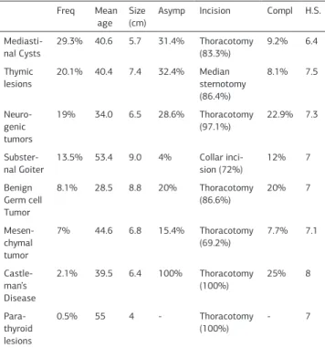
Most of the lesions (55.4%) were located in the anterior me-diastinum, 28.8% were located in posterior meme-diastinum, and 15.8% were located in the middle mediastinum. The most com-mon benign mediastinal lesions were mediastinal cysts (29.3%), thymic lesions (20.1%), and neurogenic tumors (19%) (Table 1). The other lesions were intrathoracic thyroid (13.5%), germ cell tumors (8.1%), mesenchymal tumors (7%), Castleman disease (2.1%), and parathyroid lesions (0.5%).
Most of the benign mediastinal masses were located in the anterior mediastinum (44.5%); 26.1% were asymptomatic. The most common masses were thymic lesions in the anterior medi-astinum, mediastinal cysts in the middle medimedi-astinum, and neu-rogenic tumors in the posterior mediastinum. General features of the benign mediastinal masses are shown in Table 2.
A 35-year-old male patient with a 6cm bronchogenic cyst in the middle mediastinum, a 48-year-old male patient with a 7cm pericardial cyst in the anterior mediastinum, and a 69-year-old male patient with an 18cm mature cystic teratoma in the ante-rior mediastinum were admitted with hemoptysis due to bron-chial rupture. A postoperative prolonged air leak occurred in the mature cystic teratoma case.
Myasthenia gravis was observed in three cases: a 33-year-old female patient with a 4cm thymic hyperplasia, a 49-year-old female patient with a 9cm thymic hyperplasia, and a 52-year-old female patient with a 1.5cm thymolipoma. No postopera-tive complications occurred in any of these patients. However, a 15-year-old female patient with a 14cm thymic hyperplasia
Table 1. Frequency of locations of benign mediastinal masses
Anterior Middle Posterior TOTAL
Mediastinal cysts 24 15 15 54 (29.3%)
Thymic lesions 33 4 - 37 (20.1%)
Neurogenic tumors 1 6 28 35 (19%)
Intrathoracic thyroid 23 1 1 25 (13.5%)
Benign Germ Cell Tumors
15 - - 15 (8.1%)
Mesenchymal Tumors 4 3 6 13 (7%)
Castleman’s disease 1 - 3 4 (2.1%)
Parathyroid Lesions 1 - - 1 (0.5%)
TOTAL 102 (55.4%) 29 (15.8%) 53 (28.8%) 184
Table 2. General features, surgical approaches, and treatment results of benign mediastinal masses
Freq Mean
age Size (cm)
Asymp Incision Compl H.S.
Mediasti-nal Cysts
29.3% 40.6 5.7 31.4% Thoracotomy
(83.3%)
9.2% 6.4
Thymic lesions
20.1% 40.4 7.4 32.4% Median
sternotomy (86.4%)
8.1% 7.5
Neuro-genic tumors
19% 34.0 6.5 28.6% Thoracotomy
(97.1%) 22.9% 7.3
Subster-nal Goiter 13.5% 53.4 9.0 4% Collar inci-sion (72%) 12% 7
Benign Germ cell Tumor
8.1% 28.5 8.8 20% Thoracotomy
(86.6%)
20% 7
Mesen-chymal tumor
7% 44.6 6.8 15.4% Thoracotomy
(69.2%) 7.7% 7.1
Castle-man’s Disease
2.1% 39.5 6.4 100% Thoracotomy
(100%) 25% 8
Para-thyroid lesions
0.5% 55 4 - Thoracotomy
(100%) - 7
Benign Mediastinal Tümörlerin Cerrahi Tedavi Sonuçları / Surgical Treatment Results of Benign Mediastinal Tumors
with accompanying autoimmune aplastic anemia required me-chanical ventilation for 24 hours postoperatively.
The most commonly preferred incision was thoracotomy, used in 61.9% of the cases in this study. The other incisions were me-dian sternotomy (25%), collar incision (9.8%), mediastinoscopy (1.6%), collar + median sternotomy (1.1%), and collar incision + right thoracotomy (0.6%).
Median sternotomy was the most common incision used for resecting anterior mediastinal masses; thoracotomy was per-formed most commonly to remove middle mediastinal tumors; and most posterior mediastinal tumors were removed via tho-racotomy. In only one case, a collar incision and right thoracot-omy was performed to resect a retrosternal goiter with ectopic thyroid in the posterior mediastinum (Table 3).
Mediastinoscopy was used for two pericardial cysts and a bronchogenic cyst; the median size was 3.3 cm. There were no complications or recurrences ater resection during the median followup period of 6.7 months (range, 2–12 months).
A 62-year-old female patient who underwent myoplasty to re-move a 10cm schwannoma in the posterior mediastinum expe-rienced postoperative hemorrhagic drainage.
Collar incision and median sternotomy was performed in two cases: a 13cm retrosternal goiter and an 8cm schwannoma that extended from the neck to the middle mediastinum. Ater resec-tion, the patient with the 13cm retrosternal goiter experienced hematoma, which was treated with lavage.
A 67-year-old male patient with an 8cm retrosternal goiter had vena cava superior syndrome, and respiratory failure occurred due to compression, for which he underwent mechanical venti-lation for 50 days preoperatively. Ater resection, a hematoma that occurred in the collar incision needed to be drained with a catheter.
Collar incision and right thoracotomy was performed in a sub-sternal goiter with posterior mediastinal ectopic thyroid case; no complications occurred.
A patient with thymic hyperplasia and myasthenia gravis had respiratory failure postoperatively and required mechanical ventilation for 24 hours. In addition, a 65-year-old female pa-tient with a 4.2cm pericardial cyst underwent right thoracotomy for total resection. In the second year of followup, a lung hernia developed through the thoracotomy incision, and the chest wall defect was repaired with mesh.
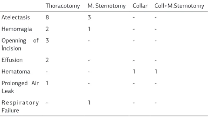
Early postoperative complications (postoperative 1–30 days) occurred in 12.5% of the cases in this study (Table 4); the most common complication was atelectasis. Other complications, in order of frequency, were hemorrhage, wound infection, hema-toma, efusion, prolonged air leak, and respiratory failure. The complications occurred in 14% of thoracotomies, 10.9% of
me-dian sternotomies, 5.6% of collar incisions, and one of the two collar incision and median sternotomy cases. When the cases were evaluated in terms of histopathologic types, the complica-tion rates were 25% for Castleman disease, 22.9% for neuro-genic tumors, 20% for germ cell tumors, 9.3% for cystic lesions, 8.1% for thymic lesions, 8% for substernal thyroids, and 7.7% for mesenchymal tumors. There was no recurrence or death in any of the 184 cases during the followup period.
Discussion
While mediastinal tumors are rare, malignant tumor rates have been increasing in recent years [1, 2]. Many studies have evalu-ated benign and malignant tumors together, and according to their results, the mediastinal masses are located, in order of frequency, in the anterior, posterior, and middle mediasti-num [3]. The most common lesions are thymoma in the ante-rior mediastinum, congenital cysts in the middle mediastinum, and neurogenic tumors in the posterior mediastinum [4]. While neurogenic tumors are the most common mediastinal tumors found in children, the most common mediastinal tumor in adults is thymoma [5].
We observed in our study that most of the benign mediastinal masses were located in the anterior mediastinum. The most common benign mediastinal masses were thymic lesions in the anterior mediastinum, cystic lesions in the middle mediastinum, and neurogenic tumors in the posterior mediastinum. In many studies, thymoma is the most common mass found in the medi-astinum. However, in our study, mediastinal cystic lesions were more common than thymomas. Neurogenic tumors were the most common benign lesions found in children, and the most common masses in adults were cystic lesions.
Complaints associated with mediastinal masses and lesions vary according to the location of the mass, complications, and secretion of hormones and cytokines. Symptomatic patients are more likely to have malignant masses. A previous study reported that only 46% of patients with benign tumors were symptomatic, compared to 85% of patients with malignancies [4]. In our study, 73.9% of the patients were symptomatic. In order of frequency, the most common symptoms were dyspnea, chest pain, cough, swelling in the neck, fever, back pain, dys-phagia, hemoptysis, myasthenia gravis, sweating, nausea, and restlessness.
Thoracic computed tomography (CT) is used to identify medi-astinal masses and their relationship to the surrounding struc-tures, as well as to determine cystic, solid, vascular, and sot-Table 3. Choice of incision and tumor location
Anterior Middle Posterior Total Percentage
Thoracotomy 42 20 52 114 61.9%
Median sternotomy 44 2 - 46 25%
Collar incision 17 1 - 18 9.8%
Mediastinoscopy 2 1 - 3 1.6%
Collar+M.Sternotomy 1 1 - 2 1.1%
Collar+Thoracotomy - - 1 1 0.6%
Table 4. Evaluation of incisions and complication frequency
Thoracotomy M. Sternotomy Collar Coll+M.Sternotomy
Atelectasis 8 3 -
-Hemorragia 2 1 -
-Openning of
İncision 3 - -
-Efusion 2 - -
-Hematoma - - 1 1
Prolonged Air Leak
1 - -
-Respiratory Failure
-Benign Mediastinal Tümörlerin Cerrahi Tedavi Sonuçları / Surgical Treatment Results of -Benign Mediastinal Tumors
tissue structures. CT was used for preoperative evaluations in all of the cases in our series. In addition, magnetic resonance, positron emission tomography CT, echocardiography, CT angi-ography, neck CT, thyroid ultrasonangi-ography, thyroid and parathy-roid scintigraphy, and esophagoscopy were used in 26.1% of the patients in this study.
If investigation results show that a mediastinal mass is likely to be benign, it can be removed surgically without biopsy [4]. Oth-erwise, transthoracic or transbronchial needle biopsy, medias-tinoscopy, anterior mediastinotomy, or video-assisted thoracic surgery can be used for diagnosis, depending on the anatomic location and radiographic appearance of the lesion. The sensi-tivity of transthoracic needle biopsy is 42–91% and the speci-icity is 96–100% for anterior mediastinal masses [6]. Surgical resection is required for patients that cannot be diagnosed with noninvasive methods. Pulmonary atelectasis, compression of adjacent structures, adhesions, and malignant transformation do not preclude the surgery [7].
Surgical resection is recommended for bronchogenic cyst cases, due to risk of malignant transformation, deinitive diagnosis, and perforation prevention, even if the patients are asymptom-atic [8]. Recurrence can occur because of incomplete resection. In this study, a bronchogenic cyst case presented with hemop-tysis due to rupture of the cyst, and no complications occurred ater surgery. We performed a subtotal resection with medias-tinoscopy for a 3cm bronchogenic cyst, and no complications or recurrence occurred. We found no malignant transformation in any of our bronchogenic cyst cases.
Myasthenia gravis is an autoimmune disease, and 15.6% of those patients have thymic hyperplasia [9]. In our series, myas-thenia gravis was observed in three patients: two thymic hyper-plasia cases and one thymolipoma. Diaz’s review showed that myasthenia gravis patients undergoing thymectomy were likely to achieve improvement and medication-free remission and to become asymptomatic, compared to patients who did not un-dergo thymectomy [10]. Thymic cysts represent 1% of medi-astinal masses [11], and while some authors have pointed out that surgery is not required if the deinitive diagnosis is thymic cyst, most publications suggest resection due to the potential of malignancy [12-15].
Most pericardial cysts are asymptomatic and are detected in-cidentally. The 2004 European Society of Cardiology guidelines suggest that the treatment for congenital and inlammatory cysts is percutaneous aspiration and ethanol sclerosis [16]. In our study, 57.9% of the cases were symptomatic, and one pa-tient had hemoptysis due to cyst perforation. Aspiration and subtotal cyst wall excision with mediastinoscopy were per-formed in two cases, and total resection with thoracotomy was performed in the other cases. Hemorrhagic drainage and wound infection occurred in the patients who underwent thoracotomy; however, none of the patients experienced recurrence.
When more than 50% of the thyroid parenchyma is located be-low the sternal notch, it is called retrosternal goiter. The stan-dard treatment for substernal goiter with compression symp-toms is surgery. If surgery is not feasible, radioiodine can be an alternative treatment choice, especially for patients with high risks of complications [17].
Rupture of mature cystic teratoma into the bronchus, pleura,
pericardium, or mediastinum occurs rarely, but it can lead to serious complications [18]. We observed in our study a mature cystic teratoma case with rupture into the bronchus and occur-rence of hemoptysis. The mass size was 17.8 cm, and a tho-racotomy was performed for resection, ater which a postop-erative prolonged air leak occurred. However, the patient was discharged on the tenth postoperative day.
Mediastinoscopy can be used for histological diagnosis and treatment of anterosuperior-located mediastinal masses, with very low morbidity and mortality rates [19]. A previous review determined that morbidity, recovery times, and discharge times were all higher with more invasive procedures compared to me-diastinoscopy. The researchers also mentioned that although total excision of the cyst wall is diicult via mediastinoscopy, removal of more than 90% of the cyst wall is necessary for absorption of luid secreted by the remnant tissue through the surrounding structures. It is important to follow up carefully with these patients to monitor them for recurrence. We per-formed mediastinoscopies for three mediastinal cystic lesions (two pericardial cysts and one bronchogenic cyst), with no com-plications or recurrence.
Video-assisted thoracoscopic excision of mediastinal masses is a safe and feasible approach that might ofer signiicant postoperative advantages over open procedures. In a previous study, the border of indication for thoracoscopic resection in cystic mediastinal tumors was described as smaller than 7 cm [20]. The researchers also suggested that mediastinal cysts with wall thicknesses less than 5 mm and no FDG accumulation might be observed without resection, due to the very unlikely chance of being a neoplasm. During the study development period, we did not have enough experience in VATS resection therefore the results were not evaluated in this study.
The incision of choice for diagnosis and treatment depends on the lesion location, its characteristics, and the patient’s clinical manifestations. We used median sternotomy more frequently for anterior mediastinal masses. Median sternotomy is more suitable for lesions located in the anterior side of the middle mediastinum, and thoracotomy is a better choice for the poste-rior region of the middle mediastinum. Thoracotomy is best for exploration of posterior mediastinal masses.
Median sternotomy complications occur in less than 3% of pa-tients. Infections oten develop due to unsuccessful bleeding control, and 0.2–3% of patients develop mediastinitis [21-22]. The most common complication of thoracotomy is hemorrhagic drainage. In our series atelectasis was the most common com-plication, and thoracotomy complications were more frequent than with other incisions.
Surgery is the management of choice for patients with benign mediastinal lesions [23]. With minimal operative risk, we can achieve a deinite histological diagnosis and total excision of the lesion.
Conclusion
medi-Benign Mediastinal Tümörlerin Cerrahi Tedavi Sonuçları / Surgical Treatment Results of medi-Benign Mediastinal Tumors
astinal tumors. However, surgery provides diagnosis and treat-ment of mediastinal lesions. In this study, we evaluated a large series of benign mediastinal lesions that underwent resection, and we compared our results with others in the literature.
Competing interests
The authors declare that they have no competing interests.
References
1. Rao Aroor A, Prakasha SR, Seshadri S, Teerthanath S, Raghuraj U. A Study of Clinical Characteristics of Mediastinal Mass. J Clin Diagn Res 2014;8(2):77–80. 2. Vaziri M, Pazooki A, Zahedi-Shoolami L. Mediastinal masses: Review of 105 cases. Acta Medica Iranica 2009;47(4):297–300.
3. Donahue JM, Nichols FC. Primary mediastinal tumors and cysts and diagnostic investigation of mediastinal masses In: General Thoracic Surgery. Eds: Shields TW, Lo Cicero III J, Reed CE, Feins RH. Philadelphia: Lippincott Williams & Wilkins; 2009.p.2195-9.
4. Duwe BV, Sterman DH, Musani AI. Tumors of the Mediastinum. Chest 2005;128(4):2893-909.
5. Juanpere S, Cañete N, Ortuño P, Martínez S, Sanchez G, Bernado L. A diagnostic approach to the mediastinal masses. Insights Imaging 2013;4(1):29–52. 6. Herman SJ, Holub RV, Weisbrod GL, Chamberlain DW. Anterior mediastinal masses: utility of transthoracic needle biopsy. Radiology 1991;180(1):167-70. 7. Eryiğit H, Ürek Ş, Örki A, Aksoy F, Kutlu CA. Sol parakardiak kitlelerin ayırıcı tanısında matür kistik teratom. Akciger 2006;12(2):92-5.
8. Cuypers P, De Leyn P, Cappelle L, Verougstraete L, Demedts M, Denefe G. Bron-chogenic cysts: a review of 20 cases. Eur J Cardio-thorac Surg 1996;10(6):393-6.
9. Akaishi T, Yamaguchi T, Suzuki Y, Nagane Y, Suzuki S, Murai H, et al. Insights into the classiication of myasthenia gravis. PLoS One 2014;9:e106757; DOI: 10.1371 /journal.pone.0106757.
10. Diaz A, Black E, Dunning J. Is thymectomy in non-thymomatous myasthenia gravis of any beneit? Interact Cardiovasc Thorac Surg 2014;18(3):381-9. 11. Bieger RC, McAdams AJ. Thymic cysts. Arch Pathol 1966;82(6):535–41. 12. Hirano S, Motohara T, Nishibe T, Narita Y, Ohkubo T, Takahashi T. Cystic medi-astinal tumor: a clinical study. J Jpn Assoc Chest Surg 1997;11(1):13-9.
13. Suster S. Multilocular thymic cyst: an acquired reactive process. Study of 18 cases. Am J Surg Pathol 1991;15(4):338-98.
14. Rastegar H, Arger P, Harken AH. Evaluation and therapy of mediastinal thymic cyst. Am Surg 1980;46(4):236-8.
15. Morresi-Hauf A, Wöckel W, Kirchner T. Thymic cyst with initial malignant trans-formation. In: Pathologe 2008;29(4):308-10.
16. Maisch B, Seferovic PM, Ristic AD, Erbel R, Rienmüller R, Adler Y, et al. Guide-lines on the diagnosis and management of pericardial diseases executive sum-mary; The Task force on the diagnosis and management of pericardial diseases of the European society of cardiology.Eur Heart J 2004;25(7):587-610.
17. Ríos A, Rodríguez JM, Canteras M, Galindo PJ, Tebar FJ, Parrilla P. Surgi-cal Management of Multinodular Goiter With Compression Symptoms. Arch Surg 2005;140(1):49-53.
18. Cheung YC, Ng SH, Wan YL, Pan TK. Ruptured mediastinal cystic tera-toma with intrapulmonary bronchial invasion: CT demonstration. Br J Radiol 2001;74(888):1148-9.
19. Burjonrappa SC, Taddeucci R, Arcidi J. Mediastinoscopy in the treatment of mediastinal cysts. JSLS 2005;9(2):142-8.
20. Hida Y, Muto J, Kaga K, Kato T, Ishikawa K, Nakada-Kubota R, et al. Indica-tion of video-assisted thoracic surgery for mediastinal mass lesions. Kyobu Geka 2012;65(11):934-8.
21. LoCicero III J. Sternotomy and Thoracotomy for Mediastinal Disease In: Shields TW, Lo Cicero III J, Reed CE, Feins RH eds. General Thoracic Surgery. Philadelphia: Lippincott Williams & Wilkins, 2009.p.2153-6.
22. Olbrecht VA, Barreiro CJ, Bonde PN, et al. Clinical outcomes of noninfectious sternal dehiscence ater median sternotomy. Ann Thorac Surg 2006;82(3):902-7. 23. Dosios T, Kuoskos E, Kyriakou V. Surgical Management of Mediastinal Lesions. Tuberk Toraks 2006;54(3):207-12.
How to cite this article: