J of Evolution of Med and Dent Sci/ eISSN- 2278-4802, pISSN- 2278-4748/ Vol. 3/ Issue 10/Mar 10, 2014 Page 2536
HISTOLOGICAL PATTERN OF OVARIAN TUMORS IN A TERTIARY CARE
HOSPITAL IN KUMAON REGION OF UTTARAKHAND: A FIVE YEAR
RETRO-PROSPECTIVE STUDY
Yuman Hasan1, Ghazala Rizvi2, Hari Shankar Pandey3
HOW TO CITE THIS ARTICLE:
Yuman Hasan, Ghazala Rizvi, Hari Shankar Pandey. Histological pattern of Ovarian Tumors in a Tertiary care Hospital in Kumaon Region of Uttarakhand: A five year Retro-Prospective Study. Journal of Evolution of Medical and Dental Sciences 2014; Vol. 3, Issue 10, March 10; Page: 2536-2542,
DOI: 10.14260/jemds/2014/2169
ABSTRACT: BACKGROUND: Ovarian cancer is the fourth leading cause of death in females around the world and therefore an area of concern for health care professionals. The further distressing aspect of these tumors is that they do not manifest any symptoms till they are clinically advanced or have attained a considerable size. AIMS: To study the histological pattern of ovarian tumors and compare it with other studies. SETTINGS AND DESIGN: Retro- prospective study. METHODS AND MATERIAL: A retro prospective study was done on 131 ovarian tumors received over a period of five years. Data including the age, clinical presentation, related history, involvement (whether unilateral or bilateral) were obtained from the histopathological requisition forms of the patients. Histopathology reports of all the cases were recorded from the data base. The histological categorization of ovarian tumors was done according to the WHO Classification of 2003. STATISTICAL ANALYSIS USED: None. RESULTS: Out of the 131 tumors studied, 87 (66.41%) were benign, 32 (24.42%) were malignant and 12 (9.16%) were borderline. Surface epithelial tumors (67.17%), including benign, borderline and malignant, contributed the bulk of the cases. Ovarian tumors were seen in younger age group and borderline tumors, both serous and mucinous, were reported more compared to other studies. Mucinous cystadenocarcinoma was the most common malignancy. CONCLUSIONS: Ovarian tumors once detected should be removed immediately without further delay. A very extensive sampling and careful histopathological examination should be the rule as the patient’s management and prognosis depends largely on the histologic type of tumor.
KEYWORDS: Ovarian tumors, histologic type, borderline tumors, malignant.
KEYMESSAGES: Ovarian cancer is among the leading causes of death in females and is notorious for not manifesting any symptoms until late. The management and prognosis depends on the histologic type of tumor. Thus, immediate removal, extensive sampling and careful histopathological examination should be the rule while dealing with such cases.
INTRODUCTION: Ovarian tumors constitute 23% of all gynecologic tumors with ovarian carcinoma being the sixth most common female cancer and the fourth leading cause of death due to cancer in women. This therefore makes this one of the major health concerns for women all over the world.1 In
eastern India, the fourth most frequent reported malignancy in females was ovarian.2 The
Surveillance Epidemiology and End Results (SEER) calculations of the lifetime risk for ovarian cancer indicate that 1 in 55 women will develop ovarian cancer over their lifetime.3
J of Evolution of Med and Dent Sci/ eISSN- 2278-4802, pISSN- 2278-4748/ Vol. 3/ Issue 10/Mar 10, 2014 Page 2537 000 women per year.4Regrettably, there is no effective screening test as the ovaries are relatively
inaccessible and precursor lesions are largely unknown. As mentioned earlier ovarian cancers often present themselves in advanced stage therefore any precursor lesion that may have been present are not recognized. Furthermore, identification of any putative precursor lesion within the ovary would generally involve the removal of that ovary and therefore the natural history of the lesion cannot be observed. Nulliparity and family history are considered as the risk factors for ovarian cancers. A higher frequency of carcinoma is seen in unmarried women and in married women with low parity.5
The relative frequency of the various ovarian tumors differs in the Western and Asian countries. Surface epithelial tumors account for 50-55% of all ovarian tumors with their malignant counterparts contributing 90% of all ovarian cancers in the Western world.
Japan on the other hand gives a figure of 46-50% and 70- 75% respectively. Germ cell tumors account for 30% of primary ovarian tumors and malignant germ cell tumors account for 3% of ovarian cancers in the western world.6 Bulk of the ovarian tumors occurs in the reproductive age
group with only less than 5% in children. 75-80% of these tumors are benign and 55-65% of benign tumors occur in women under 40 years of age. Serous tumors are uncommon in the first two decades whereas mucinous are more often seen between fourth to sixth decade. In patients under 20 years, 60% are germ cell tumors.
Diverse histopathology is seen in ovarian tumors which reflects the different cell of origin of these the tumors. This is also the basis of the classification of these tumors. Determining the various histologic patterns in ovarian tumors is important not only for diagnosis but also for the prognosis. The prognosis can be predicted from the degree of differentiation of these tumors. The stage and laterality can help to determine the nature; sex cord stromal tumors are mostly confined to a single ovary.7
Our hospital being the sole Government Medical College in this region, patients come not only from the adjoining and far flung areas in the hills but from plains as well specially those who cannot afford the luxury of expensive private nursing homes and hospitals. We cater to both rural as well as urban population in our area.
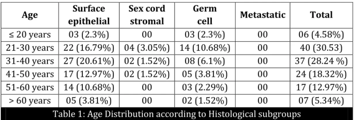
J of Evolution of Med and Dent Sci/ eISSN- 2278-4802, pISSN- 2278-4748/ Vol. 3/ Issue 10/Mar 10, 2014 Page 2538 RESULTS: The total number of patients over a period of five years was 117, out of which 14 patients had bilateral tumors. Therefore the total number of tumors studied in this period were 131. Out of these 87(66.41%) were benign, 32(24.42%) were malignant and 12(9.16%) were borderline. Surface epithelial tumors (67.17%), including benign, borderline and malignant, contributed the bulk of the cases.
Age Surface epithelial
Sex cord stromal
Germ
cell Metastatic Total
≤ 20 years 03 (2.3%) 00 03 (2.3%) 00 06 (4.58%)
21-30 years 22 (16.79%) 04 (3.05%) 14 (10.68%) 00 40 (30.53) 31-40 years 27 (20.61%) 02 (1.52%) 08 (6.1%) 00 37 (28.24 %) 41-50 years 17 (12.97%) 02 (1.52%) 05 (3.81%) 00 24 (18.32%) 51-60 years 14 (10.68%) 00 03 (2.29%) 00 17 (12.97%) > 60 years 05 (3.81%) 00 02 (1.52%) 00 07 (5.34%)
Table 1: Age Distribution according to Histological subgroups
Maximum number of surface epithelial tumors was seen in the age group of 31-40 years and minimum were seen below 20 and over 60 years. Germ cell tumors were seen in a younger age group as expected. Overall the maximum number of tumors was seen in the age group of 21-30 years (Table 1). Among the surface epithelial tumors benign serous cystadenoma was the most commonly occurring tumor (29.54%) with 31-40 years being the age group with maximum tumors. Mucinous cystadenocarcinoma was most common among malignant surface epithelial tumors. (Figure 1)
Germ cell tumors were the next most occurring histological type seen (26.71%). Mature cystic teratoma accounted for maximum number of cases (n=32). Only two yolk sac tumors were seen. No metastatic tumors were seen in our study. Mature cystic teratoma was most common among benign (25.80%) and mucinous cystadenocarcinoma (53.12%) among the malignant tumor ovarian tumors in our study.
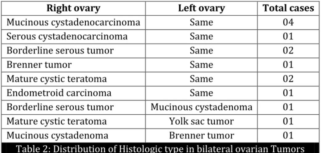
J of Evolution of Med and Dent Sci/ eISSN- 2278-4802, pISSN- 2278-4748/ Vol. 3/ Issue 10/Mar 10, 2014 Page 2539 In 14 cases bilateral ovarian tumors were seen, in which eleven cases had the same type of tumor in both the ovaries. Three cases showed different type in both the ovaries (Table 2)
Right ovary Left ovary Total cases
Mucinous cystadenocarcinoma Same 04
Serous cystadenocarcinoma Same 01
Borderline serous tumor Same 02
Brenner tumor Same 01
Mature cystic teratoma Same 02
Endometroid carcinoma Same 01
Borderline serous tumor Mucinous cystadenoma 01
Mature cystic teratoma Yolk sac tumor 01
Mucinous cystadenoma Brenner tumor 01
Table 2: Distribution of Histologic type in bilateral ovarian Tumors
DISCUSSION: In our study 66.41% of ovarian tumors were benign and 24.42% were malignant and 9.16% were borderline tumors. Similar studies in India carried out by Pilli et al8 showed 75.2%
benign ovarian tumors, 21.9% malignant and 2.8% borderline tumors. Whereas this figure was 66.03% for benign, 31.60% for malignant and 2.35% for borderline tumors in study by Prabhakar et al.9 In Nepal R Jha et al reported 83.9% benign, 16.1% malignant and 2.8% borderline ovarian
tumors.10 In Pakistan a similar study by Ahmad et al showed 59.18% benign and 40.81% malignant
tumors.11 Compared to other studies where borderline tumors are 2-3%, our study showed a higher
figure of 9.16%. A comparison of the results of our study with other similar studies conducted is shown in Table 3.
Type of ovarian tumor
Pilli et al
Prabhakar et al
Jha et al
Ahmad et al
Present study Benign tumors 75.2% 66.03% 83.9% 59.18% 66.41% Malignant tumors 21.9% 31.60% 16.1% 40.81% 24.42% Borderline tumors 2.8% 2.35% 2.8% 3.27% 9.16%
Table 3: Comparison with other studies
Among the histological types, most prevalent category encountered in our study was surface epithelial tumors, which was also seen in other studies in the West12 as well as the neighboring
countries. The distribution of various ovarian tumors based on the histological type has been compared with other studies in Table 4.
Histologic type
Pudasaini et al
Pilli et al
Ahmad et al
Jha et al
Present study Surface epithelial 69.5% 70.9% 63.50% 52.2% 67.17%
J of Evolution of Med and Dent Sci/ eISSN- 2278-4802, pISSN- 2278-4748/ Vol. 3/ Issue 10/Mar 10, 2014 Page 2540 Sex cord stromal 2.5% 6.7% 5.84% 3.1% 6.10%
Metastatic 8.5% 0.7% 2.45% 2.4% 0%
Table 4: Comparison of Histologic type of tumor
In our study mature cystic teratoma was most common benign tumor which is similar to the study by Jha et al and Ahmad et al but Pilli et al and Pudasaini et al have reported serous cystadenoma as the most common benign tumor. Thanikasalam et al13 in Malaysia showed teratomas
as highest occurring benign tumor in Malays and Chinese whereas serous cystadenoma was most common occurrence in Indians. Most common malignant tumor was mucinous cystadenocarcinoma in our study which was similar to study by Pilli et al but in contrast to Ahmad et al where serous cystadenocarcinoma was found to be most common (Table 5).
Histologic type Pilli et al Ahmad et al Present Study
Serous Cystadenoma 42.9% 34.26% 19.84%
Borderline serous - 1.63% 3.05%
Serous cystadenocarcinoma - 12.51% 3.05%
Mucinous cystadenoma 25.5% 10.76% 13.74%
Borderline mucinous - 1.63% 6.10%
Mucinous cystadenocarcinoma - 6.43% 12.97%
Endometroid carcinoma 0.7% 4.91% 4.58%
Clear cell Carcinoma 0.35% 0.81% 0.76%
Brenner - 0.46% 3.05%
Granulosa cell tumor 6.7% 2.80% 5.64%
Malignant steroid tumor - - 0.8%
Mature cystic teratoma 17% 20.81% 25.80%
Dysgerminoma 2.48% 2.69% 0.8%
Yolk sac tumor 1.77% 0.93% 1.61%
Table 5: Comparison of the distribution of Histologic type of tumor
The peak incidence of ovarian tumors in our study was seen in 31-40 years age group which is similar to reports by Pudasaini et al and Kayastha et al.14 Maximum cases of benign serous
cystadenocarcinoma were also seen in the same age group but mucinous cystadenocarcinoma was seen in older age group of 41-50 years. This is different from the study by Ahmad et al where borderline as well as malignant mucinous tumors were in the younger age group but similar to study by Jha et al where the mean age was 50.7 years for mucinous cancers. Borderline malignancy was seen more compared to other studies.
J of Evolution of Med and Dent Sci/ eISSN- 2278-4802, pISSN- 2278-4748/ Vol. 3/ Issue 10/Mar 10, 2014 Page 2541 CONCLUSION: Epithelial tumors were most common tumors in our study which is similar to the data reported in the neighboring countries as well. Germ cell tumors were the next most common tumors out of which mature cystic teratoma was the most prevalent. Among the malignant tumors mucinous cystadenocarcinoma contributed the maximum number of cases. In our study maximum tumors were seen in the younger age group. Borderline tumors (both serous and mucinous included) were seen in a higher percentage than other studies. Maximum cases of borderline tumors were seen between 20-40 years. Therefore any ovarian tumor in this age group should be surgically removed immediately and sampled thoroughly to rule out borderline malignancy.
Though ovarian tumors can be diagnosed clinically as well as by radiological means, a thorough histopathological examination is mandatory to find out the origin and type of tumor as the management and prognosis of the patient is dependent to a large extent on the histological type apart from the stage and grade of the tumor.
REFERENCES:
1. Tortolerol L, Mitchell FM, Rhodes HE. Epidemiology and screening of ovarian cancer. Obstet and Gynecol Clin North Am 1994; 21:63-75.
2. Sen U, Sankararnarayanan R, Mandal S, Ramana AV, Parkin DM, Siddique M. Cancer patterns in Eastern India: The first report of Kolkata Cancer Registry. Int J Cancer 2002; 100:86-91.
3. Piver MS. Prophylactic Oophorectomy: Reducing the U.S Death Rate from Epithelial Ovarian Cancer. A Continuing Debate. Oncologist1996; 1: 326-30.
4. Murad A. Ovulation induction and ovarian tumors, the debate continues. J Pak Med Assoc 1998; 48:353-6.
5. Piver MS, Baker TR, Jishi ME et al. A report of 658 families from the Gilds Radner Familial Ovarian Cancer Registry, 1981-1991. Cancer, 1993; 71:582-88.
6. Tavassoli FA, Devilee P. WHO Classification of tumors. Pathology and Genetics, Tumors of Breast and Female Genital Organs. IARC Press: Lyon 2003.
7. Yasmin S, Yasmin A, Asif M. Clinicohistological pattern of Ovarian in Peshawar Region. J Ayub Med Coll Abbotabad 2008; 20(4): 11-13.
8. Pilli GS, Suneeta KP, Dhanded AV, Yenni VV. Ovarian tumors: a study of 282 cases. J Indian Med Assoc 2002; 100:420, 432-4, 447.
9. Prabhakar BR, Maingi K. Ovarian tumors – prevalence in Punjab. Indian J Pathol Microbiol 1989; 32: 276-81.
10.Jha R, Karki S. Histologic pattern of ovarian tumors and their age distribution. Nepal Med Coll J 2008; 10:81-5.
11.Ahmad Z, Kayani N, Hasan SH, Muzaffar S, Gill MS. Histological pattern of ovarian neoplasm. J Pak Med Assoc 2000; 50:416-9.
12.Huusom LD, Frederiksen K, Hogdall EV, Glud E, Christensen L, Hogdall CK, et al. Association of reproductive factors, oral contraceptive use and selected lifestyle factors with the risk of ovarian borderline tumors: a Danish case-control study. Cancer Causes Control 2006; 17:821-9. 13.Thanikasalam K, Ho CM, Adeed N, Shahidan MN, Azizah WK. Pattern of ovarian tumors among
Malaysian women at General Hospital, Kuala Lumpur. Med J Malaysia 1992; 47:139-46. 14.Kayastha S. Study of ovarian tumors in Nepal Medical College Teaching Hospital.
J of Evolution of Med and Dent Sci/ eISSN- 2278-4802, pISSN- 2278-4748/ Vol. 3/ Issue 10/Mar 10, 2014 Page 2542
AUTHORS:
1. Yuman Hasan 2. Ghazala Rizvi 3. Hari Shankar Pandey
PARTICULARS OF CONTRIBUTORS:
1. Third Year Post Graduate Student, Department of Pathology, Government Medical College, Haldwani, Uttarakhand. 2. Associate Professor, Department of Pathology,
Government Medical College, Haldwani, Uttarakhand.
3. Associate Professor, Department of Pathology, Government Medical College, Haldwani, Uttarakhand.
NAME ADDRESS EMAIL ID OF THE CORRESPONDING AUTHOR:
Dr. Yuman Hasan,
III Year Post Graduate Student, Department of Pathology, Flat – 74, S. R. Hostel,
Government Medical College, Haldwani, Uttarakhand – 263139. E-mail: yuman.h@gmail.com