Congenital lung malformations*
Malformações pulmonares congênitasCristiano Feijó Andrade, Hylas Paiva da Costa Ferreira, Gilberto Bueno Fischer
Abstract
Congenital lung malformations are rare and vary widely in their clinical presentation and severity, depending mostly on the degree of lung involvement and their location in the thoracic cavity. They can manifest at any age and can be the source of significant morbidity and mortality in infants and children. Individuals with congenital lung malformations can present with respiratory symptoms at birth or can remain asymptomatic for long periods. Recently, there has been an increase in the early diagnosis of these malformations, a change that is attributable to the routine use of prenatal ultrasound. The clinical manifestation of these malformations varies from respiratory distress in the immediate postnatal period to an incidental finding on chest X-rays. Early diagnosis and prompt treatment offer the possibility of absolutely normal lung development. The treatment of asymptomatic patients with lung malformations is controversial, because the prognosis of these diseases is unpredictable. The management of these lesions depends on the type of malformation and symptoms. Because of the risk of complications, most authors recommend resection of the lesion at the time of diagnosis. Lobectomy is the procedure of choice and yields excellent long-term results. This article describes the principal congenital lung malformations, their diagnosis, and the controversies regarding treatment.
Keywords: Cystic adenomatoid malformation of lung, congenital; Bronchopulmonary sequestration; Pulmonary surgical procedures; Diagnosis.
Resumo
As malformações congênitas do pulmão são raras e variam muito na sua forma de apresentação clínica e gravidade, dependendo principalmente do grau de envolvimento pulmonar e de sua localização na cavidade torácica. Elas podem se manifestar em qualquer idade e podem ser fonte de importante morbidade e mortalidade em lactentes e crianças. Os indivíduos com malformações congênitas do pulmão podem apresentar sintomas respiratórios ao nascimento, enquanto outros podem permanecer assintomáticos por longos períodos. Atualmente, com o uso rotineiro da ultrassonografia pré-natal, vem ocorrendo um aumento no diagnóstico mais precoce dessas malformações. A manifestação clínica dessas malformações varia desde uma disfunção respiratória pós-natal imediata a um achado acidental na radiografia de tórax. O diagnóstico precoce e o tratamento imediato oferecem a possibilidade de um desenvolvimento pulmonar absolutamente normal. Quando assintomáticos, a conduta para o tratamento dos pacientes com malformações pulmonares ainda é controversa, uma vez que o prognóstico dessas afecções é imprevisível. O manejo dessas lesões depende do tipo de malformação e de sintomas. Devido ao risco de complicação, a maioria dos autores sugere a ressecção da lesão no momento em que essa é identificada. A lobectomia é o procedimento de escolha, fornecendo excelentes resultados a longo prazo. Este artigo descreve as principais malformações pulmonares congênitas, seu diagnóstico e controvérsias quanto o tratamento.
Descritores: Malformação adenomatoide cística congênita do pulmão; Sequestro broncopulmonar; Procedimentos cirúrgicos pulmonares; Diagnóstico.
* Studied carried out at the Santo Antônio Children’s Hospital, Santa Casa Sisters of Mercy Hospital Complex, Porto Alegre, Brazil. Correspondence to: Cristiano Feijó Andrade. Hospital da Criança Santo Antônio, Serviço de Cirurgia Torácica Pediátrica, Rua Annes Dias, 285, Centro, CEP 90020-090, Porto Alegre, RS, Brasil.
Tel 55 51 3214-8674. E-mail: [email protected] Financial support: None.
type of pulmonary sequestration designated bronchopulmonary foregut malformation, in which an abnormal lung is connected to the gastrointestinal tract.(16) Extralobar pulmonary sequestration, also known as accessory lung,(5) is covered by visceral pleura and separated from the functioning lung, whereas intralobar pulmonary sequestration can be completely covered by normal lung tissue or by a segment of the visceral pleura of the lung lobe within which the sequestration occurs.(11-14)
Pulmonary sequestrations are responsible for 1.1-1.8% of all pulmonary resections.(14) In rare cases, the extralobar and intralobar forms can occur simultaneously.(15) The etiology of pulmonary sequestration has yet to be fully clarified. Although there is a consensus that the extralobar form is congenital, it is still debated whether the intralobar form is congenital or acquired.(11) There is a theory that intralobar pulmonary sequestration is either congenital, arising from a lung bud that originates in the foregut, or a result of a sequence of events, including bronchial obstruction, pneumonia, local pleurisy, and parasitization of pulmonary ligament arteries.(17) No chromosomal abnormalities have been associated with pulmonary sequestration.(10)
The ratio between intralobar pulmonary sequestration and extralobar pulmonary sequestration is 3:1; intralobar pulmonary sequestration is equally distributed between the genders,(18) and extralobar pulmonary sequestration is more prevalent in the male gender (80% of cases).(10,15,18)
Extralobar pulmonary sequestrations are generally located in the lower lobes of the left lung (80%),(4,13,15,19) in close proximity to the costophrenic sulcus (Figure 1), and are less common than are intralobar pulmonary sequestrations, which account for approximately 75% of all cases of pulmonary sequestrations. (9-11,13,15,18,20) Approximately 10% of all extralobar
pulmonary sequestrations are located below the diaphragm.(18) In intralobar pulmonary sequestrations, there is no lung segment predominance(10); however, some authors have reported a greater frequency of intralobar pulmonary sequestrations in the posterior basal segment of the left lower lobe.(5,20) Such sequestrations can go undetected throughout
Introduction
Lung malformations constitute a spectrum of lesions that originate in the embryonic period. In the literature, there is no consensus regarding the nomenclature for such anomalies. Some authors eschew the use of the term congenital lung malformations(1) because they consider the terms “malformation” and “congenital” to be synonymous. Other authors lump malformations together by using the term “congenital thoracic malformations” to designate all such anomalies. (2) However, “congenital lung malformations”
is the term that is most widely used in the literature,(3-5) and that is the term that will be used in the present review.
Although there has been a significant increase in the rate at which malformations are diagnosed in the prenatal and neonatal periods, most lung malformations are identified at autopsy.(5,6) The annual incidence of congenital lung malformations ranges from 30 to 42 cases per 100,000 population, or 0.06% to 2.2% of all patients admitted to general hospitals.(6,7)
Patients with congenital lung malformations can present with respiratory symptoms at birth or can remain asymptomatic for long periods. When the diagnosis is delayed, such patients present with some type of complication, such as pulmonary infections. Therefore, it is difficult to obtain real, consistent data regarding the prevalence of congenital lung malformations(5-7); however, it has been estimated that 10% of cases are identified at birth, whereas 14% are diagnosed by 15 years of age.(8) The present review addresses the principal congenital lung malformations seen in clinical practice. Although pulmonary arteriovenous malformations (AVMs) are not restricted to the lung parenchyma, they have also been included in the review.
Pulmonary sequestration
extralobar pulmonary sequestration is composed of irregular bronchi and bronchioles, as well as by alveoli that are twice to five times their normal size.(18) Dilated lymphatic vessels in the subpleural region are found in 85% of cases.(18) Intralobar pulmonary sequestration is characterized by multiple cysts of various sizes that, upon microscopic examination, show lung parenchyma replete with inflammatory tissue and fibrosis, with vestiges of bronchi and bronchioles covered by fibrous, lymphocyte-infiltrated tissue.(18)
The diagnosis of pulmonary sequestration can be established in the prenatal period (at approximately gestational week 18) by ultrasound, which reveals a homogeneous, echodense, well-defined mass with anomalous the lifetime of the individual(18) or can manifest
as recurrent lower lobe infections.(18)
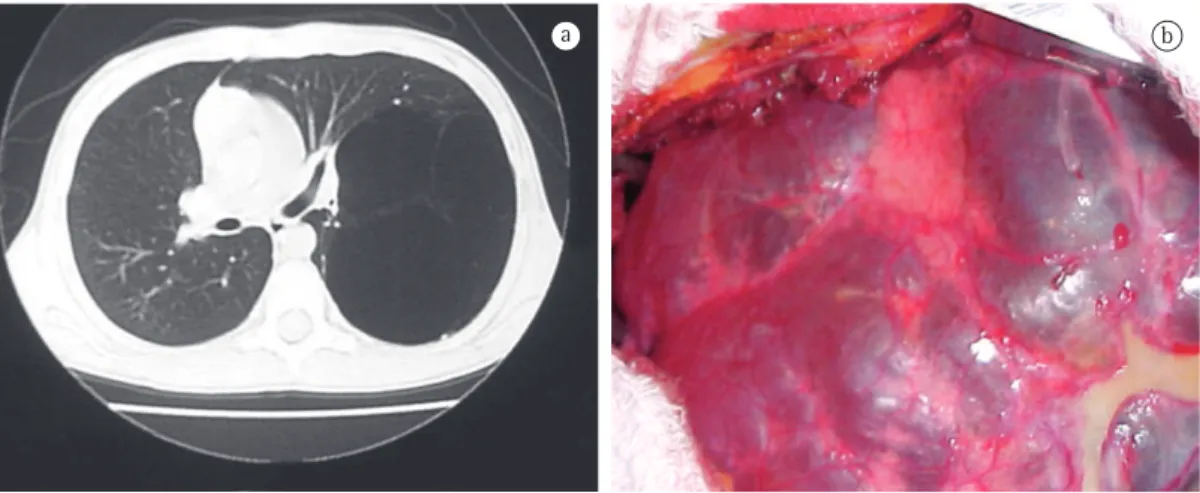
Both types of pulmonary sequestration receive their blood supply from the systemic circulation (Figure 2), generally via the descending thoracic aorta or the abdominal aorta.(4,11-13,18,19) In approximately 15% of cases, blood supply is provided by a different systemic artery.(11,13,18,19) In intralobar pulmonary sequestration, venous drainage is into the pulmonary veins,(18) whereas it is into the systemic venous system (azygos vein or portal vein) in extralobar pulmonary sequestration.(10,18,19)
Extralobar pulmonary sequestration is macroscopically characterized by an oval or pyramidal lesion of 0.5-1.5 cm that can be larger in cases of recurrent infections.(18) Histologically,
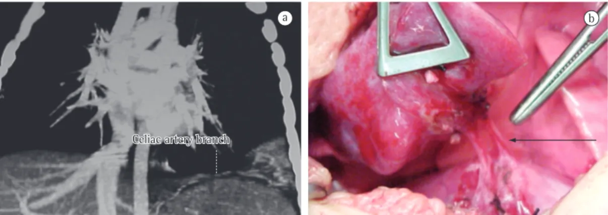
Figure 1 - Extralobar pulmonary sequestration. In a, CT scan of the chest showing lung tissue receiving arterial supply from a branch of the celiac artery. In b, intraoperative view of sequestrectomy showing the vascular pedicle (arrow). Note the consolidated appearance of the lung tissue.
in up to 50% of cases(10,11,20) or by other types of lung malformation, such as type II CCAM. (5,13,18) Case reports of concurrent extralobar and intralobar pulmonary sequestrations suggest that the two forms share a common embryonic origin.(10)
The treatment of choice is surgical resection of the lesion through sequestrectomy or pulmonary lobectomy(3,4,11,12,14,15,20) at the time of diagnosis principally because of the possibility of recurrent infections, hemorrhage, malignant transformation, and other complications. However, there is still controversy as to when resection should be performed.(8,13-15,18,20,23) Many authors recommend that asymptomatic patients with extralobar sequestration remain under observation only, because such lesions rarely cause symptoms and because there have been no reports of malignant degeneration. However, in such lesions, a mixed component, with a morphology similar to that of CCAM, cannot be ruled out. The surgical treatment of choice in cases of extralobar pulmonary sequestration is sequestrectomy in which the pedicle is carefully resected and ligated; sequestrectomy can also be performed through video-assisted thoracoscopy, a procedure that has been shown to be safe and to have a low rate of postoperative complications.(8,10-14,18,19,23,25) Sequestrectomy is also the resection of choice in cases of intralobar pulmonary sequestration, in order to preserve a greater quantity of normal lung tissue that continues to develop until approximately 8 years of age.(8) Unfortunately, it is not always possible to delineate the sequestered tissue within the affected lobe, in which case pulmonary lobectomy is required.(11,14,18,20,23)
The rate of surgical complications is relatively low in cases of pulmonary sequestration. One study(24) reported that, among 540 pulmonary resections for pulmonary sequestration, there were only 5 deaths, all of which were due to uncontrollable hemorrhage in the intraoperative period. To date, there have been no reports of intraoperative deaths.(3,8,18)
CCAM
In 2002, Stocker(26) proposed a new term to designate CCAM—congenital pulmonary airway malformation—principally because neither cysts nor adenomatoid malformations are always observed. However, for the present review, we vascularization.(11,15,21) After birth, a chest
X-ray finding of an elongated or cystic lesion, adjacent or posterior to the cardiac silhouette and described as a triangular, well-defined mass, is suggestive of the diagnosis.(11,15,18)
In the prenatal period, it can be difficult to diagnose pulmonary sequestration by ultrasound, because extralobar sequestration can be mistaken for a normal lung (the tissues having similar characteristics), whereas intralobar sequestration can be incorrectly diagnosed as congenital cystic adenomatoid malformation (CCAM).(5) Nuclear magnetic resonance imaging can be useful in clarifying the diagnosis in such cases. (22) Extralobar pulmonary sequestration accounts
for approximately 23% of the pulmonary lesions that are diagnosed in the prenatal period.(18) Of those, up to 68% can disappear over the course of the prenatal radiological follow-up period.(11) A CT scan of the chest reveals the sequestered lung tissue and its vascularization, as well as other associated malformations, if present.(11,15) The test of choice for the postnatal diagnosis is CT angiography.(11,13,15,18) Until recently, conventional angiography was the gold standard for demonstrating the arterial supply and venous drainage of such lesions. Currently, CT angiography is used instead of conventional radiography because the former is less invasive and more efficient in diagnosing such lesions, showing the vascularization of the sequestered tissue in great detail and therefore allowing safer surgical planning.(10-12,19,23)
Patients who are asymptomatic at birth can subsequently develop cough, hemoptysis, and recurrent pneumonia,(11,15,19) the last being the most common finding,(18) or can remain asymptomatic and be diagnosed incidentally (as occurs in 15.5% of intralobar sequestrations and 10% of extralobar sequestrations).(24) Other patients can develop complications, such as hemoptysis, massive hemothorax, cardiovascular complications, fungal/bacterial infections, benign tumors, or malignant degeneration. (4,11,14,18)
carcinoma and to pleuropulmonary blastoma), pneumothorax, hemoptysis, and hemothorax. (4,35,36) A small number of patients present with
a more aggressive form, which is represented by an extensive, rapidly growing lesion that can lead to hydrops fetalis caused by mediastinal shift, inferior vena cava obstruction, and cardiac compression, resulting in intrauterine death.(4,15,33) In such cases, drainage can be performed in utero by means of thoracentesis or thoracoamniotic shunt placement with cyst aspiration.(15,21) The combination of CCAM and certain other malformations, such as pulmonary sequestration, has been demonstrated in various studies, raising the hypothesis that type II CCAM and extralobar pulmonary sequestration have the same embryonic origin.(13,15,18,20,27,30,34,35) However, it is much less common for CCAM to be accompanied by malformations such as facial defects, heart defects, neural tube defects, renal dysplasia/agenesis, and omphalocele.(15,27,34)
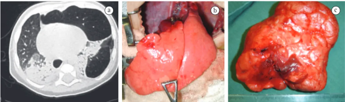
The lesions vary widely in size and can affect an entire lobe or part of it, as well as an entire lung. The lesion can affect both sides of patients with equal frequency and can affect any lung lobe, although it occurs more frequently in the lower lobes and rarely affects more than one lobe (85-95% in only one lobe; Figure 3), being slightly more common in males than in females.(4,15,32) Although CCAM is connected with the tracheobronchial tree and derives its blood supply from the pulmonary circulation, the affected lung area shows reduced vascularization. (15) The incidence of CCAM is approximately 1 case per 10,000-35,000 pregnancies,(4,15,37,38) and CCAM has not been associated with race, age, maternal exposure to any given factor, or genetic factors.(32)
One group of authors(39) conducted a detailed study of CCAMs based on 38 cases and divided CCAMs into 3 types, on the basis of the histological characteristics of the malformations. Initially, CCAMs were classified as types I, II, and III, on the basis of the size of their cysts and histological characteristics,(4,5,15,28,30,31) the frequency for types I, II, and III being 50-70%, 20-40%, and approximately 10%, respectively.(32) Subsequently, two other types of CCAM, types 0 and IV, were added to the classification.(26)
Type I CCAM (Figure 3) is characterized by a lung mass, generally confined to a lobe, containing a single cyst or multiple cysts over chose to adopt the most common term, i.e.,
CCAM. Of all congenital lung malformations, 25-30% are CCAMs,(15) and approximately 30% of all patients with CCAM are at risk of respiratory failure at birth.(27) It has been shown that CCAMs are hamartomatous lesions,(27) with focal dysplasia and anomalous development,(4,28) and are characterized by a multicystic mass of lung tissue with proliferation of bronchial structures and lung tissue showing aberrant, differentiated architecture, with various degrees of cyst formation.(5,26,29-32) The possible mechanisms by which the lesion develops and the exact period of time over which it develops have yet to be determined; however, there is evidence that CCAMs occur between gestational weeks 5 and 22 and result in a wide variety of pathological and radiological presentations.(15)
Although there are other theories for the origin of CCAM, some authors believe that it is due to an anomalous development in lung maturation,(29,30) whereas others consider CCAM to represent focal pulmonary dysplasia because skeletal muscle can be identified within the cyst walls and carcinomas can develop.(29) According to one group of authors, CCAM probably results from arrested bronchial maturation and, at the same time, overgrowth of mesenchymal elements near gestational week 5 or 6.(4,20,33) Histologically, cartilage is absent, reflecting bronchial malformation; however, patients with CCAM present with small bronchial communications, which are very likely responsible for the infections, and hyperinflation.(27,33) Cystic formations are covered with columnar or cuboidal epithelium.
Some of the microscopic findings in CCAM distinguish it from other pulmonary lesions, as well as from normal lung. Such findings include polypoid projections of the mucosa, an increase in smooth muscle and elastic tissue within cyst walls, absence of cartilage in the masses, presence of mucus-secreting cells, and absence of inflammation.(15,26)
of all CCAMs,(5,15,32,39) and can affect an entire lung or, more rarely, both lungs.(5) The cut-off surface of type III CCAM is firm and generally does not present with cystic lesions; when such lesions are present, they are no larger than 0.5 cm in diameter. Microscopic examination reveals irregularities in the bronchial structure, which is covered with small air spaces covered by cuboidal epithelium,(15,27) similar to an immature lung without bronchi.(5,15,39) Type III CCAM is more common in males than in females.(15) The mass effect of type III CCAMs generally causes mediastinal shift in the fetus and results in contralateral pulmonary hypoplasia; it can also cause hydrops fetalis as a result of caval system obstruction and cardiac compression.(15)
In 1985, one group of authors proposed a new classification, based on prenatal ultrasound, and created two new categories: macrocystic lesions, consisting of one or more cysts larger than 5 cm in diameter; and microcystic lesions, larger and more solid, containing multiple cysts smaller than 5 mm in diameter.(40)
More recently, Stocker proposed two new types of CCAM,(26) based on the type of tissue of origin of the malformation and on the stage of development of the tracheobronchial tree. (26) One of those types, type 0, originates from tracheal or bronchial tissue and presents as a component equivalent to acinar dysplasia, with incidence of 1-3% and cysts of up to 0.5 cm in diameter covered by pseudostratified epithelium. (5) Type 0 CCAM affects all lung lobes and is largely incompatible with life, as well as being associated with severe cardiac abnormalities; it is seen in preterm and term infants who are 2 cm (3-10 cm) in diameter,(5,15,27,32,39) covered
by pseudostratified ciliated epithelium, with areas of mucus-secreting epithelium covered by fibromuscular tissue,(5,15,27,32,39) and generally filled with air or mucus. This type of lesion results from injury during the canalicular or pseudoglandular period of lung development. (32) The adjacent lung tissue is normal and is generally compressed by the malformation, which has the effect of an intrathoracic mass(15,35) and can lead to respiratory failure in the neonatal period. Patients with type I CCAM generally present with symptoms in the first week of life. In extremely rare cases, type I CCAM patients can remain asymptomatic into adulthood. The combination of type I CCAM and other malformations is also rare.(15)
In general, type II CCAMs are diagnosed in the first year of life and affect only one lobe, presenting as multiple cysts of small diameter (0.5-2.0 cm) covered by bronchiolar epithelium (cuboidal or columnar) and separated by alveolar septa.(5,15,27,32,39) Type II CCAMs are similar to intralobar pulmonary sequestration; the two entities are differentiated by their histology and, in many cases, occur concomitantly in the same patient, probably arising during the canalicular period of lung development. (32) This type of CCAM has been associated with various other malformations, such as esophageal atresia, tracheoesophageal fistula, bilateral renal agenesis, and intestinal atresia, as well as with bone and central nervous system abnormalities.(5,15)
Type III CCAM is the least common of the three, accounting for approximately 5-10%
period, probably through a mechanism of apoptosis and lack of blood supply.(15,29,33,37)
Patients with symptomatic CCAM can present with a history of recurrent pneumonia, cyanosis, respiratory difficulty, and pneumothorax.(15,28) In the prenatal phase, surgical intervention is still the best treatment option for the microcystic forms, which are associated with hydrops fetalis. However, there are certain contraindications to surgery, including chromosomal abnormalities, multiple pregnancy, presence of other anatomical abnormalities, and presence of maternal or psychosocial factors. The surgical treatment of such lesions, when performed in the fetal period through open surgery, shows a survival rate of approximately 50%,(21) whereas a surgical procedure performed during cesarean delivery, while the child is still connected to the mother by the umbilical cord—a procedure designated ex utero intrapartum therapy—shows a survival rate of nearly 90%.(42) In some cases, the use of steroids (betamethasone and dexamethasone) can prevent CCAM from developing further, thus preventing hydrops fetalis.(21) The management of asymptomatic lesions is controversial; the exact length of the observation period, the type of resection, and the extent of the resection have yet to be determined.(5,8,15,33,38) However, most authors recommend resection because of the risk of infection or neoplastic transformation that such lesions carry, principally in cases of bronchioloalveolar carcinoma, myxosarcoma, pleuropulmonary blastoma, and rhabdomyosarcoma.(4,5,8,15,20,33,36-38) Recently, one group of authors studied 129 cases of CCAM and found that the incidence of pleuropulmonary blastoma among the 54 patients who underwent surgery was 4%.(43) Although such tumors are rare, over 8% are associated with CCAM. In symptomatic patients, there is consensus that the lesion must be resected, generally by means of pulmonary lobectomy, which avoids residual disease and recurrence in the remaining tissue. (33,34,37,38) One group of authors reported that only 10% of the asymptomatic patients will develop complications and that a conservative approach is an alternative for such patients.(44) Resection of the area of the lesion exclusively is not recommended.(33,38) Lobectomy is the procedure of choice for patients in whom the lesion is confined to a single lobe. However, when more than one lobe is affected, neither cyanotic at birth and survive only a few hours.(26)
The other type of CCAM proposed by Stocker,(26) type IV, has a distal acinar origin, with large peripheral cysts of up to 7 cm in diameter, and accounts for 10-15% of all cases of CCAM; type IV CCAM cases are often erroneously classified as type I.(15,26) Patients with type IV CCAM can present with respiratory dysfunction caused by pneumonia or pneumothorax and are rarely asymptomatic or diagnosed on the basis of incidental findings.(26)
Although ultrasound is accurate in detecting CCAM, diagnostic errors can occur. The differential diagnosis includes bronchogenic cysts, enteric cysts, pulmonary sequestration, congenital lobar emphysema, and mediastinal masses.(41) Although chest X-ray was the first truly accurate method for identifying pulmonary lesions, routine chest X-rays do not rule out the presence or confirm the resolution of CCAM. (33) Chest X-ray typically shows multiple, round,
thin-walled cysts filled with air or secretion; it can also show large lesions with mediastinal shift, lung herniation across the midline, and the mass effect of the lesion, as well as nonspecific consolidation.(27,32) For the diagnosis of CCAM, a CT scan of the chest is mandatory; it determines the size of the cysts, reveals associated anomalies, defines the anatomical extent of the malformation more accurately, and reveals lesions that, on chest X-rays or ultrasound of the chest, appear to be resolved.(27,28,32-34)
The histology of congenital lobar emphysema is generally normal, with mild alveolar dilation, without malformation or destruction(5) of alveolar septa. In some cases, the number of alveoli can truly increase, thus characterizing the lung or lobe as hyperplastic.(5,51)
The clinical presentation of congenital lobar emphysema varies widely, from mild lung disease to acute respiratory failure at birth and recurrent pulmonary infections,(46,50) most patients being asymptomatic in the immediate postnatal period.(46) According to one group of authors,(46) although the vast majority of patients present with symptoms before age 6 months, some can remain asymptomatic for years.(46,47) Congenital lobar emphysema is twice as common in males as it is in females(47,49) and affects, in order of frequency, the left upper lobe (in 40-50% of cases),(50,51) the middle lobe (in 30-40%), and the right upper lobe (in 20%),(49,52) although bilateral involvement has also been described. (49) One third of all patients are symptomatic at
birth, and 50% are diagnosed in the first month of life. Up to 10% of patients with congenital lobar emphysema present with associated abnormalities, the most common being cardiac abnormalities.(4) Most patients present with moderate degrees of respiratory dysfunction in the first days of life. Respiratory dysfunction worsens progressively with the passing of days as a result of increased lung hyperinflation. Patients with congenital lobar emphysema tend to present with cyanosis, which can progress to frank respiratory failure.(50)
In the prenatal period, congenital lobar emphysema can be diagnosed by ultrasound, and there have been case reports of spontaneous volume reduction and resolution, occasionally even in utero.(22,46)
Congenital lobar emphysema is generally diagnosed at birth, on the basis of a profile of respiratory failure, or through routine chest X-rays taken during routine medical visits; a routine chest X-ray reveals a hyperinflated lung lobe, sometimes accompanied by lung herniation across the midline, mediastinal shift, and areas of atelectasis in the adjacent lung parenchyma.(4) The affected lobe can appear opacified because of amniotic fluid retention.(4) The acquisition of CT scans of the chest plays an essential role in the diagnosis of congenital lobar emphysema because it allows the evaluation of bronchial bilobectomy nor pneumonectomy should be
performed; normal areas should be preserved, although there is a greater risk of prolonged air leak.(3,44) In general, the surgical procedure can be safely performed in infants between 3 and 9 months of age, with favorable clinical evolution and no need for prolonged mechanical ventilation in the postoperative period.(4,15,33,37,38) However, some authors have recommended that surgery be performed in the neonatal period, near the fifth day of life, because neonates tolerate surgical trauma better.(27,37,38) Some authors have recently reported that video-assisted thoracoscopic resection is safe and easy to perform, and that postoperative recovery is favorable.(25,33,37,45) Thoracotomy and video-assisted surgery are well-tolerated by children; there is generally no need for blood transfusions, and the rates of complications and deaths are low, the mean length of postoperative hospital stay being 2.3 days.(15,25,27,33,45) Postoperative follow-up evaluation of pulmonary function generally shows good results, with less severe radiological changes, such as mediastinal shift and hyperinflation of the remaining lobe; the latter can be explained by compensatory lung growth and by the fact that the number of alveoli increases until the age of 8 years.(8,15,27,44)
Congenital lobar emphysema
AVM
Various terms have been used to designate AVMs, including arteriovenous fistula, pulmonary arteriovenous aneurysm, pulmonary hemangioma, pulmonary cavernous angioma, and pulmonary telangiectasia.(53,54) The mean age at diagnosis is approximately 41 years,(55,56) and the incidence of AVM is 2-3 cases per 100,000 population.(57) Only 25-50% of patients with AVM present with signs and symptoms, which include cyanosis, digital clubbing, dyspnea, fatigue, and polycythemia.(53-58) Males and females are equally likely to have an AVM. (55) Of all patients with AVM, 8-20% present
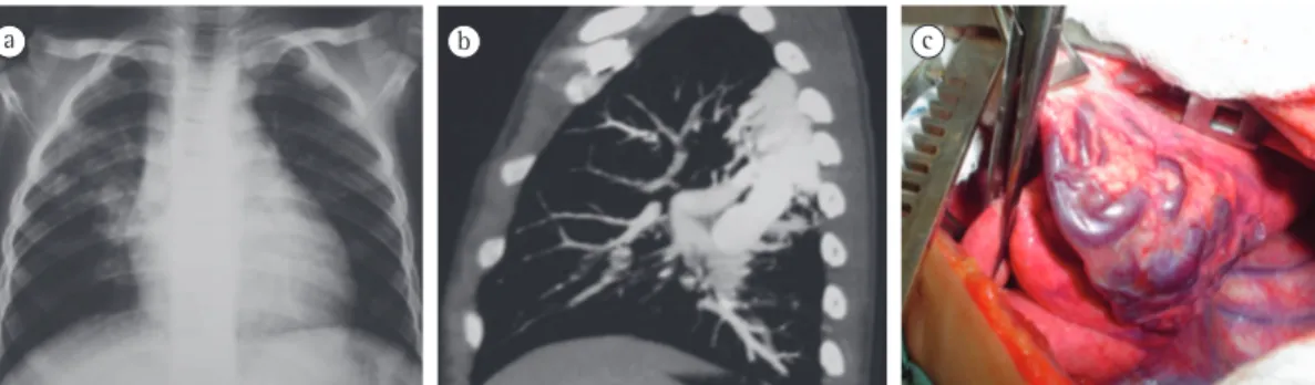
with bilateral AVM, and 33-50% present with multiple AVMs,(55-57) the incidence being higher in the middle and left lower lobes (Figure 5).(54,57)
Complications of AVMs are related to connections of the pulmonary capillary bed, with loss of the filtering function of the lung; this allows emboli and bacteria to directly exit the systemic circulation, which results in embolism or brain abscesses.(53,54,57,58) It can also result in bleeding of these abnormal vessels, leading to hemoptysis or hemothorax.(54)
The most common presentation of AVM is an abnormal and direct communication between pulmonary arteries and veins through small aneurysms,(53,58) in which right-to-left shunt occurs, although normal cardiac function is maintained in most cases. As children with AVM grow, they present with decreased oxygen saturation and polycythemia.(54,55,57) Patients with AVM generally present with a normal obstructions and anatomical changes,(4,52)
thereby defining the limits of the affected lobe and indicating the location of its blood vessels. The differential diagnosis of congenital lobar emphysema includes pneumatoceles, pneumothorax, pulmonary atelectasis, and pulmonary hypoplasia.(47)
Congenital lobar emphysema is generally treated surgically, and lobectomy through thoracotomy or video-assisted lobectomy(52) is the surgical treatment of choice in symptomatic children.(4,46,47) Although it is logical to recommend surgery for symptomatic patients and observation for asymptomatic patients, the approach to oligosymptomatic children remains controversial.(46) A small proportion of patients have been treated conservatively, with good results.(47,50) Prenatal and postnatal spontaneous resolution suggests that congenital lobar emphysema is a partially reversible process or that the growth of the normal lung parenchyma compensates for the nonfunctioning congenital lobar emphysema.(46) Fiberoptic bronchoscopy is recommended in order to rule out mucus plugging, foreign body, and changes in the airway.(3,50) In patients with congenital lobar emphysema, pulmonary lobectomy results in a minimal loss of lung volume in the remaining parenchyma, and, because of compensatory lung growth during childhood, there is no long-term difference in respiratory function.(5,8,46) Surgical treatment is safe in such patients, most case series having reported low rates of morbidity and mortality.(47)
of dyspnea/hypoxemia; prevention of hemoptysis; and, first and foremost, prevention of neurological complications. Embolization and surgical resection are the principal methods for the treatment of AVM.(53,56,58)
The treatment of choice for AVM is conservative lung resection; when the AVM is peripheral, wedge resection is the preferred procedure, whereas large lesions might require lobectomy or pneumonectomy,(53-55,57,59) embolization being reserved for patients in whom the surgical treatment is contraindicated or for those with multiple lesions.(53,57,58) Embolization yields excellent initial results, and resolution is achieved in 80-100% of cases; however, embolization has a high recurrence rate.(53,54,57) There have been reports of lung transplantation as a form of treatment for diffuse AVMs.(57) According to one group of authors,(60) embolization has its limitations and specific indications, primarily because of the risk of migration of the embolization coils in the long term, which underscores the recommendation for surgery rather than embolization. The mortality rate among untreated symptomatic patients ranges from 4% to 22%, and it can be as high as 40%.(53,57)
Final considerations
Congenital lung malformations constitute a heterogeneous group of lung diseases. The number of cases that are diagnosed early has increased thanks to new diagnostic techniques, such as CT, prenatal ultrasound, CT angiography, and nuclear magnetic resonance imaging. A CT scan of the chest is the gold standard for the echocardiogram, and chest X-rays reveal
opacification in the affected area.(57)
The histological presentation of AVM varies widely, from diffuse telangiectasia to complex structures consisting of an aneurysmal sac dilated by the confluence of arteries and veins. (53) Approximately 95% of those arteriovenous anastomoses originate from the pulmonary arteries.(53) However, various other arterial origins have been diagnosed, the most common being those that originate from the bronchial arteries, internal mammary artery, or descending aorta.(53) Such malformations tend to increase in size, especially when there is more than one, and rarely resolve spontaneously.(53) The connection(s) between pulmonary arteries and veins can be single, multiple, or similar to diffuse telangiectasia; they can also be unilateral or bilateral.(57) Large fistulas are generally localized and single, whereas small fistulas are generally numerous.(57)
Of the patients with hereditary hemorrhagic telangiectasia (Osler-Weber-Rendu syndrome), 45-88% present with pulmonary AVMs.(54-57)
Angiography is the method of choice for the diagnosis of AVM, as well as for the delineation of the arterial supply and venous drainage of AVM.(53-55,57) More recently, three-dimensional magnetic resonance imaging has been shown to be effective and accurate in diagnosing AVM, with the advantage of being a noninvasive test.(53,54,57) The differential diagnoses of AVMs include CCAM, bronchial atresia, pulmonary sequestration, cystic neoplasms, bronchogenic cysts, and esophageal duplications.(56)
The treatment of AVM has three basic objectives(53,57,58): improvement of the symptoms
13. Jesch NK, Leonhardt J, Sumpelmann R, Gluer S, Nustede R, Ure BM. Thoracoscopic resection of intra- and extralobar pulmonary sequestration in the first 3 months of life. J Pediatr Surg. 2005;40(9):1404-6. 14. Van Raemdonck D, De Boeck K, Devlieger H, Demedts
M, Moerman P, Coosemans W, et al. Pulmonary sequestration: a comparison between pediatric and adult patients. Eur J Cardiothorac Surg. 2001;19(4):388-95. 15. Azizkhan RG, Crombleholme TM. Congenital cystic
lung disease: contemporary antenatal and postnatal management. Pediatr Surg Int. 2008;24(6):643-57. 16. Stocker JT, Drake RM, Madwell JE. Cystic and congenital
lung disease in the newborn. Perspect Pediatr Pathol. 1978;4:93-8.
17. Stocker JT, Malczak HT. A study of pulmonary ligament arteries. Relationship to intralobar pulmonary sequestration. Chest. 1984;86(4):611-5.
18. Corbett HJ, Humphrey GM. Pulmonary sequestration. Paediatr Respir Rev. 2004;5(1):59-68.
19. Suda T, Hasegawa S, Negi K, Hattori Y. Video-assisted thoracoscopic surgery for extralobar pulmonary sequestration. J Thorac Cardiovasc Surg. 2006;132(3):707-8.
20. Shanmugam G, MacArthur K, Pollock JC. Congenital lung malformations--antenatal and postnatal evaluation and management. Eur J Cardiothorac Surg. 2005;27(1):45-52.
21. Adzick NS. Management of Fetal Lung Lesions. Clin Perinatol. 2009;36(2):363-76.
22. Liu YP, Chen CP, Shih SL, Chen YF, Yang FS, Chen SC. Fetal cystic lung lesions: Evaluation with magnetic resonance imaging. Pediatr Pulmonol. 2010;45(6):592-600. 23. Tanaka T, Ueda K, Sakano H, Hayashi M, Li TS, Zempo
N. Video-assisted thoracoscopic surgery for intralobar pulmonary sequestration. Surgery. 2003;133(2):216-8. 24. Savic B, Birtel FJ, Tholen W, Funke HD, Knoche R. Lung
sequestration: report of seven cases and review of 540 published cases. Thorax. 1979;34(1):96-101.
25. Koontz CS, Oliva V, Gow KW, Wulkan ML. Video-assisted thoracoscopic surgical excision of cystic lung disease in children. J Pediatr Surg. 2005;40(5):835-7.
26. Stocker JT. Congenital pulmonary airway malformation: a new name and an expanded classification of congenital cystic adenomatoid malformation of the lung. Hystopathology. 2002;41(suppl 2):424-58. 27. Waszak P, Claris O, Lapillonne A, Picaud JC, Basson E,
Chappuis JP, et al. Cystic adenomatoid malformation of the lung: neonatal management of 21 cases. Pediatr Surg Int. 1999;15(5-6):326-31.
28. Tawil MI, Pilling DW. Congenital cystic adenomatoid malformation: is there a difference between the antenatally and postnatally diagnosed cases? Pediatr Radiol. 2005;35(1):79-84.
29. Bratu I, Flageole H, Chen MF, Di Lorenzo M, Yazbeck S, Laberge JM. The multiple facets of pulmonary sequestration. J Pediatr Surg. 2001;36(5):784-90. 30. McLean SE, Pfeifer JD, Siegel MJ, Jensen ER, Schuler
PM, Hirsch R, et al. Congenital cystic adenomatoid malformation connected to an extralobar pulmonary sequestration in the contralateral chest: common origin? J Pediatr Surg. 2004;39(8):e13-7.
31. Laberge JM, Puligandla P, Flageole H. Asymptomatic congenital lung malformations. Semin Pediatr Surg. 2005;14(1):16-33.
diagnosis of congenital lung malformations in the postnatal period, whereas obstetric ultrasound is considered an excellent method for diagnosing congenital lung malformations in the prenatal period.
There is currently a tendency to perform minimally invasive pulmonary resections through video-assisted thoracoscopy because these procedures yield good results, including less postoperative pain and earlier recovery, as well as good cosmetic results. There is also a tendency to recommend such procedures increasingly earlier, i.e., in the immediate postnatal period for critical cases or near 3 months of age for stable patients. Because of the potential complications of congenital lung malformations, surgical treatment should not be delayed until adulthood.
References
1. Costa Junior AS. Surgical treatment of lung malformations in pediatric patients. J Bras Pneumol. 2010;36(4):521-2.
2. Bush A. Prenatal presentation and postnatal management of congenital thoracic malformations. Early Human Development. 2009;85(11):679-84. 3. Ferreira HP, Fischer GB, Felicetti JC, Camargo Jde J,
Andrade CF. Surgical treatment of congenital lung malformations in pediatric patients. J Bras Pneumol. 2010;36(2):175-80.
4. Reiss I, Van de Ven CP, Tibboel D. Congenital lung malformations. Intensivmed. 2008;45(1):12-8.
5. Wright C. Congenital malformations of the lung. Curr Diagn Pathol. 2006;12(3):191-201.
6. Sylvester KG, Albanese GT. Bronchopulmonary Malformations. In: Ashcraft KW, Holcomb III GW, Murphy JP, editors. Pediatric Surgery. 4 ed. Philadelphia: Elsevier Saunders; 2005. p. 276-89.
7. Costa Junior AS, Perfeito JA, Forte V. Surgical treatment of 60 patients with pulmonary malformations: what have we learned? J Bras Pneumol. 2008;34(9):661-6. 8. Papagiannopoulos K, Hughes S, Nicholson AG,
Goldstraw P. Cystic lung lesions in the pediatric and adult population: surgical experience at the Brompton Hospital. Ann Thorac Surg. 2002;73(5):1594-8. 9. de Lagausie P, Bonnard A, Berrebi D, Petit P, Dorgeret
S, Guys JM. Video-assisted thoracoscopic surgery for pulmonary sequestration in children. Ann Thorac Surg. 2005;80(4):1266-9.
10. Halkic N, Cuenoud PF, Corthesy ME, Ksontini R, Boumghar M. Pulmonary sequestration: a review of 26 cases. Eur J Cardiothorac Surg. 1998;14(2):127-33. 11. Gezer S, Tastepe I, Sirmali M, Findik G, Turut H, Kaya
S, et al. Pulmonary sequestration: a single-institutional series composed of 27 cases. J Thorac Cardiovasc Surg. 2007;133(4):955-9.
cystic adenomatoid malformations of the lung. J Pediatr Surg. 2008;43(1):35-9.
46. Mei-Zahav M, Konen O, Manson D, Langer JC. Is congenital lobar emphysema a surgical disease? J Pediatr Surg. 2006;41(6):1058-61.
47. Thakral CL, Maji DC, Sajwani MJ. Congenital lobar emphysema: experience with 21 cases. Pediatr Surg Int. 2001;17(2-3):88-91.
48. Nayar PM, Thakral CL, Sajwani MJ. Congenital lobar emphysema and sequestration--treatment by embolization. Pediatr Surg Int. 2005;21(9):727-9. 49. Maiya S, Clarke JR, More B, Desai M, Parikh D. Bilateral
congenital lobar emphysema: how should we proceed? Pediatr Surg Int. 2005;21(8):659-61.
50. Ulku R, Onat S, Ozcelik C. Congenital lobar emphysema: differential diagnosis and therapeutic approach. Pediatr Int. 2008;50(5):658-61.
51. Mani H, Suarez E, Stocker JT. The morphologic spectrum of infantile lobar emphysema: a study of 33 cases. Paediatr Respir Rev. 2004;5(Supplement 1):S313-S20. 52. Gluer S, Reismann M, Ure BM. Congenital lobar
emphysema. Ann Thorac Surg. 2008;85(2):665. 53. Thung KH, Sihoe AD, Wan IY, Lee TW, Wong R, Yim AP.
Hemoptysis from an unusual pulmonary arteriovenous malformation. Ann Thorac Surg. 2003;76(5):1730-3. 54. Marianeschi SM, McElhinney DB, Reddy VM. Pulmonary
arteriovenous malformations in and out of the setting of congenital heart disease. Ann Thorac Surg. 1998;66(2):688-91.
55. Mitchell RO, Austin EH, 3rd. Pulmonary arteriovenous malformation in the neonate. J Pediatr Surg. 1993;28(12):1536-8.
56. Butter A, Emran M, Al-Jazaeri A, Bouron-Dal Soglio D, Bouchard S. Pulmonary arteriovenous malformation mimicking congenital cystic adenomatoid malformation in a newborn. J Pediatr Surg. 2006;41(5):e9-11. 57. Fraga JC, Favero E, Contelli F, Canani F. Surgical
treatment of congenital pulmonary arteriovenous fistula in children. J Pediatr Surg. 2008;43(7):1365-7. 58. Lee DW, White RI, Jr., Egglin TK, Pollak JS, Fayad PB,
Wirth JA, et al. Embolotherapy of large pulmonary arteriovenous malformations: long-term results. Ann Thorac Surg. 1997;64(4):930-9; discussion 9-40. 59. Ravasse P, Maragnes P, Petit T, Laloum D. Total
pneumonectomy as a salvage procedure for pulmonary arteriovenous malformation in a newborn: report of one case. J Pediatr Surg. 2003;38(2):254-5.
60. Puskas JD, Allen MS, Moncure AC, Wain JCJ, Hilgenberg AD, Wright C, et al. Pulmonary arteriovenous malformations: therapeutic options. Ann Thorac Surg. 1993;56(2):253-8.
32. Herrero Y, Pinilla I, Torres I, Nistal M, Pardo M, Gómez N. Cystic Adenomatoid Malformation of the Lung Presenting in Adulthood. Ann Thorac Surg. 2005;79:326-9.
33. Khosa JK, Leong SL, Borzi PA. Congenital cystic adenomatoid malformation of the lung: indications and timing of surgery. Pediatr Surg Int. 2004;20(7):505-8. 34. Sapin E, Lejeune V, Barbet JP, Carricaburu E, Lewin F,
Baron JM, et al. Congenital adenomatoid disease of the lung: prenatal diagnosis and perinatal management. Pediatr Surg Int. 1997;12(2-3):126-9.
35. Fitzgerald DA. Congenital cyst adenomatoid malformations: resect some and observe all? Paediatr Respir Rev. 2007;8(1):67-76.
36. West D, Nicholson AG, Colquhoun I, Pollock JC. Bronchioloalveolar Carcinoma in Congenital Cystic Adenomatoid Malformation of Lung. Ann Thorac Surg. 2007;83(2):687-9.
37. Butterworth SA, Blair GK. Postnatal spontaneous resolution of congenital cystic adenomatoid malformations. J Pediatr Surg. 2005;40(5):832-4. 38. Laberge JM, Bratu I, Flageole H. The management of
asymptomatic congenital lung malformations. Paediatr Respir Rev. 2004;5 Suppl A:S305-12.
39. Stocker JT, Madewell JE, Drake RM. Congenital cystic adenomatoid malformation of the lung. Classification and morphologic spectrum. Hum Pathol. 1977;8(2):155-71.
40. Adzick NS, Harrison MR, Glick PL, Golbus MS, Anderson RL, Mahony BS, et al. Fetal cystic adenomatoid malformation: prenatal diagnosis and natural history. J Pediatr Surg. 1985;20(5):483-8.
41. Epelman M, Kreiger PA, Servaes S, Victoria T, Hellinger JC. Current Imaging of Prenatally Diagnosed Congenital Lung Lesions. Semin Ultrasound CT MR. 2010;31(2):141-57.
42. Hedrick HL, Flake AW, Crombleholme TM, Howell LJ, Johnson MP, Wilson RD, et al. The ex utero intrapartum therapy procedure for high-risk fetal lung lesions. J Pediatr Surg. 2005;40(6):1038-43; discussion 44. 43. Nasr A, Himidan S, Pastor AC, Taylor G, Kim PCW.
Is congenital cystic adenomatoid malformation a premalignant lesion for pleuropulmonary blastoma? J Pediatr Surg. 2010;45(6):1086-9.
44. Kim HK, Choi YS, Kim K, Shim YM, Ku GW, Ahn KM, et al. Treatment of Congenital Cystic Adenomatoid Malformation: Should Lobectomy Always Be Performed? . Ann Thorac Surg. 2008;86(1):249-53.
About the authors
Cristiano Feijó Andrade
Thoracic Surgeon. Porto Alegre Hospital de Clínicas, Federal University of Rio Grande do Sul, and Santo Antônio Children’s Hospital, Santa Casa Sisters of Mercy Hospital Complex, Porto Alegre, Brazil.
Hylas Paiva da Costa Ferreira
Assistant Professor of Emergency Medicine. Universidade Federal do Rio Grande do Norte – UFRN, Federal University of Rio Grande do Norte – Natal, Brazil.
Gilberto Bueno Fischer