* Study carried out at the Faculdade de Medicina da Universidade de São Paulo – FMUSP, University of São Paulo School of Medicine, São Paulo, Brazil. 1. Medical Student. Faculdade de Medicina da Universidade de São Paulo – FMUSP, University of São Paulo School of Medicine, São Paulo, Brazil. 2. PhD in Science. Faculdade de Medicina da Universidade de São Paulo – FMUSP, University of São Paulo School of Medicine, São Paulo, Brazil. 3. PhD in Medical Pathology. Instituto do Coração InCor, Heart Institute, São Paulo, Brazil.
4. Masters in Statistics. Pontificia Universidade Católica de São Paulo – PUCSP, Pontifical Catholic University of São Paulo, São Paulo, Brazil.
5. Associate Professor in the Department of Pulmonary Pathology. Faculdade de Medicina da Universidade de São Paulo – FMUSP, University of São Paulo School of Medicine, São Paulo, Brazil.
Correspondence to: Edwin Roger Parra. Av. Dr. Arnaldo, 455, Sala 1143, CEP 01246-903, São Paulo, SP, Brasil. Tel 55 11 3061-7427. E-mail: [email protected]
Submitted: 2 February 2007. Accepted, after review: 20 June 2007.
Pulmonary histopathological alterations in patients
with acute respiratory failure: an autopsy study*
Alterações histopatológicas pulmonares em pacientes com insuficiência respiratória aguda: um estudo em autópsias
Alexandre de Matos Soeiro1, Edwin Roger Parra2, Mauro Canzian3, Cecília Farhat4, Vera Luiza Capelozzi5
Abstract
Objective: To present the pulmonary histopathological alterations found in the autopsies of patients with acute respiratory failure (ARF) and determine whether underlying diseases and certain associated risk factors increase the incidence of these histopathological patterns.
Methods: Final autopsy reports were reviewed, and 3030 autopsies of patients > 1 year of age with an underlying disease and associated risk factors were selected. All had developed diffuse infiltrates and died of ARF-related pulmonary alterations. Results: The principal pulmonary histopathological alterations resulting in immediate death were diffuse alveolar damage (DAD), pulmonary edema, lymphocytic interstitial pneumonia (LIP) and alveolar hemorrhage. The principal underlying diseases were AIDS, bronchopneumonia, sepsis, liver cirrhosis, pulmonary thromboembolism, acute myocardial infarction (AMI), cerebrovascular accident, tuberculosis, cancer, chronic kidney failure and leukemia. The principal associated risk factors were as follows: age ≥ 50 years; arterial hypertension; congestive heart failure; chronic obstructive pulmonary disease; and diabetes mellitus. These risk factors and AIDS correlated with a high risk of developing LIP; these same risk factors, if concomitant with sepsis or liver cirrhosis, correlated with a risk of developing DAD; thromboembolism and these risk factors correlated with a risk of developing alveolar hemorrhage; these risk factors and AMI correlated with a risk of developing pulmonary edema.
Conclusion: Pulmonary findings in patients who died of ARF presented four histopathological patterns: DAD, pulmonary edema, LIP and alveolar hemorrhage. Underlying diseases and certain associated risk factors correlated positively with specific histopathological findings on autopsy.
Keywords: Respiratory insufficiency; Autopsy; Lung diseases, interstitial; Pulmonary edema; Hemorrhage.
Resumo
Objetivo: Apresentar alterações histopatológicas pulmonares encontradas em autópsias de pacientes falecidos por insuficiência respiratória aguda (IRA) e verificar se doenças de base e específicos fatores de risco associados aumentam a incidência dessas alterações. Métodos: Foram revisados laudos finais de autopsias e selecionadas 3.030 autopsias de pacientes > 1 ano de idade, com infiltrado pulmonar radiológico, porta-dores de doença de base e fatores de risco associados, que morreram por alterações pulmonares decorrentes de IRA. Resultados: As principais alterações histopatológicas pulmonares causadoras de morte imediata foram: dano alveolar difuso (DAD); edema pulmonar; pneumonia intersticial linfocítica (PIL) e hemorragia alveolar. As principais doenças de base encontradas foram: AIDS; broncopneumonia; sepse; cirrose hepática; tromboembolismo pulmonar; infarto agudo do miocárdio (IAM); acidente vascular cerebral; tuberculose; câncer; insuficiência renal crônica e leucemia. Os principais fatores de risco associados foram: idade ≥ 50 anos; hipertensão arterial; insuficiência cardíaca congestiva; doença pulmonar obstrutiva crônica e diabetes mellitus. Pacientes com esses fatores de risco e AIDS apresentaram alta probabilidade de desenvolver PIL; pacientes com esses mesmos fatores, de desenvolver DAD, se portadores de sepse ou cirrose hepática; pacientes com trom-boembolismo e os mesmos fatores de risco, de desenvolver hemorragia alveolar; pacientes com esses fatores de risco e IAM, de desenvolver edema pulmonar. Conclusão: Os achados pulmonares em pacientes com óbito por IRA apresentaram quatro padrões histopatológicos: DAD, edema pulmonar, PIL e hemorragia alveolar. Doenças de base e específicos fatores de risco associados correlacionaram-se positivamente com determinados padrões histopatológicos detectados à autópsia.
high complexity. From 1990 to 2000, 18,899 autopsies were performed, translating to an annual mean of 1889 autopsies; within the last five years, autopsies were performed in 60% of all cases of in-hospital death.(4,5) At our institution, all autopsies
are performed by a resident pathologist, supervised by a senior pathologist who also checks the histo-logical analysis to prepare the final reports. The final reports contain an abridged description of the principal clinical, laboratorial and radiological data of the patients. After reviewing the final reports, we selected all patients over 1 year of age who presented ARF and diffuse infiltrates on chest X-ray. We also obtained the following data for each patient: age, gender, major underlying disease, pulmonary cause of immediate death and associated comorbidities.
Of the autopsies performed during the period examined (1990 to 2000), 3030 (16%) met our study criteria. The remaining 15,869 autopsies were excluded for being performed in the following circumstances: intrauterine or perinatal death; death due to malformation; and in individuals not diagnosed with ARF.
The descriptive analysis of the data included median, minimum and maximum values. The prob-ability of patients with underlying diseases and comorbidities developing specific histopatholog-ical pattern (odds ratio) and dying of ARF-related pulmonary alterations was determined by logistic regression. All statistical procedures were performed using the program Statistical Package for the Social Sciences, version 10.0 (SPSS Inc., Chicago, IL, USA). The level of statistical significance was set at 5%.
Results
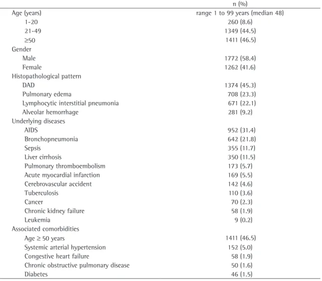
Table 1 summarizes the data related to the study population.
The age of the patients ranged from 1 to 99 years (median, 48 years): 286 (9.6%) were between 1 and 20 years of age; 1333 (43.9%) were between 21 and 49 years of age; and 1411 (46.5%) were age 50 or older. Of the autopsies included in the sample, 1772 (58.4%) were performed in males, and 1262 (41.6%) were performed in females.
The examination of the final reports of the 3030 autopsies allowed the following pulmonary histopathological alterations to be identified as the immediate cause of ARF-related death: DAD; pulmonary edema; lymphocytic interstitial
pneu-Introduction
Acute respiratory failure (ARF) is a major cause of death in patients with a variety of primary underlying diseases. In addition, comorbidities and mortality have been reported to be higher than 40-50% in patients with ARF, especially in those with diffuse infiltrates on chest X-rays.(1-3) The
differential diagnosis of pulmonary alterations in these patients includes interstitial pneumonia, recur-rence of the underlying disease and diffuse alveolar damage (DAD), as well as other processes such as pulmonary edema and pulmonary embolism, or any combination of these.(4,5) Prompt investigation
and diagnosis are essential to improving patient survival.(6-8) Clinical and radiographic findings of
ARF are nonspecific,(9-11) and computed tomography
has been shown to be useful in the assessment of these patients,(12) primarily in determining when
biopsy is indicated. In this context, the complexity of clinical presentations makes diagnosis a constant challenge for the clinician. Despite recent advances, most types of diagnostic support are still expensive. Clinicians often initiate treatment in order to avoid the rapid progression of the disease and to spare the patient from more invasive procedures. It is there-fore important to determine the leading causes of death in this population to establish correct prophy-lactic actions, which is the least expensive and most strategic way to prevent pulmonary alterations and to avoid the need for lung biopsy.(12)
The aim of this study was to present the pulmo-nary histopathological alterations identified in autopsies of patients whose chest X-rays revealed diffuse infiltration and who later died from ARF, as well as to determine whether underlying diseases and associated comorbidities increase the risk of developing specific histopathological patterns.
Methods
(152/5%); having congestive heart failure (58/1.9%); presenting chronic obstructive pulmonary disease (50/1.6%); and having diabetes (46/1.5%).
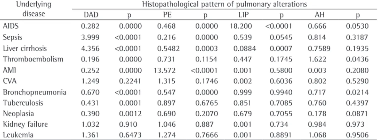
Table 2 shows the results of the logistic regres-sion used to predict the probability of developing specific histopathological findings in ARF for patients with certain associated risk factors and underlying diseases. A high probability of devel-oping the LIP histopathological pattern was found for patients with those risk factors and AIDS (OR = 18.20; p < 0.0001). Patients aged 50 years or older with systemic arterial hypertension, conges-tive heart failure, chronic obstrucconges-tive pulmonary disease or diabetes were more likely to develop the DAD histopathological pattern if they also had monia (LIP) and alveolar hemorrhage (Figure 1). We
found that DAD was present in 1371 cases (45.2%), pulmonary edema in 708 (23.3%), LIP in 671 (22.1%) and alveolar hemorrhage in 281 (9.2%). The prin-cipal underlying diseases were AIDS (in 952/31.4% of the cases), bronchopneumonia (in 642/21.8%), sepsis (in 355/11.7%), liver cirrhosis (in 350/11.5%), pulmonary thromboembolism (in 173/5.7%), acute myocardial infarction (AMI, in 169/5.5%), cere-brovascular accident (in 142/4.6%), tuberculosis (in 110/3.6%), cancer (in 70/2.3%), chronic kidney failure (in 58/1.9%) and leukemia (in 9/0.2%). The principal associated risk factors were as follows: being age 50 or older (1411/46.5% of the cases); suffering from systemic arterial hypertension
Table 1 - Data of the study population.
n (%)
Age (years) range 1 to 99 years (median 48)
1-20 260 (8.6)
21-49 1349 (44.5)
≥50 1411 (46.5)
Gender
Male 1772 (58.4)
Female 1262 (41.6)
Histopathological pattern
DAD 1374 (45.3)
Pulmonary edema 708 (23.3)
Lymphocytic interstitial pneumonia 671 (22.1)
Alveolar hemorrhage 281 (9.2)
Underlying diseases
AIDS 952 (31.4)
Bronchopneumonia 642 (21.8)
Sepsis 355 (11.7)
Liver cirrhosis 350 (11.5)
Pulmonary thromboembolism 173 (5.7)
Acute myocardial infarction 169 (5.5)
Cerebrovascular accident 142 (4.6)
Tuberculosis 110 (3.6)
Cancer 70 (2.3)
Chronic kidney failure 58 (1.9)
Leukemia 9 (0.2)
Associated comorbidities
Age ≥50 years 1411 (46.5)
Systemic arterial hypertension 152 (5.0)
Congestive heart failure 58 (1.9)
Chronic obstructive pulmonary disease 50 (1.6)
Discussion
The present study represents one of the largest autopsy studies in patients with underlying diseases and associated risk factors who developed pulmo-nary alterations and died of ARF. The autopsies included in this study were of patients treated at the Hospital das Clínicas, a tertiary health care facility receiving a large volume of patients with diseases of high complexity, especially ARF and terminal diseases. It is not surprising that, within the last 15 years, ARF has been a leading cause of morbidity and mortality.(8,9,13-15) Since its
appear-ance as a widespread clinical problem, we have been closely associated with research into the sepsis (OR = 3.99; p < 0.0001) or liver cirrhosis
(OR = 4.35; p < 0.0001). Those associated risk factors and pulmonary thromboembolism corre-lated significantly with a risk of developing alveolar hemorrhage (OR = 1.62; p = 0.04). A significant probability of developing pulmonary edema as the specific histopathological pattern was found for patients with the same risk factors concomitant with AMI (OR = 13.57; p < 0.0001). Patients presenting cerebrovascular accident, bronchopneumonia, tuberculosis, neoplasia, chronic kidney failure and leukemia presented consequent protection and had an equal, albeit lesser, chance of developing any of the specific histopathological patterns: DAD, pulmo-nary edema, LIP and alveolar hemorrhage.
a
500 m
b
500 m
c
500 m
d
500 m
obliterative fibrosis, neoformation of septa and moderately organizing fibrosis (characterizing DAD) were prevalent morphological findings in patients who presented sepsis or liver cirrhosis as the under-lying disease.(22-24) Therefore, DAD deserves special
mention in this investigation. The first aspect is related to the association between DAD and patients under mechanical ventilation at the time of death, thus raising the question of whether DAD might be secondary to ventilator-induced lung injury rather than related to the underlying disease. This very important question might not have been answered in this study, since our data were collected retro-spectively from postmortem pathological reports, which do not always include information about mechanical ventilation. In addition, when DAD is secondary to barotraumas, the morphological pattern is characterized almost exclusively by thin hyaline membranes, whereas in cases of sepsis and liver cirrhosis all of the DAD cases presented more prominent hyaline membranes and included marked alterations in the alveolar septa as well as exten-sive alveolar collapse. One group of authors studied epidemiology and outcomes in patients with DAD treated in European ICUs.(24) They found that acute
lung injury occurred in 463 (7.1%) of the 6522 admissions evaluated and in 16.1% of the mechani-cally ventilated patients. In addition, 65.4% cases occurred in the ICU.
cause and treatment of ARF. In the present study, we found that 16% of the patients aged 1 or older who were autopsied died from ARF, defined as any of several acute pulmonary pathologies producing life-threatening impairment of pulmonary function in individuals with previously normal lungs. Some authors observed similar results in adult patients,(8)
as did others in pediatric patients treated in an inten-sive care unit (ICU).(9) We also found that ARF was
more common in patients 50 years of age or older. As described by other authors,(8,11,13-18) infections
were another common complication of ARF in our study. Our study specifically showed that patients who developed ARF had underlying diseases such as AIDS (31.4%), sepsis (11.7%), cirrhosis (11.5%), pulmonary thromboembolism (5.7%), AMI (5.5%), cerebrovascular accident (4.6%), tuberculosis (3.6%), cancer (2.3%), chronic kidney failure (1.9%) and leukemia (0.2%). These findings were very similar to those of other studies in the literature.(8,11,18-21)
Histologically, ARF is related to increased permeability of the alveolar capillary membrane, leading to edema, pertinacious exudates, hyaline membranes, congestion, and hemorrhage.(21) The
most specific, prevalent histopathological finding as a cause of ARF in our sample was DAD, which was identified in 1371 (45.2%) of the autopsies evaluated. Diffuse involvement, uniform temporal appearance of alveolar collapse, hyaline membranes,
Table 2 - Results of the logistic regression used to predict the probability (odds ratio) of developing specific histopathological patterns leading to acute respiratory failure in patients with various underlying diseases and presenting specific risk factors (being ≥ 50 years of age, systemic arterial hypertension, congestive heart failure, chronic obstructive pulmonary disease or diabetes).
Underlying disease
Histopathological pattern of pulmonary alterations
DAD p PE p LIP p AH p
AIDS 0.282 0.0000 0.468 0.0000 18.200 <0.0001 0.666 0.0530
Sepsis 3.999 <0.0001 0.216 0.0000 0.539 0.0545 0.814 0.3187
Liver cirrhosis 4.356 <0.0001 0.5482 0.0003 0.0884 0.0007 0.7589 0.1935
Thromboembolism 0.196 0.0000 0.731 0.1154 0.447 0.1745 1.622 0.0436
AMI 0.252 0.0000 13.572 <0.0001 0.001 0.5800 0.003 0.2080
CVA 1.249 0.2241 1.315 0.1746 0.002 0.6036 0.802 0.5290
Bronchopneumonia 0.670 <0.0001 0.547 0.0000 0.999 0.9940 0.717 0.0214
Tuberculosis 0.431 0.0001 0.897 0.6765 0.851 0.7085 0.760 0.4397
Neoplasia 0.390 0.0012 0.690 0.2070 0.679 0.7055 0.178 0.0871
Kidney failure 1.032 0.910 1.046 0.887 0.001 0.734 0.984 0.973
Leukemia 1.361 0.6473 1.274 0.7666 0.001 0.8891 1.068 0.9506
evaluation. Such diagnostic disagreement can vary from 10 to 90%, depending on the disease and the population involved.(6-9,11,18) These discrepancies
might be attributable to different clinical manifesta-tions of a single disease and to poor quality medical care.(9) However, recent studies have demonstrated
the value of autopsy in medical education, in the evaluation of medical procedures, in physiopa-thology, in epidemiology, in public health and in the evaluation of the treatment employed.(1,6-8,10,11,26-29)
Nevertheless, and specifically in pediatrics, autopsy can detect genetic diseases, allowing better coun-seling of the family.(7,9)
Regarding issues such as whether to take into account the value of autopsy in medical educa-tion and medical procedures, and considering that many of the postmortem findings might have influ-enced treatment during the life of the patient and prevented the death of other patients, our study has limitations. First and foremost, this was a retro-spective study of medical reports, in which the quality of information can be limited, despite the recognized reputation of our institution in medical teaching and research. Final reports usually contain an abridged description of principal clinical, labora-torial and radiological data related to the patients. The second limitation of our study is related to interobserver variability, even taking into account that, at our institution, all autopsies are performed by the resident pathologist supervised by a senior pathologist who also checks the histological analysis in order to prepare the final reports. Different opin-ions can be held by different observers. In addition, the accuracy of autopsy findings also depends on the interest and skill of the pathologist.
Despite the limitations of this retrospective cohort study, the large sample of autopsies analyzed allows us to conclude that DAD, pulmonary edema, LIP and alveolar hemorrhage were the major histopathological patterns found in patients who died from ARF. Patients with underlying diseases and specific risk factors presented a higher risk of developing these specific histopathological findings on autopsy and should be evaluated to avoid an unfavorable outcome due to ARF.
Further investigations are warranted in order to evaluate the prevalence of ARF in prospective samples of patients and to determine the various mechanisms by which ARF develops in cases of differing etiologies. Despite the decline of autopsy In the present study, pulmonary edema was
the principal morphological substrate of ARF in 708 (23.3%) patients. Nevertheless, in patients who developed AMI, a significant association with death from pulmonary edema (OR = 13.57) was found for risk factors such as being 50 years of age or older, suffering from systemic arterial hyperten-sion, presenting chronic obstructive pulmonary disease and having diabetes. In fact, pulmonary edema was previously reported by other authors in 57% of patients with cardiovascular or infectious diseases who had undergone surgery(14). Another
group of authors described pulmonary edema as a complication of ARF in 70% of patients without cardiovascular disease.(20)
We found that, in 22.1% of the patients who developed ARF during the course of underlying disease, LIP was characterized by broadened, edema-tous alveolar septa with mononuclear inflammatory infiltrate consisting of lymphocytes, histiocytes, plasma cells and neutrophils. As expected, this increased the risk of death in patients presenting the risk factors previously mentioned and having AIDS as the underlying disease (OR = 18.20).
Alveolar hemorrhage was found in the lungs of 281 patients (9.2%) and was considered the imme-diate cause of ARF, representing a significant risk of death (OR = 1.62) for patients with pulmonary thromboembolism and specific risk factors. One group of authors described pulmonary emboli in 5% of patients who died from ARF.(13) Two other groups
reported pulmonary emboli in 14 and 20%, respec-tively, of patients who died from ARF.(14,25)
Our study showed the value of autopsy in deter-mining pulmonary histopathological alterations in patients with underlying diseases and specific risk factors who developed ARF. Many questions have arisen regarding the value of autopsy in view of the technological advances that have improved the sensitivity and specificity of new diagnostic methods.(6-11) As a consequence, the number of
autopsies performed at various institutions around the world has decreased in recent decades. Several reasons are given for this decline: a lack of interest on the part of clinicians and pathologists; emotional pressure from the family; and the legal conse-quences that can follow surprising findings.(7,9,12,26)
in recent decades, our findings suggest that its performance can provide complementary informa-tion that is important for achieving quality control and improving treatment protocols.
References
1. Barbas CS, Capelozzi VL, Hoelz C, Magaldi RB, Souza R, Sandeville ML, et al. Impacto de biópsia pulmonar a céu aberto na insuficiência respiratória aguda refratária. J Bras Pneumol. 2006;32(5):418-23.
2. Milberg JA, Davis DR, Steinberg KP, Hudson LD. Improved survival of patients with acute respiratory distress syndrome (ARDS): 1983-1993. JAMA. 1995;273(4):306-9.
3. Behrendt CE. Acute respiratory failure in the United States: incidence and 31-day survival. Chest.2000;118(4):1100-5. 4. Xavier AC, Siqueira SA, Costa LJ, Mauad T, Nascimento
Saldiva PH. Missed diagnosis in hematological patients-an autopsy study. Virchows Arch. 2005;446(3):225-31. 5. Bernardi FD, Saldiva PH, Mauad T. Histological examination
has a major impact on macroscopic necropsy diagnoses. J Clin Pathol. 2005;58(12):1261-4.
6. Blosser SA, Zimmerman HE, Stauffer JL. Do autopsies of critically ill patients reveal important findings that were clinically undetected? Crit Care Med. 1998;26(8):1332-6. 7. Kumar P, Taxy J, Angst DB, Mangurten HH. Autopsies in
children: are they still useful? Arch Pediatr Adolesc Med. 1998;152(6):558-63.
8. Mort TC, Yeston NS. The relationship of pre mortem diagnoses and post mortem findings in a surgical intensive care unit. Crit Care Med. 1999;27(2):299-303.
9. Castellanos Ortega A, Ortiz Melón F, García Fuentes M, Prieto Valderrey F, Santidrián Miguel JP, Mazorra Macho F. [The evaluation of autopsy in the pediatric intensive unit] [Article in Spanish]. An Esp Pediatr. 1997;46(3):224-8. 10. Fernandez-Segoviano P, Lázaro A, Esteban A, Rubio JM,
Iruretagoyena JR. Autopsy as quality assurance in the intensive care unit. Crit Care Med. 1988;16(7):683-5. 11. Stevanovic G, Tucakovic G, Dotlic R, Kanjuh V. Correlation
of clinical diagnoses with autopsy findings: a retrospective study of 2,145 consecutive autopsies. Hum Pathol. 1986;17(12):1225-30.
12. Canzian M, Soeiro AM, Taga MF, Barbas CS, Capelozzi VL. Correlation between surgical lung biopsy and autopsy findings and clinical data in patients with diffuse pulmonary infiltrates and acute respiratory failure. Clinics. 2006;61(5): 425-32
13. Gerain J, Sculier JP, Malengreaux A, Rykaert C, Thémelin L. Causes of deaths in an oncologic intensive care unit: a clinical and pathological study of 34 autopsies. Eur J Cancer. 1990;26(3):377-81.
14. Tóth T, Szöts I, Juhász M, Benedek G. The importance of pulmonary complications as a cause of death in surgical patients. Int Surg. 1984;69(1):35-7.
15. Motsay GJ, Lillehei RC. Acute respiratory distress syndrome in adults. Definition, etiology and treatment. Int Surg. 1973;58(5):304-7.
16. Friederici HH, Sebastian M. Autopsies in a modern teaching hospital. A review of 2,537 cases. Arch Pathol Lab Med. 1984;108(6):518-21.
17. Thornton CM, O’Hara MD. A regional audit of perinatal and infant autopsies in Northern Ireland. Br J Obstet Gynaecol. 1998;105(1):18-23.
18. Demling RH, Nerlich M. Acute respiratory failure. Surg Clin North Am. 1983;63(2):337-55.
19. Clowes GH Jr. Pulmonary abnormalities in sepsis. Surg Clin North Am. 1974;54(5):993-1013.
20. Vito L, Dennis RC, Weisel RD, Hechtman HB. Sepsis presenting as acute respiratory insufficiency. Surg Gynecol Obstet. 1974;138(6):896-900.
21. Kobzik L. The lung. In: Cotran RS, Kumar V, Collins T, Robbins SL, editors. Robbins pathologic basis of disease. 6th ed. Philadelphia: Saunders; 1999. p. 626-679.
22. Katsurada K, Yamada R, Sugimoto T. Respiratory insufficiency in patients with severe head injury. Surgery. 1973;73(2):191-9.
23. Flick MR. Pulmonary edema and acute lung injury. In: Murray JF, Nadel JA, editors. Textbook of respiratory medicine. 2nd ed. Philadelphia: Saunders; 1994. p. 1725-62.
24. Dyck DR, Zylak CJ. Acute respiratory distress in adults. Radiology. 1973;106(3):497-501.
25. Moser KM, LeMoine JR, Nachtwey FJ, Spragg RG. Deep venous thrombosis and pulmonary embolism. Frequency in a respiratory intensive care unit. JAMA. 1981;246(13):1422-4. 26. Smith CJ, Scott SM, Wagner BM. The necessary role of
the autopsy in cardiovascular epidemiology. Hum Pathol. 1998;29(12):1469-79.
27. Terrabuio Junior AA, Parra ER, Farhat C, Capelozzi VL. Autopsy-proven causes of death in lungs of patients immunocompromised by secondary interstitial pneumonia. Clinics. 2007;62(1):69-76.
28. Andrade ZR, Garippo AL, Saldiva PH, Capelozzi VL. Immunohistochemical and in situ detection of cytomegalovirus in lung autopsies of children immunocompromised by secondary interstitial pneumonia. Pathol Res Pract. 2004;200(1):25-32.