Radiographic alterations in patients presenting human
immunodeficiency virus/tuberculosis coinfection:
correlation with CD4
+T cell counts*
ROSEMERI MAURICI DA SILVA1, LÍGIA DA ROSA2, RENATA NUNES LEMOS2
*Study carried out at the Universidade do Sul de Santa Catarina (Unisul, University of Southern Santa Catarina) - Florianópolis, Santa Catarina, Brazil.
1. PhD in Pulmonology and Professor of Medicine at the Universidade do Sul de Santa Catarina (Unisul, University of Southern Santa Catarina) - Florianópolis, Santa Catarina, Brazil
2. Medical Academician at the Universidade do Sul de Santa Catarina (Unisul, University of Southern Santa Catarina) -Florianópolis, Santa Catarina, Brazil
Correspondence to: Rosemeri Maurici da Silva. Rua Moçambique, 852, Rio Vermelho - CEP 88058-000, Florianópolis, SC, Brazil. Phone: 55 48 3228.5333. E-mail: [email protected]
Submitted: 14 February 2005. Accepted, after review: 1 September 2005.
ABSTRACT
Objective: To look for correlations between radiological patterns and CD4+ T cell counts in patients coinfected with
tuberculosis and human immunodeficiency virus. Methods: Patients included were selected from among those presenting human immunodeficiency virus/tuberculosis coinfection and admitted to the Nereu Ramos Hospital, located in Florianópolis, Brazil, between January of 2000 and December of 2003. Results: A total of 87 patients were included. The mean age was 34 + 8 years, and 6.8% were non-Caucasian. The mean CD4+ T cell count was 220.2 cells/mm3 (median, 144 cells/
mm3), and 56.4% of the patients presented less than 200 cells/mm3. We identified the following radiographic patterns
and related them to the CD4+ T cell counts: the alveolar pattern in 50.6% of the cases (56.8% CD4+ T cells < 200); the
interstitial pattern in 32.2% (53.6% CD4+ T cells < 200); pleural effusion in 24.1% (47.6% CD4+ T cells < 200);
cavitation in 24.1% (57.1% CD4+ T cells < 200); enlarged mediastinal or hilar lymph nodes in 11.5% (90% CD4+ T cells
< 200); and a normal pattern in 11.5% (60% CD4+ T cells < 200). The mean CD4+ T cell counts for the radiologic
patterns isolated were as follows: 235.2/mm3 (alveolar consolidation); 208.8/mm3 (interstitial); 243.3/mm3 (pleural effusion);
265/mm3 (cavitation); 115.1/mm3 (enlarged mediastinal or hilar lymph nodes) (p < 0.05); and 205.5/mm3 (presenting no
radiological alterations). As noted, mediastinal/hilar lymph node enlargement was the only pattern that correlated with the degree of cell-mediated immunity in a statistically significant way. Conclusion: With the exception of mediastinal/ hilar lymph node enlargement, the radiographic patterns were randomly distributed in relation to the CD4+ T cell counts.
INTRODUCTION
The advent of acquired immunodeficiency syndrome (AIDS), recognized in 1981, had a profound impact on the global problem of tuberculosis (TB), changing its epidemiology and, principally, making it more difficult to control.(1-2)
Infection with the human immunodeficiency virus (HIV) is one of the greatest risk factors for the development of the disease in individuals who have previously been infected with the bacillus. Immunocompetent people infected with TB have a 10% chance of developing the active form of the disease over the course of their lifetimes, whereas among HIV-positive individuals co-infected with TB, this risk is from 8% to 10% per year. In addition, TB is one of the first and most significant complications among those infected with HIV, occurring prior to other common infections.(3-4)
In the year 1999, 10.7 million people were identified as being co-infected with TB/HIV. This corresponds to 0.18% of the global population. In Brazil, of the 40.7 million individuals infected with TB, approximately 300,000 are estimated to be co-infected with HIV.(3)
The CD4+ T cell counts in peripheral blood have
prognostic implications for the evolution of HIV infection, whereas the measurement of cellular immunocompetence is more useful in the follow-up evaluation of patients suffering from this syndrome.(1,5) There is a decrease in the numbers of
these cells during HIV infection. This results in great susceptibility to a first TB infection or to reactivation of a previous TB infection.
The radiologic alterations of the patients coinfected with TB/HIV depend on the CD4+ T cell
counts. When the cell counts are below 200 cells/ mm3, most of the presentations are atypical. At the onset of HIV infection, that is, when CD4+ T cell
counts are greater than 200 cells/mm3, the radiologic presentation of TB is similar to that of immunocompetent patients, with a typical reactivation pattern and alveolar consolidation areas at the apex, in posterior segments of the upper lobes and superior segments of the lower lobes, which are frequently accompanied by cavitation.(6-12) Significant
radiographic differences have been documented between patients in more advanced phases of HIV infection, when CD4+ T cell counts fall below 200
cells/mm3, and immunocompetent patients.(6-8,10-11)
The objective of this study was to analyze whether CD4+ T cell counts are truly associated with
radiographic variations in pulmonary TB.
METHODS
A cross-sectional study was carried out at the Nereu Ramos Hospital, in Florianópolis (in the state of Santa Catarina). We analyzed all of the clinical charts of individuals over the age of fourteen admitted between January of 2000 and December of 2003 with HIV/pulmonary TB coinfection.
The patients who had not been submitted to chest X-rays or CD4+ T cell counts were excluded
from the study, as were those who presented some pulmonary comorbidity.
Individuals who tested positive in the serologic testing for HIV were defined as patients with HIV/ AIDS. Those who presented identification of Mycobacterium tuberculosis in their sputum samples, bronchoalveolar lavage, and/or pulmonary and pleural biopsies, were defined as TB patients. The chest X-rays of the patients were analyzed by a specialist in radiology with experience at a referral center for TB. The analysis was performed at two distinct time points, without previous knowledge of the CD4+ T cell counts, with the
objective of evaluating the intra-observer agreement. In the event of disagreement between the first and the second evaluation, a second radiologist was consulted. The analysis both professionals agreed upon was defined as the standard pattern.
The chest X-rays were classified as presenting one of the following alteration patterns: alveolar consolidation; interstitial; pleural effusion; cavitation; or mediastinal/hilar lymph node enlargement, and t h e i r r e s p e c t i v e a s s o c i a t i o n s . L a t e r a l a n d anteroposterior radiographs were taken using a Siemens device at 120 kV and 3-6 mAs.
A registration form was filled out for each participant. The values of the variables of interest were filed and entered into a database using the Epidata program. The statistical analysis was performed using the Epi Info program. The chi-square test and Fisher's exact test were used to compare the intervals of CD4+ T cell counts in
The present study was submitted to and approved by the Ethics Committee of the Nereu Ramos Hospital.
RESULTS
A total of 105 patients were evaluated, and 18 of them were excluded from the study (10 due to the lack of a chest X-ray and 8 due to a pulmonary comorbidity). Therefore, the study sample consisted of 87 patients. The mean age of the participants was 34 years ± 8 years. The prevalence of non-Caucasians was 6.8%, and 83.9% of the participants were males.
The mean CD4+ T cell count was 220.2 cells/mm3.
The median was 144 cells/mm3, the maximum was
985 cells/mm3, and the minimum was 7 cells/mm3.
The prevailing radiologic alteration was the interstitial pattern, followed by the alveolar consolidation pattern and by the combination of alveolar consolidation and cavitation (Table 1).
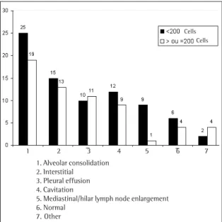
After evaluating the radiologic alterations isolated, we observed that the alveolar consolidation pattern predominated, followed by the interstitial pattern. Cavitations were found in the same number of cases in which pleural effusion was described. The distribution of the radiologic alterations by CD4+ T cell counts, using 200 cells/mm3 as a cutoff
point, is demonstrated in Figure 1. The mean CD4+
T cell counts for each radiologic pattern are presented in Figure 2.
The distribution pattern of the mediastinal/hilar lymph node enlargement was the only variable that presented statistical significance (p < 0.05), both regarding the distribution of CD4+ T cell counts as
well as the mean count related to each pattern. The radiologic analysis was concordant in 81.6% of the cases.
DISCUSSION
When accompanied by HIV, thoracic TB can present various radiologic patterns. No statistically significant differences were found when the
Radiographic alterations n %
Interstitial 14 16.1
Alveolar consolidation 12 13.8
Alveolar consolidation+ cavitation 12 13.8
Normal 10 11.5
Alveolar consolidation+ pleural effusion 8 9.8
Pleural effusion 4 4.6
Alveolar consolidation + cavitation + 3 3.4 mediastinal/hilar lymph node enlargement
Alveolar consolidation+ interstitial 2 2.3 Interstitial + pleural effusion 2 2.3
Interstitial + cavitation 2 2.3
Alveolar consolidation + interstitial 2 2.3 + pleural effusion
Alveolar consolidation+ pleural effusion 2 2.3 + cavitation
Interstitial + pleural effusion + 2 2.3 mediastinal/hilar lymph node enlargement
Mediastinal/hilar lymph node enlargement 1 1.1 Interstitial + mediastinal/hilar 1 1.1 lymph node enlargement
Derrame pleural + linfonodomegalia 1 1.1 mediastinal e/ou hilar
Alveolar consolidation+ interstitial 1 1.1 + cavitation
Alveolar consolidation+ interstitial 1 1.1 + mediastinal/hilar lymph node enlargement
Alveolar consolidation+ interstitial + cavitation 1 1.1 + mediastinal/hilar lymph node enlargement
Other 6 6.9
Total 87 100
TABLE 1
Radiographic alterations in 87 patients with HIV/ Aids and pulmonary tuberculosis
Figure 1 - Distribution of the radiologic alterations isolated, by CD4+ T cell count *p<0.05
1 2 3 4 5 6 7 1. Alveolar consolidation
2. Interstitial 3. Pleural effusion 4. Cavitation
5. Mediastinal/hilar lymph node enlargement 6. Normal
7. Other
radiologic alterations were compared with the degree of immunosuppression, except in regard to the mediastinal/hilar lymph node enlargement, which was significantly prevalent in patients with severe immunosuppression.
As previously stated, the mean CD4+ T cell count
was 220.2 cells/mm3. However, this value could have
been influenced by the extreme values of 7 and 985 cells/mm3. Therefore, the median (144 cells/
mm3) is a more appropriate way to represent the
sample. This study was carried out in a referral hospital for the treatment of infectious diseases, and this cell count reflects the pattern of the patients who seek medical assistance in this facility (those who are more susceptible to the various types of opportunistic infections).
The radiologic alterations found in the patients were distributed into various patterns. According to the comprehensive analysis of the radiologist, the interstitial pattern was the most common, in 16.1% of the patients, followed by the alveolar consolidation, and the alveolar consolidation associated with cavitation, both with a 13.8% frequency.
In the etiological diagnosis of patients with pulmonary disease and HIV, the presence of an interstitial pattern has a wide nonspecific differential diagnosis. In addition to TB, it comprises other diseases such as pneumocystosis, histoplasmosis, cryptococcosis, and cytomegalovirus infection. In addition, an alveolar consolidation pattern found
in a patient with HIV represents probable bacterial pneumonia, independently of the CD4+ T cell count.
However, in patients who present severe immunosuppression, pulmonary TB should be considered.(6)
In a study carried out in Africa, the interstitial and alveolar consolidation patterns were found in 12% and 9.8%, respectively, of the patients who presented pulmonary TB and HIV. In that study, cavitation represented the most common finding, although in a smaller proportion among the HIV-positive than in the HIV-negative patients (33% versus 78%).(13)
Approximately 45% of the X-rays analyzed presented multiple patterns, which made it difficult to correlate them with the CD4+ T cell count
intervals. Therefore, we decided to classify the patients into two categories, according to whether or not they presented isolated radiologic alterations, independently of the associations among them.
When the chest X-rays were analyzed as to whether or not they presented the respective alterations, we found that the alveolar consolidation pattern was predominant and was present in 50.6% of the chest X-rays of the patients studied, 56.8% of whom presented a CD4+ T cell count below 200
cells/mm3 (p > 0.05).
When the CD4+ T cell counts are greater than
200 cells/mm3 and alveolar consolidation is found, TB is not included in the differential diagnosis. Nevertheless, when these counts drop to less than
Figure 2 - Distribution of the radiologic alterations isolated, by mean CD4+ T cell count, in 87 patients with HIV/AIDS
and tuberculosis
Mediastinal/hilar lymph node enlargement
Normal
Interstitial
Alveolar consolidation
Pleural effusion
200 cells/mm3, this possibility should be
considered.(7) Comparing positive and
HIV-negative patients, the alveolar consolidation pattern occurred in 9.8% of the HIV-positive patients, compared with 3.8% of the HIV-negative patients.(13)
The interstitial pattern was the second most frequent in the sample studied. This pattern can be found in patients coinfected with TB/HIV, although it is not common.(6,10)
The prevalence of pleural effusion found was 24.1%, 47.6% of which presented CD4+ T cell
counts below 200 cells/mm3 (p > 0.05). Pleural
effusion is common in patients with pulmonary TB and HIV, and a greater proportion is found in patients whose counts are greater than 200 cells/mm3.(5,10,14)
Comparing the prevalence of pleural effusion in patients with TB, whether positive or HIV-negative, we observed that it occurred in 25.3% of the cases, 76.3% of which were HIV-positive. Comparing the occurrence of this finding with the degree of immunosuppression, we observed that 36.8% of the patients presented normal or moderate immune status, whereas 63.2% presented a high degree of immunosuppression.(15)
Cavitation, which is the most common radiologic alteration in patients with pulmonary TB and preserved immunity, was found in 24.1% of the cases in our study, and 57.1% of the patients presented CD4+ T cell counts lower than 200 cells/mm3. The
absence of statistical significance in this finding reveals that the cavitation can occur even at severe levels of immunosuppression.
In the initial stages of HIV infection, the form of pulmonary TB presented was similar to that seen in the patients without HIV, with a typical pattern of cavitation. However, when the CD4+ T cell counts
begin to drop, the cavitation ceases to occur.(10) With
CD4+ T cell counts between 200 and 500 cells/mm3,
the most frequent pattern of pulmonary TB is cavitation, becoming less so when these counts drop below 200 cells/mm3.(7) What we observed in our
study was that cavitation, together with pleural effusion, was the third most commonly found pattern. Therefore, the cavitation occurred with less frequency. However, it did not present any correlation with the immunosuppression level. The cavitation was the most common finding in the chest X-rays of patients whose CD4+ T cell counts were
equal to or greater than 200 cells/mm3 and was
considerably more common among such patients
than among those presenting counts below this value (20% versus 7%).(14)
Mediastinal/hilar lymph node enlargement was present in 11.5% of the 87 patients studied, and only 10% of those patients presented CD4+ T cell
counts greater than 200 cells/mm3 (p < 0.05). The mean CD4+ T cell count in those patients was 115.1
cells/mm3. When compared with the mean counts
of patients presenting other alterations, this difference was found to be statistically significant (p < 0.05).
Lymph node enlargement is a frequent manifestation of TB in HIV-positive patients.(10) The
onset of adenopathy can be related to the drop in cellular immunity.(7) Lymph node enlargement is seen
in only 7% of patients with CD4+ T cell counts equal
to or greater than 200 cells/mm3, compared with
30% of patients presenting counts below this value (p = 0.01).(14) Approximately 64.5% of the patients
who present lymph node enlargement present moderate or severe immunologic involvement.(15)
Mean CD4+ T cell counts in patients with mediastinal/
hilar lymph node enlargement were lower (115.1 cells/ mm3) than those observed in patients presenting
other alterations (p < 0.05).
Patients with pulmonary TB who are HIV-positive can also present normal chest X-rays.(10) We observed this pattern in 11.5% of the patients studied, 60% of whom presented CD4+ T cell counts
below 200 cells/mm3.
In one study, normal chest X-rays were seen in 12% of seventeen HIV-positive patients with pulmonary TB. However, no HIV-negative TB patients presented normal X-rays.(11)
The difficulty in establishing a typical pattern for pulmonary TB in patients with HIV/AIDS is a challenge for physicians, who are confronted with the increasingly common problem of hard-to-control TB, which is inconsistent with the usual diagnostic findings. This random distribution of the radiologic alterations in these patients reinforces the idea that chest X-ray, as an isolated method, is inconclusive in the diagnosis of pulmonary TB in HIV-positive patients, as well as in ruling out this diagnostic possibility.
REFERENCES
2. Duncan BB, Schimidt MI, Giugliani ERJ. Tuberculose. In: Palombini BC, Hetzel JL, Correa da Silva LC, editores. Medicina ambulatorial: condutas clínicas em atenção primária à saúde. 2a ed. Porto Alegre: Artes Médicas; 1996. p. 352-8.
3. Brasil. Ministério da Saúde. Fundação Nacional de S a ú d e . C e n t r o d e R e f e r ê n c i a P r o f . H é l i o F r a g a . Sociedade Brasileira de Pneumologia e Tisiologia. Controle da Tuberculose: uma proposta de integração ensino-serviço [CD-ROM]. 5a ed. Rio de Janeiro; 2002. 4. Davis L, Beck JM, Shellito J. Update: HIV infection and p u l m o n a r y h o s t d e f e n s e s . S e m i n R e s p i r I n f e c t . 1993;8(2):75-85.
5. Boiselle PM, Aviram G, Fishman JE. Update on lung disease in AIDS. Semin Roentgnol. 2002;37(1):54-71. 6. Haramati LB, Jenny-Avital ER. Approach to the diagnosis of pulmonary disease in patients infected with the human imunodeficiency virus. J Thorac Imaging. 1998;13(4):247-60.
7. S h a h R M , Ka j i A V, O s t r u m B J , F r i e d m a n A C . Interpretation of chest radiographics in AIDS patients: usefulness of CD4+ lymphocyte counts. Radiographics.
1997;17(1):47-58; discussion 59-61. Erratum in: Radiographics. 1997;17(3):804.
8. Keiper MD, Beumont M, Elshami A, Langlotz CP, Miller WT Jr. CD4+ T lymphocyte count and the radiographic
presentation of pulmonary tuberculosis. A study of the relationship between these factors in patients with human immunodeficiency virus infection. Chest. 1995;107(1):74-80.
9. Post FA, Wood R, Pillay GP. Pulmonary tuberculosis in HIV infection: radiographic appearance is related to CD4+
T-lymphocyte count. Tuberc Lung Dis. 1995;76(6):518-21.
1 0 . Naidich DP, McGuinness G. Pulmonary manifestations of AIDS. CT and radiographic correlations. Radiol Clin North Am. 1991;29(5):999-1017.
11 . P i t c h e n i k A E , R u b i n s o n H A . T h e r a d i o g r a p h i c appearance of tuberculosis in patients with the acquired immune deficiency syndrome (AIDS) and pre-AIDS. Am Rev Respir Dis. 1985;131(3):393-6.
12. Lawn SD, Evans AJ, Sedgwick PM, Acheampong JW. Pulmonary tuberculosis: radiological features in West Africans coinfected with HIV. Br J Radiol. 1999;72(856):339-44.
1 3 . Tshibwabwa-Tumba E, Mwinga A, Pobee JO, Zumla A. Radiological features of pulmonary tuberculosis in 963 HIV-infected adults at three Central African hospitals. Clin Radiol. 1997;5(11):837-41.
1 4 . Perlman DC, El-Sadr WM, Nelson ET, Matts JP, Telzak EE, Salomon N, et al. Variation of chest radiographic patterns in pulmonary tuberculosis by degree of human immunodeficiency virus-related immunosuppression. The Terry Beirn Community Programs for Clinical Research on AIDS (CPCRA). The AIDS Clinical Trials Group (ACTG). Clin Infect Dis. 1997; 25(2):242-6. 1 5 . Kawooya VK, Kawooya M, Okwera A. Radiographic