Laparoscopic Surgery for Renal Stones: Is it Indicated in the
Modern Endourology Era?
Andrei Nadu, Oscar Schatloff, Roy Morag, Jacob Ramon, Harry Winkler
Department of Urology, The Chaim Sheba Medical Center, Tel Hashomer, Israel and The Sackler School of Medicine, Tel Aviv University, Israel
ABSTRACT
Purpose:To report the outcomes of laparoscopic surgery combined with endourological assistance for the treatment of renal stones in patients with associated anomalies of the urinary tract. To discuss the role of laparoscopy in kidney stone disease.
Materials and Methods: Thirteen patients with renal stones and concomitant urinary anomalies underwent laparoscopic stone surgery combined with ancillary endourological assistance as needed. Their data were analyzed retrospectively including stone burden, associated malformations, perioperative complications and outcomes.
Results: Encountered anomalies included ureteropelvic junction obstruction, horseshoe kidney, ectopic pelvic kidney, fussed-crossed ectopic kidney, and double collecting system. Treatment included laparoscopic pyeloplasty, pyelolithotomy,
DQGQHSKUROLWKRWRP\FRPELQHGZLWKÀH[LEOHQHSKURVFRS\DQGVWRQHUHWULHYDO,QWUDRSHUDWLYHFRPSOLFDWLRQVZHUHORVWVWRQHV
in the abdomen diagnosed in two patients during follow up. Mean number of stones removed was 12 (range 3 to 214). Stone free status was 77% (10/13) and 100% after one ancillary treatment in the remaining patients. One patient had a postoperative urinary leak managed conservatively. Laparoscopic pyeloplasty was successful in all patients according to clinical and dynamic renal scan parameters.
Conclusions:,QFDUHIXOO\VHOHFWHGSDWLHQWVODSDURVFRSLFDQGHQGRXURORJLFDOWHFKQLTXHVFDQEHVXFFHVVIXOO\FRPELQHGLQ DRQHSURFHGXUHVROXWLRQWKDWGHDOVZLWKFRPSOH[VWRQHGLVHDVHDQGUHSDLUVXQGHUO\LQJXULQDU\DQRPDOLHV
Key words: kidney; laparoscopy; nephrolithiasis; genitourinary abnormalities Int Braz J Urol. 2009; 35: 9-18
INTRODUCTION
(QGRXURORJLFDOWHFKQLTXHVKDYHUHYROXWLRQ -ized the treatment of urinary stones to the point they have rendered open stone surgery anachronistic (1). Procedures like open ureterolithotomy, open neph-rolithotomy, or open pyelolithotomy have become anecdotal. However, patients with large stone burdens and associated renal malformations are prone to
re-TXLUHPXOWLSOHHQGRXURORJLFDOSURFHGXUHVLQRUGHUWR
have their stones retrieved and anomalies repaired.
Thus in these selected patients, open stone surgery can
FHUWDLQO\EHFRQVLGHUHGUHDVRQDEOH,QFHQWHUVZLWK HVWDEOLVKHG H[SHULHQFH LQ DGYDQFHG UHFRQVWUXFWLYH
laparoscopy, this can be a feasible option if the goals of stone clearance and correction of malformations
FRXOGEHDFKLHYHGLQDVLQJOHSURFHGXUH,QFRUSRUDWLQJ ODSDURVFRSLFWHFKQLTXHVFRQIHUWKHVHSDWLHQWVVXUJLFDO HI¿FDF\FRPELQHGZLWKWKHDGYDQWDJHVRIPLQLPDOO\
invasive surgery.
sur-Laparoscopic Management of Complex Renal Stones
gery in combination with endourological procedures involving a variety of cases of renal stones in the setting of underlying urinary tract malformations.
MATERIALS AND METHODS
The data of twenty-nine patients who un-derwent laparoscopic procedures for kidney stones between January 2004 and May 2007 were retrospec-tively analyzed. Retrieved data included indications for intervention, stone burden, associated malforma-tions, perioperative complications and outcomes in terms of functional results and stone free status. Fifteen patients underwent laparoscopic nephrec-tomy due to non functioning kidneys and were not included in the present study. One patient underwent laparoscopic pyelolithotomy without harboring
un-GHUO\LQJPDOIRUPDWLRQVDQGWKXVZDVDOVRH[FOXGHG
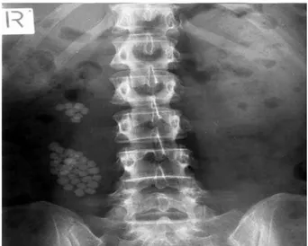
The remaining thirteen patients underwent laparo-scopic stone removal and reconstructive procedures combined with ancillary endourological assistance as needed. Preoperative stone scenario and associated anomalies are detailed in Table-1 and Figures-1 to 3. All cases were discussed with the endourology unit and considered unlikely that stones and underlying
DQRPDOLHVFRXOGEHHI¿FLHQWO\DGGUHVVHGZLWKDVLQJOH
endourological procedure. Three patients had previ-ous unsuccessful endourological procedures, with
LQDELOLW\WRJDLQSHUFXWDQHRXVDFFHVVDQGVLJQL¿FDQW
residual stones after nephroscopy among main causes of failure.
Patients underwent preoperative anatomical and functional evaluation with non contrast CT scan and either intravenous pyelography or diuretic renal scans in cases of suspected ureteropelvic junction (UPJ) obstruction. Postoperative assessment consisted
RI[UD\VXOWUDVRXQGDQGRU&7DVDSSURSULDWH$
diuretic renal scan was performed in patients who underwent concomitant pyeloplasty.
6XUJLFDOSURFHGXUHVDQGWKHLUWHFKQLTXHDUH
summarized below. Combinations were used to deal
ZLWKVSHFL¿FSDWLHQWQHFHVVLWLHV
Table 1 – Patients characteristics.
N of Patients Preoperative Diagnosis
6 UPJO + multiple pelvic and calyceal stones 2 UPJO + horseshoe kidney + multiple stones
1 UPJO + two stones 3 cm each in the renal pelvis + a calyceal diverticulum with a 2 cm stone 1 UPJO + ectopic pelvic kidney + 2 cm upper pole stone
1 UPJO + situs inversus + crossed-fussed ectopic kidney + tens of stones in lower moiety
1 83-2SHOYLFNLGQH\REVWUXFWLYHK\GURFDO\[PXOWLSOHSHOYLFDQGFDO\FHDOVWRQHV
1 Double collecting system + 9 cm complete staghorn stone in the lower moiety
UPJO = ureteropelvic junction obstruction.
SURGICAL TECHNIQUE
Laparoscopic Pyeloplasty and Pyelolithotomy / Flexible Nephroscopy
The operative room setting includes one lapa-roscopic cart and one endourological cart to enable simultaneous laparoscopy and nephroscopy. Using a four port transperitoneal approach, the ureter is
identi-¿HGDQGIROORZHGFUDQLDOO\WKH83-LVH[SRVHGDQGWKH
renal pelvis dissected. The renal pelvis is opened above the UPJ, the ureter is spatulated on its lateral aspect and dismembered. Stones in the renal pelvis are removed with an atraumatic grasper and placed in a laparoscopic
EDJ$ÀH[LEOHF\VWRQHSKURVFRSHLVSDVVHGWKURXJK
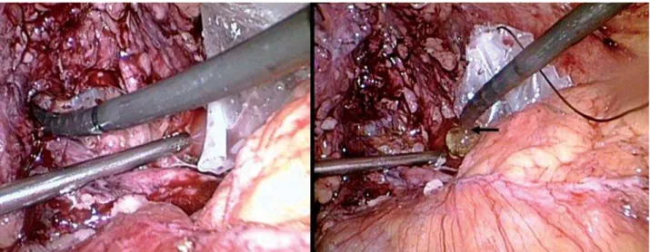
one of the 10 mm ports and guided laparoscopically through the opening in the renal pelvis. The kidney is systematically inspected and calyceal stones removed with a basket or fragmented with Holmium:YAG laser lithotripsy. A double J stent is introduced in an ante-grade fashion, the renal pelvis is reduced as needed, and ureteropelvic anastomosis is performed with two (one posterior and one anterior) 4/0 polyglactin run-ning sutures. A percutaneous drain is placed and the bag with stones removed (Figure-4).
Figure 3 – Retrograde pyelography of a patient with an ectopic pelvic kidney, ureteropelvic junction obstruction and a 2 cm stone in the upper pole (arrow). Intraoperatively, infundibular stricture was encountered and the patient underwent laparoscopic pyelo-plasty, and “cut to the light” nephrolithotomy.
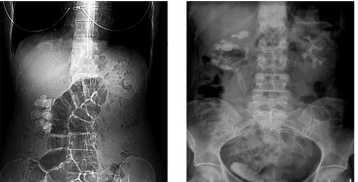
Figure 2 – Left: Complete 9 cm staghorn stone in the lower moiety of a complete duplicated pelviocalyceal system. Right: Intravenous urography performed 3 months after laparoscopic anatrophic nephrolithotomy showing dye descending through both right excretory systems.
Laparoscopic Nephrolithotomy
$³FXWIRUWKHOLJKW´WHFKQLTXHZDVXVHGZKHQ
calyceal stones were associated with infundibular
Laparoscopic Management of Complex Renal Stones
ÀH[LEOHF\VWRQHSKURVFRSHLVEURXJKWDWWKHREVWUXFWHG FDO\[FRQWDLQLQJWKHVWRQHDQGDVPDOOODSDURVFRSLF
nephrotomy is made in the kidney as indicated by the endoscopic light. The stone is removed and the kidney sutured with one layer 2/0 polyglactin (Figure-5).
Laparoscopic Anatrophic Nephrolithotomy The kidney is dissected from the surrounding
IDWWKHYDVFXODUHOHPHQWVLGHQWL¿HGDQGFODPSHGHQ
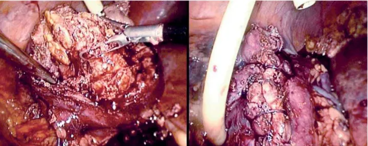
bloc with a laparoscopic Satinsky clamp. The renal parenchyma and collecting system are incised longitu-dinally on the postero-lateral aspect of the kidney; the staghorn stone is mobilized with graspers, removed and placed in an endobag. A 16 F Foley catheter is placed as a nephrostomy tube by making a small in-cision in the kidney away from the nephrotomy line. The kidney is sutured with a running 2/0 polyglactin single layer that includes renal capsule, parenchyma, Figure 4 – Left: Flexible nephroscopy through the laparoscopic pyelothomy. Right: Basket extraction of encountered stones (arrow).
,QWHUPLWWHQWVXFWLRQSUHYHQWVÀXLGDFFXPXODWLRQLQWKHDEGRPHQ
DQGFROOHFWLQJV\VWHPLQD³QRNQRW´WHFKQLTXHZLWK
the aid of Lapra-Ty clips (Ethicon-Johnsons & John-sons), Figure-6.
RESULTS
Mean age at surgery was 36 years (range 18-56), ASA score 2 (range 1-2), and average number of stones removed was 12 (range 3 to 214). Laparo-scopic pyeloplasty combined with pyelolithotomy and
ÀH[LEOHQHSKURVFRS\ZDVSHUIRUPHGLQWHQSDWLHQWV
laparoscopic pyeloplasty with pyelolithotomy and endoscopic-assisted nephrolithotomy was performed
in two; and laparoscopic anatrophic nephrolithotomy was performed in one patient (Table-2).
A double J stent and percutaneous drain was left postoperatively in all patients. Additionally, a nephrostomy drain using a 16 Fr Foley catheter was placed in two patients. All the procedures were com-pleted laparoscopically with no conversions to open
VXUJHU\,QWUDRSHUDWLYHFRPSOLFDWLRQVLQFOXGHGWZR
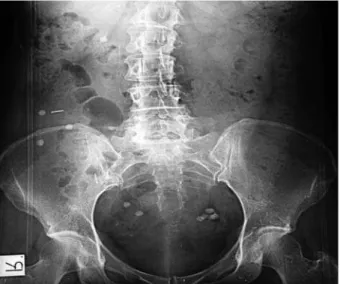
patients with lost stones in the abdomen diagnosed during follow up (Figure-7) and variable degrees of
LUULJDWLRQÀXLGDFFXPXODWLRQVHFRQGDU\WRQHSKURV -copy in several others. One patient had a postoperative
XULQDU\OHDNLQWKHFRQWH[WRIDQLQGZHOOLQJGRXEOH-stent, which was not replaced at the time of surgery, Figure 6 – Left: Laparoscopic anatrophic nephrolithotomy, stone extraction. Right: One layer LapraTy™ assisted suturing of the kidney.
Table 2 – Procedures performed and their indication.
Procedure Indication
/3FRPELQHGZLWK/3/DQGÀH[LEOHQHSKURVFRS\ Patients with UPJO and associated pelvic and calyceal stones
2. LP combined with LPL and endoscopic-assisted LNL 3DWLHQWVZLWK83-2DQGVWRQHVLQDQREVWUXFWHGK\GURFDO\[ or calyceal diverticulum
3. Laparoscopic anatrophic nephrolithotomy Patient with double collecting system and complete stag-horn of the lower moiety
Laparoscopic Management of Complex Renal Stones
and most probably chronically obstructed. Leakage was discovered during early postoperative period
DV XULQDU\ H[WUDYDVDWLRQ WKURXJK WKH SHUFXWDQHRXV
drain. Cystoscopic replacement of the double J stent effectively treated the complication and the patient was discharged during the following days without evidence of further leakage.
Stone free status was obtained in ten pa-tients (77%), and the remaining three were rendered stone free after one ancillary procedure (shockwave lithotripsy - SWL) in two patients and retrograde nephroscopy in another).
,QDOOWZHOYHSDWLHQWVZLWK83-REVWUXFWLRQWKH
pyeloplasty was considered successful according to clinical (disappearance of pain) and diuretic renal scan parameters. The mean washout half-life time in the diuretic renal scan improved from 43 to 22 minutes.
Warm ischemia time for laparoscopic anat-rophic nephrolithotomy was 43 minutes; the patient was rendered stone free and renal function remained within preoperative values.
COMMENTS
Endourology has revolutionized the treat-ment of urinary stones. Notwithstanding, underlying
DQDWRPLFDQRPDOLHVH[WUHPHO\ODUJHVWRQHEXUGHQV
RUDFRPELQDWLRQRIWKHPFDQVLJQL¿FDQWO\GHFUHDVH
the success rate of endourological procedures (3,4). The surgical approach in these special cases should
HI¿FLHQWO\DGGUHVVWKHODUJHVWRQHEXUGHQDQGDVVRFL -ated malformations in a single procedure. Although open surgery is an alternative, laparoscopic surgery might be a feasible option additionally conferring the advantages of minimally invasive surgery.
Micali et al. reported 17 patients who
under-ZHQWODSDURVFRSLFVWRQHH[WUDFWLRQLQFOXGLQJZLWK
renal calculi and 9 with associated anomalies (UPJ
REVWUXFWLRQZLWKVWRQHVL]HXSWRFP¿IWHHQSDWLHQWV
were eventually rendered stone free and one patient had a postoperative urinoma. These authors concluded that indications for laparoscopy included stones
as-VRFLDWHG ZLWK DQDWRPLFDO DEQRUPDOLWLHV UHTXLULQJ
reconstruction and calculi for which endourological procedures had failed (5). Ramakumar et al. reported 90% three month stone-free rate in 19 patients who underwent laparoscopic pyelolithotomy and pyelo-plasty (6). Similar results were recently reported by Srivastava et al. (7) and Stein et al. (8) with 75 and 80% stone-free rates respectively.
1DPELUDMDQHWDOUHSRUWHGWKHLUH[SHULHQFH
with eighteen patients who underwent different lapa-roscopic procedures with concomitant stone removal, including pyeloplasties, partial nephrectomies, and calyceal ablations. Stone-free status was achieved in 93% of cases. The authors concluded that laparoscopy
LVHIIHFWLYHIRUFRPSOH[UHQDOVWRQHVDQGWKDWWKHQHHG
for open surgery should be rare in the future (9). Me-ria et al. compared laparoscopic pyelolithotomy to percutaneous nephrolithotomy (PNL) in 32 patients with pelvic stones without underlying malformations.
6WRQHIUHHUDWHVZHUHQRWVLJQL¿FDQWO\GLIIHUHQWYV
82%). However, the laparoscopic group had higher operative time, urological complications (12% urine
OHDNDQGFRQYHUVLRQWRRSHQVXUJHU\ZDVUHTXLUHGLQ
two patients. They concluded that indications for each
WHFKQLTXHPXVWEHGHWHUPLQHGDOWKRXJK31/UHPDLQV WKHJROGVWDQGDUGIRUODUJHSHOYLFVWRQHV,WLVRXU
belief and current practice that SWL, retrograde and
SHUFXWDQHRXVWHFKQLTXHVDUHWKH¿UVWDSSURDFKHVWR WUHDWNLGQH\VWRQHVGXHWRWKHLUH[FHOOHQWUHVXOWVDQG
minimal morbidity. The role of laparoscopy is not to replace any of these options, but to compliment them in situations where decreased success or increased Figure 7 – Lost stones in the abdomen incidentally discovered
PRUELGLW\LVH[SHFWHGVSHFLDOO\DVUHJDUGVODUJHVWRQH EXUGHQVFRH[LVWLQJZLWKXQGHUO\LQJPDOIRUPDWLRQV$V
showed in our series and as reported by other authors
ODSDURVFRSLFDQGHQGRXURORJLFDOWHFKQLTXHV
can be successfully combined in the same procedure to improve the stone free rate and simultaneously resolve synchronous anomalies.
Tunc et al. published a study on 150 patients with stones in anomalous kidneys treated with SWL,
LQFOXGLQJGXSOH[KRUVHVKRHVPDOURWDWHG
14 pelvic, and 4 crossed ectopic. The overall stone-free rate was 68%, with the worst results obtained in crossed ectopic kidneys with stone clearance of only 25% (13). Pure percutaneous approach has also been reported in anomalous kidneys. Although highly effective with an overall stone-free rate of 83%, the anterior displacement of the collecting sys-tem together with an unpredictable vascular supply and interposition of bowel between the kidney and the abdominal wall makes the procedure technically
GHPDQGLQJ0RUHRYHULWUHTXLUHVDKLJKO\SUHFLVH
imaging system (i.e. CT guided) to minimize risk of visceral damage during kidney puncture and tract creation (14). Pure endoscopic management has also been accomplished for anomalous kidneys. Weizer et al. reported a 75% stone-free rate (15), meanwhile Braz et al. reported an 81% stone-free rate at three
PRQWKVKRZHYHUUHTXLUHGDQFLOODU\WUHDWPHQWV
(16). Due to urinary stasis, these patients suffer from poor spontaneous stone passage, with persistence or growth of residual fragments in 60% of cases.
/DSDURVFRSLF WHFKQLTXHV VHHP HVSHFLDOO\
useful for stones located in anomalous kidneys. Our overall stone-free rate was 77% (10/13), and reached a 100% after one ancillary treatment (i.e. SWL or neph-roscopy). Additionally, anomalies to be addressed (i.e. UPJ obstruction) were successfully repaired with optimal functional outcome.
There are several operative pitfalls that need special consideration when combining laparoscopy with endourological procedures. The operating room and the space around the operating table become lim-ited when the laparoscopic and endourological towers need to be brought to work simultaneously. The laser cart and endourological instrumentation table pose ad-ditional ergonomic problems. One serious limitation
LVWKHGLI¿FXOW\WRREWDLQÀXRURVFRSLFLPDJHVGXULQJ
VWRQHUHPRYDO,QWURGXFWLRQRID&DUPEHFRPHVD
challenge in the described set-up and even if possible
WKHLPDJHVREWDLQHGLQWKHLQVXIÀDWHGDEGRPHQRID
patient in lateral decubitus are far from informative.
'HÀDWLQJWKHDEGRPHQPD\LPSURYHWKHLPDJLQJEXW
is unpractical and time consuming, and even after this,
LPDJHVDUHRISRRUTXDOLW\GXHWRSDWLHQWSRVLWLRQDQG IUHHÀXLGLQWKHDEGRPHQWKXVVHULRXVO\OLPLWLQJWKH
ability to identify small residual stones.
,UULJDWLQJÀXLGWKDWÀRZVIUHHO\LQWRWKHDEGR -men during the nephroscopy is of some concern and might be a limiting factor. Although some of it can
EHVXFWLRQHGVWLOOODUJHTXDQWLWLHVRIÀXLGDFFXPXODWH
and occupy the space of the pneumoperitoneum. As
ÀXLGDFFXPXODWHVEHWZHHQERZHOORRSVLWFDQQRWEH GLUHFWO\ DVSLUDWHG:H RYHUFDPH WKLV GLI¿FXOW\ E\
placing the patient in a “head down” position for two
PLQXWHVDOORZLQJWKHÀXLGWRDFFXPXODWHXQGHUWKH
diaphragm where it becomes easily aspirated with the laparoscopic suction.
,GHQWLI\LQJVWRQHVLQVLGHREVWUXFWHGFDO\FHV FDQEHFRPHDUHDOFKDOOHQJH,QWUDRSHUDWLYHXOWUDVRXQG
can be useful in these situations (9) however; it poses additional restrictions to the already cumbersome operative scenario. We found a solution relying on
³FXWIRUWKHOLJKW´WHFKQLTXHZKHUHWKHOLJKWRIWKH HQGRVFRSHPDUNVWKHVWRQHFRQWDLQLQJFDO\[DQGWKH ORFDWLRQ IRU WKH SDUHQFK\PDO LQFLVLRQ ,Q WKH WZR
cases performed, the light of the endoscope precisely delineated the place for the nephrotomy (Figure-5).
Kaouk et al. created a porcine model to ad-dress the feasibility of laparoscopic anatrophic
neph-UROLWKRWRP\'HJHUHWDOUHSRUWHGWKH¿UVWFDVH
in humans (18), and recently Simforoosh et al. (19)
UHSRUWHGWKHLUVHULHVRI¿YHSDWLHQWVZLWKEHLQJ UHQGHUHGVWRQHIUHH,QWHUHVWLQJO\QRSRVWRSHUDWLYH XULQDU\H[WUDYDVDWLRQRFFXUUHGDOEHLWQRLQWHUQDOVWHQW
was placed. We performed a laparoscopic anatrophic
QHSKUROLWKRWRP\LQD\RXQJSDWLHQWZLWKDGXSOH[
system and a complete staghorn of the lower moiety with optimal results and no perioperative complica-tions. The kidney was repaired with one running suture including parenchyma and collecting system
ZLWKQRSRVWRSHUDWLYHXULQDU\H[WUDYDVDWLRQ
An interesting aspect of laparoscopic
Laparoscopic Management of Complex Renal Stones
have them fall out of the renal pelvis or the endobag, and locating them in the abdomen is challenging and time consuming. There is no report in the urological literature regarding this issue; however there are well described complications of lost stones in the abdomen after laparoscopic cholecystectomy. They include
in-WUDSHULWRQHDODQGDEGRPLQDOZDOODEVFHVV¿VWXODDQG
prolonged fever (20). Regarding the infectious status of struvite staghorn stones, lost stones should remain of concern. However, two patients in our series had lost stones in the abdomen and after completing more than two years of follow-up, they remain completely asymptomatic.
We are aware of the limitations of this pa-per, which consist of a small, retrospective series of patients. However, considering the limited data
SXEOLVKHGXSWRGDWHZHEHOLHYHRXUH[SHULHQFHFRQ -tributes to the developing of this novel and poorly studied approach.
CONCLUSIONS
Although classical endourological procedures should remain as the gold standard for the great major-ity of renal stones, however patients with large stone
EXUGHQVDQGXQGHUO\LQJPDOIRUPDWLRQVPLJKWEHQH¿W
from a combined laparoscopic and endourological
RQHSURFHGXUHVROXWLRQWKDWGHDOVZLWKFRPSOH[VWRQH
disease and repairs associated anomalies.
CONFLICT OF INTEREST
None declared.
REFERENCES
1. Matlaga BR, Assimos DG: Changing indications of open stone surgery. Urology. 2002; 59: 490-3; discus-sion 493-4.
$OLYL]DWRV*6NRODULNRV$,VWKHUHVWLOODUROHIRURSHQ
surgery in the management of renal stones? Curr Opin Urol. 2006; 16: 106-11.
3. Grasso M, Conlin M, Bagley D: Retrograde ureteropy-eloscopic treatment of 2 cm. or greater upper urinary
tract and minor Staghorn calculi. J Urol. 1998; 160: 346-51.
6HPHUFL%9HULW$1D]OL2,OEH\22]\XUW&&LNLOL
N: The role of ESWL in the treatment of calculi with anomalous kidneys. Eur Urol. 1997; 31: 302-4. 5. Micali S, Moore RG, Averch TD, Adams JB, Kavoussi
LR: The role of laparoscopy in the treatment of renal and ureteral calculi. J Urol. 1997; 157: 463-6. 6. Ramakumar S, Lancini V, Chan DY, Parsons JK, Kavoussi
LR, Jarrett TW: Laparoscopic pyeloplasty with concomi-tant pyelolithotomy. J Urol. 2002; 167: 1378-80. 7. Srivastava A, Singh P, Gupta M, Ansari MS, Mandhani
A, Kapoor R, et al.: Laparoscopic pyeloplasty with concomitant pyelolithotomy--is it an effective mode
RIWUHDWPHQW"8URO,QW
8. Stein RJ, Turna B, Nguyen MM, Aron M, Hafron
-0 *LOO ,6 HW DO /DSDURVFRSLF S\HORSODVW\ ZLWK FRQFRPLWDQWS\HOROLWKRWRP\WHFKQLTXHDQGRXWFRPHV
J Endourol. 2008; 22: 1251-5.
1DPELUDMDQ7-HVFKNH6$OETDPL1$EXNRUD)/HHE
K, Janetschek G: Role of laparoscopy in management
RIUHQDOVWRQHVVLQJOHFHQWHUH[SHULHQFHDQGUHYLHZ
of literature. J Endourol. 2005; 19: 353-9.
10. Meria P, Milcent S, Desgrandchamps F, Mongiat-Artus P, Duclos JM, Teillac P: Management of pelvic stones larger than 20 mm: laparoscopic transperitoneal py-elolithotomy or percutaneous nephrolithotomy? Urol
,QW
11. Fariña Pérez LA, Cambronero Santos J, Meijide Rico F, Zungri Telo ER: Laparoscopic pyelolithotomy in a pelvic kidney. Actas Urol Esp. 2004; 28: 620-3. 12. Whelan JP, Wiesenthal JD: Laparoscopic pyeloplasty
ZLWK VLPXOWDQHRXV S\HOROLWKRWRP\ XVLQJ D ÀH[LEOH
ureteroscope. Can J Urol. 2004; 11: 2207-9.
13. Tunc L, Tokgoz H, Tan MO, Kupeli B, Karaoglan U,
%R]NLUOL,6WRQHVLQDQRPDORXVNLGQH\VUHVXOWVRI
treatment by shock wave lithotripsy in 150 patients.
,QW-8URO
14. Matlaga BR, Shah OD, Zagoria RJ, Dyer RB, Streem SB, Assimos DG: Computerized tomography guided access for percutaneous nephrostolithotomy. J Urol. 2003; 170: 45-7.
15. Weizer AZ, Springhart WP, Ekeruo WO, Matlaga BR, Tan YH, Assimos DG, et al.: Ureteroscopic manage-ment of renal calculi in anomalous kidneys. Urology. 2005; 65: 265-9.
.DRXN-+*LOO,6'HVDL00%DQNV./5DMD66
Skacel M, et al.: Laparoscopic anatrophic nephroli-thotomy: feasibility study in a chronic porcine model. J Urol. 2003; 169: 691-6.
18. Deger S, Tuellmann M, Schoenberger B, Winkelmann B, Peters R, Loening SA: Laparoscopic anatrophic nephrolithotomy. Scand J Urol Nephrol. 2004; 38: 263-5.
6LPIRURRVK1$PLQVKDUL¿$7DELEL$1RRU$OL]DGHK
A, Zand S, Radfar MH, et al.: Laparoscopic anatrophic nephrolithotomy for managing large staghorn calculi.
%-8,QW
0HPRQ0$'HHLN5.0DI¿75)LW]JLEERQV5--U
The outcome of unretrieved gallstones in the peritoneal cavity during laparoscopic cholecystectomy. A pro-spective analysis. Surg Endosc. 1999; 13: 848-57.
Accepted after revision: October 24, 2008
Correspondence address:
Dr. Oscar Schatloff Department of Urology
The Chaim Sheba Medical Center
7HO+DVKRPHU,VUDHO )D[
E-mail: [email protected]
EDITORIAL COMMENT
Authors present interesting series of patients with urinary stone disease managed mainly by lapa-roscopic method. The presented cases have some kind of anomaly necessitating laparoscopic approach rather than using routine endourological procedures like percutaneous nephrostolithotomy or urethroscopic means.
Laparoscopy is gaining more popularity in managing urinary stone disease (1-3). This is especial-ly true when associated anomalies like ureteropelvic
MXQFWLRQREVWUXFWLRQRUIXVLRQDQRPDOLHVH[LVWV
Even though results of laparoscopic approach in managing stone disease in the present series and other reported series seems satisfactory (1-5), longer follow-up and more cases are necessary to better
HOXFLGDWH WKH H[DFW UROH RI ODSDURVFRS\ LQ WRGD\¶V
management of stone disease.
REFERENCES
6LPIRURRVK1$PLQVKDUL¿$7DELEL$1RRU$OL]DGHK
A, Zand S, Radfar MH, et al.: Laparoscopic anatrophic nephrolithotomy for managing large staghorn calculi.
%-8,QW
2. Basiri A, Simforoosh N, Ziaee A, Shayaninasab H, Moghaddam SM, Zare S: Retrograde, antegrade and laparoscopic approaches for the management of large,
SUR[LPDOVWRQHV$UDQGRPL]HGFOLQLFDOWULDO-(QGRX -rol. 2008; 22: 2677-80.
3. Meria P, Milcent S, Desgrandchamps F, Mongiat-Artus P, Duclos JM, Teillac P: Management of pelvic stones larger than 20 mm: laparoscopic transperitoneal py-elolithotomy or percutaneous nephrolithotomy? Urol
,QW
Laparoscopic Management of Complex Renal Stones
Percutaneous nephrolithotomy in patients with kidney malformations. J Endourol. 2007; 21: 520-4.
5. Simforoosh N, Basiri A, Danesh AK, Ziaee SA,
Shari-¿DJKGDV)7DELEL$HWDO/DSDURVFRSLFPDQDJHPHQW
EDITORIAL COMMENT
Laparoscopy is an established modality in management of renal stones in selected situations. On most occasions, laparoscopy is nephron sparing - namely - pyelolithotomy as compared with percu-taneous nephrostolithotomy (PCNL).
The authors have successfully used laparos-copy combined with endourological procedures in anomalous kidneys, mainly ureteropelvic junction obstruction. This is a retrospective study. Larger and prospective studies will help in developing concrete guidelines.
,WLVWREHQRWHGWKDWDUHQDODQJLRJUDPRU
CT angiogram is a complimentary investigation in planning reconstructive laparoscopic surgery in anomalous kidneys.
The authors have not done an infundibulo-plasty in cases of infundibular stenosis with calyceal diverticulum and; the collecting system was not closed separately in patients with staghorn calculus where
DQDQDWURSKLFS\HOROLWKRWRP\ZDVGRQH,WZRXOGEH LQWHUHVWLQJWRVHHWKHFRQ¿JXUDWLRQRIWKHFROOHFWLQJ
system using a CT scan during follow-up.
,Q PRVW FHQWHUV S\HORSODVW\ LV GRQH ZLWK
interrupted sutures. The authors have used running sutures for the anterior and posterior walls.
Meria et al. had shown similar results with different modalities (PCNL and laparoscopy) in management of stone disease in non-anomalous kid-neys; however the complication rate in was higher in laparoscopy compared to ESWL and PCNL. Hence, they concluded that ESWL or PCNL should be the
¿UVWRSWLRQLQQRQDQRPDORXVNLGQH\V
Practical problems of C arm usage;
ultra-VRXQGXVDJHDQGIUHHÀRZRILUULJDWLQJÀXLGLQWRWKH
peritoneal cavity with laparoscopy are problems to be looked into.
Combining laparoscopy with PCNL and pure endoscopy gives a better result in stone disease.
Dr. Manickam Ramalingam Department of Urology K.G. Hospital and Post Graduate Institute Coimbatore, India E-mail: [email protected] of ureteral calculi: a report of 123 cases. Urol J. 2007; 4: 138-41.