Anti-
Plasmodium
Response in the Malaria Mosquito
Anopheles gambiae
Martin K. Rono1,2,3¤a, Miranda M. A. Whitten1,2,3¤b, Mustapha Oulad-Abdelghani4, Elena A. Levashina1,2,3, Eric Marois1,2,3*
1INSERM, U963, Strasbourg, France,2CNRS, IBMC, UPR9022, Strasbourg, France,3Universite´ de Strasbourg, UMR 963, Strasbourg, France,4INSERM, U964/CNRS, UMR 7104/Universite´ de Strasbourg, IGBMC, Illkirch, France
Abstract
When taking a blood meal on a person infected with malaria, femaleAnopheles gambiaemosquitoes, the major vector of human malaria, acquire nutrients that will activate egg development (oogenesis) in their ovaries. Simultaneously, they infect themselves with the malaria parasite. On traversing the mosquito midgut epithelium, invadingPlasmodiumookinetes are met with a potent innate immune response predominantly controlled by mosquito blood cells. Whether the concomitant processes of mosquito reproduction and immunity affect each other remains controversial. Here, we show that proteins that deliver nutrients to maturing mosquito oocytes interfere with the antiparasitic response. Lipophorin (Lp) and vitellogenin (Vg), two nutrient transport proteins, reduce the parasite-killing efficiency of the antiparasitic factor TEP1. In the absence of either nutrient transport protein, TEP1 binding to the ookinete surface becomes more efficient. We also show that Lp is required for the normal expression ofVg, and for laterPlasmodiumdevelopment at the oocyst stage. Furthermore, our results uncover an inhibitory role of the Cactus/REL1/REL2 signaling cassette in the expression ofVg, but not ofLp. We reveal molecular links that connect reproduction and immunity at several levels and provide a molecular basis for a long-suspected trade-off between these two processes.
Citation:Rono MK, Whitten MMA, Oulad-Abdelghani M, Levashina EA, Marois E (2010) The Major Yolk Protein Vitellogenin Interferes with the Anti-Plasmodium Response in the Malaria MosquitoAnopheles gambiae. PLoS Biol 8(7): e1000434. doi:10.1371/journal.pbio.1000434
Academic Editor:David S. Schneider, Stanford University, United States of America
ReceivedDecember 17, 2009;AcceptedJune 10, 2010;PublishedJuly 20, 2010
Copyright:ß2010 Rono et al. This is an open-access article distributed under the terms of the Creative Commons Attribution License, which permits unrestricted use, distribution, and reproduction in any medium, provided the original author and source are credited.
Funding:This work was supported by grants from the Centre National de la Recherche Scientifique (UPR 9022 du CNRS), the Institut National de la Sante´ et de la Recherche Me´dicale (U963 INSERM), the Fondation pour la Recherche Me´dicale (FRM) to EM, the Schlumberger Foundation for Education and Research (FSER) to EAL and MMAW, from the EMBO Young Investigator Program (to EAL), and from the EC FP6th Networks of Excellence ‘‘BioMalPar’’ (to EAL and MR), EC FP7 HEALTH Collaborative Project ‘‘MALVECBLOK’’, EC FP7 Capacities Specific Programme Research Infrastructures ‘‘INFRAVEC’’, and EC FP7 Networks of Excellence ‘‘EVIMalar’’. EAL is an international research scholar of the Howard Hughes Medical Institute. The funders had no role in study design, data collection and analysis, decision to publish, or preparation of the manuscript.
Competing Interests:The authors have declared that no competing interests exist.
Abbreviations:dsRNA, double-stranded RNA; ELISA, enzyme-linked immunosorbent assay; GFP, green fluorescent protein; GST, glutathione S-transferase; HDL, high-density lipoprotein; hpi, hours post infection; Imd, Immune deficiency; IP, immuno-precipitation; KBr, potassium bromide; kDa, kiloDalton; Lp, Lipophorin (AGAP001826); LDL, low-density lipoprotein; LRR, leucine-rich repeat; MALDI, matrix-assisted laser desorption/ionization; PCR, polymerase chain reaction; qRT, quantitative real-time; RNAi, RNA interference; SDS-PAGE, sodium dodecyl sulfate polyacrylamide gel; TEP1, thioester-containing protein 1; TOF, time-of-flight; Vg, Vitellogenin (AGAP004203); TOR, Target of Rapamycin
* E-mail: [email protected]
¤a Current address: KEMRI - Wellcome Trust Research Programme, Kilifi, Kenya
¤b Current address: Department of Pure and Applied Ecology, School of the Environment and Society, Swansea University, Swansea, United Kingdom
Introduction
Malaria is a mosquito-borne parasitic disease affecting annually an estimated 250 million people, of which close to 1 million (mostly children in sub-Saharan Africa) succumb to the disease (World Health Organization fact sheet#94, April 2010; http:// www.who.int/mediacentre/factsheets/fs094/en/index.html). Sev-eral Plasmodium species cause malaria, the most deadly beingP. falciparumtransmitted mainly by theAnopheles gambiaemosquito. As mosquito females require a blood meal to produce eggs, feeding on a malaria-infected host simultaneously activates oogenesis and triggers immune responses to malaria parasites. In the midgut, ingestedPlasmodiumgametocytes differentiate within minutes into gametes. After fertilization, zygotes rapidly transform into ookinetes, i.e. motile cells that traverse the midgut epithelium
complement factor C3 [2,5,6]. Depletion of TEP1 by RNA interference (RNAi) renders mosquitoes hypersusceptible to Plasmodiuminfections, resulting in abnormally high infection levels. Two leucine-rich repeat (LRR) proteins, LRIM1 and APL1C, act as TEP1 control proteins to stabilize the mature form of TEP1 in the hemolymph [7,8] and show the same RNAi phenotype as TEP1inP. bergheiinfections [9–12]. The depletion of either protein results in precocious deposition of TEP1 on self tissues and completely aborts its binding to the ookinetes [7]. Therefore, it appears that LRR proteins regulate maintenance of mature TEP1 in circulation; however, the factors that control TEP1 targeting to the parasite surface remain unknown.
Simultaneously to the midgut crossing by ookinetes, the physiology of the mosquito is profoundly modified by a blood meal in preparation for the laying of a clutch of eggs. Within 2 to 3 d after a blood meal, the massive ovary growth allows maturation of 50–150 oocytes, a process called vitellogenesis (reviewed in [13]). The blood meal provides the mosquito with amino acids and lipids that are transferred through midgut cells to the hemolymph and signal via the Target of Rapamycin (TOR) pathway to initiate massive synthesis of nutrient transport proteins in the mosquito fat body [14]. These transport proteins include the lipid transporter lipophorin (Lp, AGAP001826) (also known as apolipoprotein II/I or retinoic and fatty acid binding protein, RFABG/P) and vitellogenin (Vg, AGAP004203), a precursor of the yolk storage protein vitellin. Both proteins are secreted into the hemolymph and transported to the ovaries. Vg is a large phospholipoglycoprotein encoded inA. gambiaeby a small family of nearly-identical genes. Insect Vg harbors potential sites for lipidation, glycosylation, and phosphorylation and is internalized by developing oocytes where it is proteolytically cleaved to generate vitellin, a nutrient source for the developing embryo (reviewed in [15,16]). Lp, encoded by a single transcript and post-translationally cleaved, is composed of two subunits of 250 and 80 kDa that together scaffold a lipidic particle. Similar to vertebrate low- and high-density lipoproteins (LDL and HDL, respectively), mosquito Lp particles contain a core of fatty acids and sterols, surrounded by an outer leaflet of phospholipids [17,18]. These particles function to deliver lipids and fatty acids to energy-consuming tissues, including rapidly growing imaginal discs in larvae, muscles, and the ovary in adult females [19]. In
addition to lipids, Lp particles serve as a vehicle for morphogen proteins in the imaginal discs ofDrosophila larvae [20]. Interest-ingly, human HDL has been shown to host a fraction of complement factor C3 [21] as well as trypanosome-killing protein complexes [22]. In mosquitoes, recent studies [23–25] have implicated Lp in both mosquito reproduction and Plasmodium survival. In particular, experimental depletion of Lp by RNAi inhibited oogenesis and also reduced the number of developing Plasmodiumoocysts in the mosquito midgut [23]. This could point to a nutritional requirement for Lp in the early stages of parasite development. Indeed, Lp has recently been detected by in vitro approaches inside developingP. gallinaceumoocysts, suggesting that it provides parasites with a source of lipids [26]. An intriguing alternative explanation is that the increasing levels of Lp following a blood meal may negatively impact mosquito immunity against parasites. Artificially blocking the physiological rise in Lp levels would then allow the immune system to exert its full strength against the parasite.
In the mosquito fat body, two distinct pathways are required for optimal expression of proteins involved in vitellogenesis: (i) the nutrient-sensing TOR pathway and (ii) a hormonal cascade that oversees production of 20-hydroxyecdysone [14,27,28]. Further-more, in Ae. aegypti mosquitoes infected with microbes and Plasmodium, the NF-kB factor REL1 positively regulates expression
of Lp and its receptor [24], suggesting that the NF-kB pathway
may also contribute to the regulation of oogenesis in addition to its known role in mosquito immunity [29–31]. However, our understanding of how oogenesis and immunity impact each other remains incomplete: on one hand depletion of Lp strongly inhibits development ofP. gallinaceum; on the other hand over-expression of Lpresulting from the depletion of the REL1 inhibitor Cactus inAe. aegypti is insufficient to rescue the complete block in parasite development [24].
Here, we investigated the role of the two major nutrient transport proteins Lp and Vg in mosquito antiparasitic responses using a common laboratory model of malaria transmission: A. gambiae mosquitoes infected with the GFP-expressing rodent parasiteP. berghei[32]. We show that similarly to Lp, Vg depletion reduces parasite survival in mosquito tissues. Strikingly however, Lp and Vg are no longer required for parasite survival if TEP1 is depleted, suggesting that the low parasite survival phenotype associated with the Lp/Vg knockdowns requires TEP1 function. We propose that Lp and Vg exert distinct non-redundant roles in reproduction and immunity: Lp is crucial for oogenesis and is required for normal Vg expression after an infectious blood meal, whereas Vg contributes to oogenesis and negatively impacts TEP1 binding to the ookinetes. We suggest that the reported negative impact of Lp depletion on ookinete survival is indirect and is mediated by reduced levels of Vg. We further demonstrate that the NF-kB factors REL1 and REL2 limit the expression ofVgafter an infectious blood meal. These results reveal an unexpected network of interactions whereby Plasmodium killing in mosquitoes is potentiated by NF-kB pathways at two levels: (i) activation of anti-Plasmodiumgenes and (ii) inhibition of the expression of the nutrient transport protein Vg.
Results
Lp and Vg Depletion Reduce Parasite Survival in aTEP1 -Dependent Manner
Lp knockdown causes a decrease in parasite loads and
simultaneously arrests oogenesis [23]. We examined whether the Lpknockdown phenotype requires the antiparasitic factor TEP1. To this end, we compared the numbers of surviving parasites in Author Summary
singleTEP1orLpknockdown mosquitoes and in doubleTEP1/Lp knockdowns by injecting double-stranded RNA (dsRNA) resulting in RNAi. Four days after dsRNA injection, mosquitoes were fed on a mouse infected with GFP-expressing parasites. Mosquitoes were dissected 8 to 10 d later to gauge prevalence of infection and mean oocyst numbers per midgut (Figure 1A, Figure S3A). As reported earlier, Lp silencing strongly reduced the number of developing oocysts. Strikingly, silencingTEP1at the same time as Lpannihilated the effect ofLpsilencing, i.e. yielded the high oocyst
numbers typically observed upon silencing of TEP1 alone.
Therefore, the low oocyst counts observed in Lp-depleted mosquitoes are not due to a nutritional dependence of ookinetes on Lp-derived lipids but are a consequence of TEP1 activity. This result also suggests that the increased parasite killing in Lp-depleted mosquitoes takes place at the ookinete stage, since TEP1 binding does not kill oocysts. Further, these results imply that the loss of Lp renders ookinetes more vulnerable to TEP1-dependent killing.
To explain these data, we initially hypothesized that Lp particles might physically sequester components of the TEP1 machinery in an inactive state, but a search for Lp-associated immune factors was unsuccessful (with the notable exception of prophenoloxidase), suggesting that TEP1-containing complexes are not carried in the hemolymph by Lp particles (see Text S1 and Figure S1).
To investigate whether the adverse effect on immunity is a specific property of Lp or may be manifested as well by other nutrient transport factors, we injected mosquitoes withdsVgand compared parasite development withdsLacZanddsTEP1-injected mosquitoes. A 4-fold reduction in mean parasite numbers was observed in thedsVggroup compared todsLacZcontrols (p,0.001, p,0.001,p,0.05, andp,0.05 depending on the replicate of this experiment; Figure 1B and Figure S3B). This effect was more profound than the effect of dsLp (Figure 1A and 1E). We then examined whether depletion of the major yolk protein would compromise oogenesis. In contrast toLpsilencing, which resulted in total abortion of ovary development, roughly 50% of mosquito females still developed eggs after silencing ofVgcompared to 80% indsLacZcontrol mosquitoes (Figure 1C), though ovaries that did mature usually contained only a few eggs bearing melanotic spots (unpublished data). When given a chance to lay, Vg-silenced females did lay a few eggs, the majority of which never hatched (unpublished data). The difference in strength between theLpand Vgsilencing phenotypes regarding egg development suggests either that Lp is more crucial than Vg for egg development or that the efficiency of Lp silencing is greater than the efficiency of Vg silencing. Residual Vg protein may allow the development of a few eggs indsVg-treated mosquitoes. It is interesting to note that the strengths of the silencing phenotypes are reversed when consid-ering parasite survival. To verify the efficiency of RNAi-mediated depletion of Lp and Vg, we used specific antibodies directed against the large and small subunits of Lp, and against Vg. RNAi silencing caused Lp and Vg protein amounts to drop below 10% of control levels (Figure S2). Subsequently, we systematically controlled for Lp and Vgsilencing efficiency and noted that Vg depletion was somewhat more variable than Lp depletion, residual Vg protein sometimes approaching 20% of control levels (unpublished data). Strikingly, this analysis revealed that the major protein bands detected in hemolymph samples by Coomassie staining of SDS-PAGE gels (or of PVDF membranes after protein transfer) correspond to the Vg and Lp signals detected by specific antibodies (Figure S2). We excised these easily visualized bands from Coomassie-stained protein gels and submitted them to MALDI mass spectrometry. The peptide mass spectra were searched against the NCBInr database. Each band
from a triplet running between 160 and 200 kDa was unequiv-ocally identified as Vg, and the bands running at ,250 and 80 kDa were unequivocally identified as the large and small subunits of Lp, respectively. In addition, a protein running at ,70 kD and showing an expression pattern identical to that of the ,200 kD Vg band (including after RNAi silencing) was identified as the N-terminal fragment of the polypeptide encoded by Vg mRNA (visible in Figures 3C, 4C, and S1). This fragment was not recognized by our Vg antibody, raised against a C-terminal Vg fragment. Its existence is consistent with the cleavage ofAe. aegypti Vg prior to secretion [33–35]. No contaminating proteins were detected at these sizes in the mass spectrometry analysis. Therefore, Lp and Vg proteins can be readily visualized after hemolymph electrophoresis and Coomassie staining of SDS-PAGE gels even without immunoblotting. The efficiency ofTEP1 silencing was also confirmed by immunoblotting (Figure 1D).
We next investigated whether Vg and Lp cooperate to sustain oogenesis and parasite development or are involved in independent processes. We performed double-knockdown experiments by simultaneously injectingdsVg-dsLp to compare to single injections ofdsVgand dsLpas controls. As expected, dsLpcompletely blocked oogenesis and the same was observed in concomitant dsLp-dsVg knockdowns (Figure 1F). Moreover, single dsVg (p= 0.0001) and double dsLp-dsVg (p,0.0001) knockdowns caused comparable reductions in oocyst counts; these reductions in oocyst numbers were stronger than in the single dsLp knockdown (p= 0.024) (Figure 1E). These results suggest that the influences of Lp and Vg on reproduction and immunity are balanced differently. Lp may be more crucial for oogenesis than Vg, whereas Vg influencesPlasmodiumsurvival more strongly than does Lp. In most experiments, the effect of VgandLpknockdowns on parasite counts did not appear to be additive (Figures 1E, 2A, and unpublished data). Although this observation is not supported by strong statistical significance, it raises the possibility that the two proteins may be involved in a single process benefiting ookinete survival in the physiological situation.
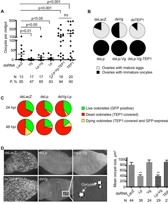
To determine whether similarly to Lp, the effect of Vg on parasite development required TEP1 function, we performed triple knockdown experiments by injecting combinations of dsTEP1, dsVg, dsLp, or controldsLacZ. Again, total inhibition of oogenesis was observed in all dsRNA combinations that included dsLp, suggesting that oogenesis is not influenced by TEP1 function but absolutely requires Lp (Figure 2B). In striking contrast, high parasite loads similar to that detected in the dsTEP1 single knockdown were obtained when TEP1 was depleted simulta-neously to Vg (unpublished data) or to both Vg and Lp (Figure 2A, Figure S3C). These findings imply that blocking the transport of lipids and vitellogenin-derived nutrients does not limit parasite survival when the immune defense is suppressed; instead, the
observed reduction in parasite numbers in dsLp and dsVg
knockdowns is dependent on TEP1. We conclude that TEP1-dependent parasite killing is more efficient when Lp and/or Vg levels are low and that the TEP1-mediated immune pressure exerted by the vector is a bigger impediment to the establishment of a Plasmodium infection than nutrient availability. If this constraint is removed via TEP1 depletion, Plasmodium parasites can effectively exploit even reduced vector resources and proceed with the formation of viable oocysts.
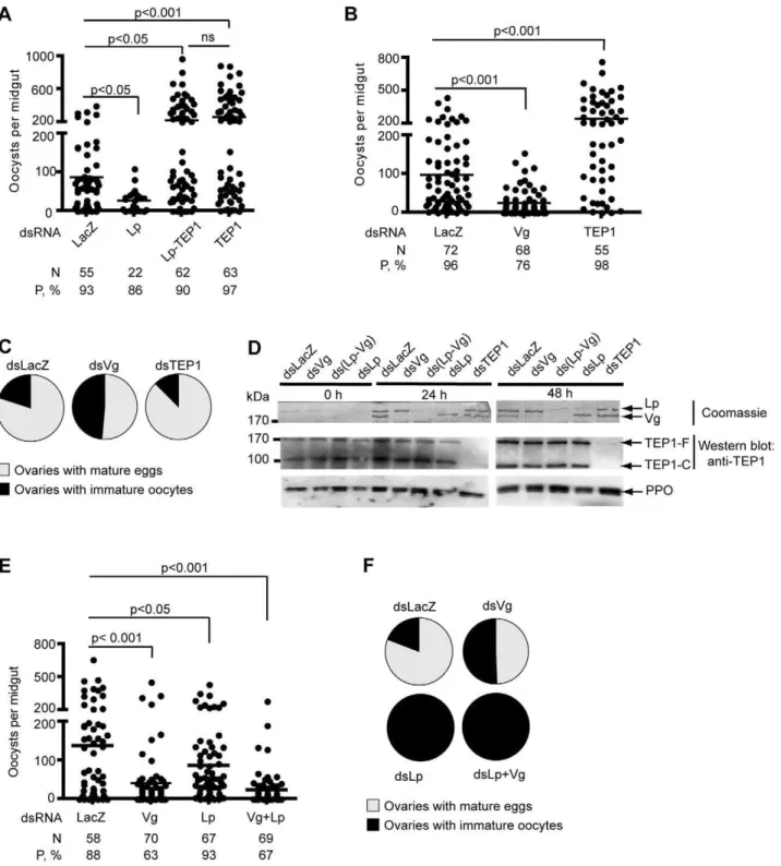
Figure 1. Effects ofLpandVgsilencing on parasite counts and oogenesis.Mosquitoes were injected with the indicated double-stranded RNA and infected withP. berghei. Parasite development was gauged 7–9 d post infection by counting GFP-expressing oocysts. Each dot represents the number of oocysts counted in one midgut. Ovaries containing mature eggs were counted 7 d post infection. Pie charts show the percentage of mosquitoes containing mature eggs (grey) versus percentage of mosquitoes containing only undeveloped oocytes (black). (A) Effect of concomitant silencing ofTEP1andLpon parasite survival. (B) Effect ofVgsilencing on parasite survival. In (A) and (B), one representative experiment out of 4 independent replicates is shown. The additional replicates are shown in Figure S3. (C) Effect ofVgsilencing on oogenesis. (D) Coomassie staining (top panel) of a PVDF membrane allows visualization of Vg and Lp in control, Vg-, and Lp-depleted mosquitoes. Hemolymph proteins were separated on a denaturing SDS-polyacrylamide gel and transferred to a PVDF membrane. Western blotting analysis of hemolymph of Vg- and Lp-depleted mosquitoes 0, 24, or 48 h after infection using anti-TEP1 antibody (bottom panel). (E, F) Parasite counts (E) and oogenesis (F) in Lp- and Vg-depleted mosquitoes.
ookinetes indsLp-anddsLp-Vg-injected mosquitoes. At early time points (24 hpi) TEP1 binding to ookinetes did not differ in the Lp or Lp-Vg -depleted versus control mosquitoes; but at 48 hpi 70% to 86% of ookinetes were TEP1 positive (i.e., either dead or moribund) in dsLp- or dsVg-Lp-injected mosquitoes versus only 41% to 68% indsLacZcontrols (Figure 2C and Table S1,p= 0.005 or less by chi-square analysis). Thus, TEP1 binding to parasites is more efficient in the absence of Lp/Vg. This strongly suggests that physiological levels of Vg and Lp interfere with the efficient binding of TEP1 to ookinetes once the invasion phase is completed.
To see if we could also detect an effect of Lp and Vg depletion at a later stage of parasite development, we examined oocyst growth. Strikingly, oocyst size 9 d after infection was markedly reduced when Lp, but not Vg, was depleted (Figure 2D). In contrast to oocyst numbers, silencingTEP1at the same time asLp did not rescue oocyst growth (unpublished data), indicating that the small oocyst size does not result from TEP1 activity in Lp-deficient mosquitoes. This supports the hypothesis that Lp contributes nutrients to oocyst development [26]. Therefore, Lp benefits Plasmodium development at two independent levels: an early effect favoring ookinete survival by protecting against TEP1-dependent killing, and a later effect favoring normal oocyst growth. The latter effect does not require Vg or TEP1 function.
Vg and Lp Do Not AffectTEP1Expression or Cleavage, but Lp Is Necessary for ProperVgExpression
Previous work [7,31] has demonstrated that boosting mosquito basal immunity via depletion of the inhibitory IkB protein Cactus
up-regulates components of the TEP1 pathway (including TEP1, LRIM1, and APL1C) and completely blocks parasite develop-ment. Therefore, we asked whether the knockdown ofVgandLp could mimic the effect of Cactus depletion and elevate TEP1 expression levels, providing an explanation to the above
observations. We silenced Lp and/or Vg and examined the
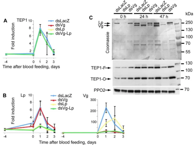
transcript levels of TEP1 before and after blood feeding using quantitative real-time polymerase chain reaction (qRT-PCR). Silencing of the two nutrient transport genes did not alterTEP1 expression (Figure 3A). We then evaluated the effect ofLpandVg silencing on TEP1 protein amounts and TEP1 cleavage in the hemolymph by immunoblotting using polyclonal anti-TEP1 antibodies. This analysis did not reveal any marked increase in the amounts of full-length or mature TEP1 protein (Figures 3C and 1D).
Surprisingly, silencing of Lpreproducibly lowered the expres-sion ofVgmRNA (Figure 3B and unpublished data). At the protein level, Lp depletion strongly reduced Vg levels at 47 h (but not
24 h) post-infectious feeding compared with the controls
(Figure 3C), confirming that Lp is indeed required for full Vg
Figure 3. Lp is required for normal Vg expression.Mosquitoes were injected withdsLp,dsVg, ordsLp+dsVg. (A, B)TEP1,Vg, andLpexpression, respectively, was measured at several time points afterP. bergheiinfection using quantitative RT-PCR. (C) Lp and Vg protein levels in mosquito hemolymph were gauged by Coomassie staining; TEP1 (full length and processed) by immunoblotting. PPO2 served as a loading control. Note that levels of Vg protein are strongly reduced at 47, but not 24, h after infection specifically indsLp-treated mosquitoes.
expression between day 1 and day 2 post-infectious blood-feeding. In contrast, the depletion of Vg had no effect onLpexpression (Figure 3B) or protein levels (Figure 3C).
Depletion ofCactusRepressesVgExpression
The unexpected observation that Lp and Vg knockdown
simultaneously arrests oogenesis and facilitates TEP1 binding to ookinetes led us to re-examine the previously observed striking phenotype ofdsCactus, which boosts basal immunity while arresting oogenesis ([31] and unpublished data). Depleting the IkB-like
repressor protein Cactus increases the activity of NF-kB factors REL1 and REL2, leading to elevated expression of TEP1 and other immune factors. Therefore, we investigated whether REL1, REL2, and Cactus influence the expression ofVgand/orLp. To this end, mosquitoes were injected with either dsRel1, dsRel2, dsCactus, or co-injected with dsRel1-dsRel2,dsRel1-dsCactus, dsRel2-dsCactus, anddsLacZcontrol. Mosquitoes were fed on an infected
mouse, and subsequently, the expression of Vg and Lp was
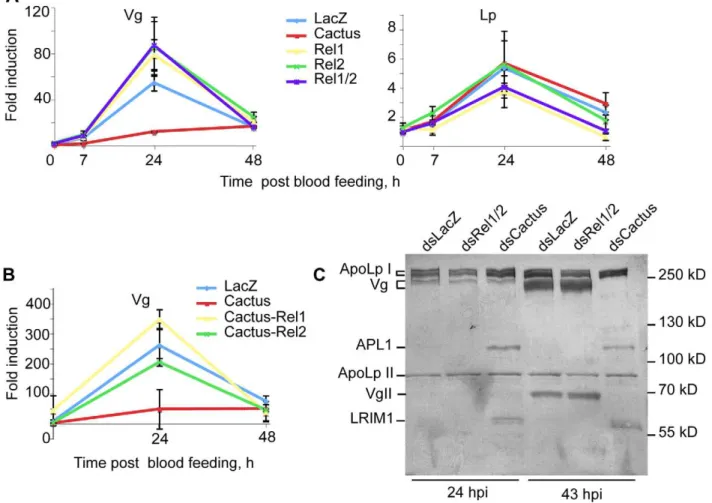
monitored by qRT-PCR. Strikingly, Vg expression was almost abolished in dsCactus mosquitoes at 24 hpi; conversely, the
depletion of REL1 or REL2 at this time point elevated Vg
expression above the levels in the dsLacZ control (Figure 4A). Interestingly, concomitant silencing ofCactus/Rel1andCactus/Rel2 restoredVgexpression to physiological levels (Figure 4B), indicat-ing that REL1 and REL2 contribute to the regulation of Vg expression. At the protein level, Vg amounts were unchanged at 24 h but strongly reduced 43 h after infectious blood feeding specifically indsCactus-injected-mosquitoes (Figure 4C), confirming the qPCR data and revealing a clear delay between mRNA and protein fluctuations. Thus, in thedsCactusbackground, whileTEP1 expression is upregulated,Vg expression is directly or indirectly repressed by REL1/2. Therefore, the Cactus protein affects TEP1 and Vg levels in opposite directions. We extended our analysis to Lp, but in contrast to the situation reported forAe. aegypti[24], its expression was unaffected by the knockdown of the NF-kB-like factors (Figure 4A). Since Vg silencing alone, unlike Cactus silencing, is not sufficient to completely block oogenesis, other molecules required by developing mosquito oocytes may be regulated by Cactus in the same manner as Vg.
Taken together, our findings uncover the complex phenotype of Cactus depletion. It leads to a lower level ofVgexpression after a blood meal, thereby contributing to the arrest in oogenesis seen in
Figure 4.Vgexpression is repressed by NF-kB factors REL1/REL2.Mosquitoes were injected with the indicated combinations ofdsCactus, dsRel1,dsRel2, and the expression levels ofVgandLpexamined by qRT-PCR at the indicated time points after infection and compared todsLacZ control. Gene expression is expressed relative to the LacZ control at time 0. (A)Vgexpression was inhibited indsCactusbut increased indsRel1/Rel2 mosquitoes. (B) Concomitant depletion of Cactus and Rel1 restored Vg expression. (C) Coomassie staining of hemolymph proteins after electrophoresis on a 7% SDS-PAGE gel and transfer to a PVDF membrane. No change in Vg protein is seen 24 h after infection, but at 43 hdsCactus completely blocks Vg expression. Protein identities are indicated to the left. The identity of the subunits of Lp and Vg, and the identity of APL1C and LRIM1 proteins were established by mass spectrometry of the Coomassie-stained bands and by immunoblotting. APL1C and LRIM1 over-expression confirms the efficiency ofCactussilencing.
Cactusknockdown mosquitoes. On the other hand, it stimulates the mosquito antiparasitic defense at least at two different levels: (i) by lowering the level of Vg, rendering TEP1-mediated killing more efficient, and (ii) by elevating the levels of TEP1 pathway proteins.
Discussion
The first indication that nutrient transport after a blood meal influences mosquito susceptibility to P. berghei was provided by Vlachou et al. [23], who demonstrated that experimental depletion of the lipid carrier protein Lp by RNAi reduces the number of developing oocysts in the mosquito midgut. Recently, these results were extended to P. falciparum [25]. However, how and at which stage of development the parasites were eliminated in Lp-deficient mosquitoes remained to be determined. We show here that the major yolk protein Vg shows a similar but more drastic knockdown phenotype than Lp onPlasmodiumsurvival and that the Lp and Vg depletion phenotypes require the function of the immune factor TEP1, which targets ookinetes for killing. Further, high numbers of parasites actually survive and turn into oocysts even in the context of Lp and/or Vg depletion, as long as TEP1 is also experimentally depleted. From these observations, we infer that physiological levels of both nutrient transport proteins following a blood meal somehow dampen the strength of the immune defense and protect ookinetes against destruction by the TEP1 pathway. The effects of Lp and Vg depletion on TEP1-mediated parasite killing are similar, and we find that Lp is required for the full induction ofVgexpression on day 2 following an infectious blood meal. We therefore propose that Lp may indirectly affect ookinete survival by influencing Vg expression, while Vg impinges either directly or more closely than Lp on the TEP1-killing mechanism.
The induction ofVgexpression after a blood meal requires both the TOR pathway and ecdysone signaling [14]. It is unclear why Lp depletion reduces the expression ofVgafter an infectious blood meal. One possible explanation is that an Lp shortage precludes ovarian follicle development, preventing the normal secretion of ecdysone by follicle cells; thus leading to the reduction in Vg expression. However, attempts to rescue theLpsilencing effect on Vg expression with exogenously provided 20-hydroxyecdysone were unsuccessful. As the lower level of Vg expression in Lp-deficientA. gambiaeis reminiscent of the situation observed in adult Ae. aegyptimosquitoes malnourished during larval life [37], it would be interesting to determine ifPlasmodiumsurvival is compromised in such malnourished mosquitoes in laboratory and field settings. The GFP-taggedP. bergheistrain used in this study provides a good model and enables analyses of vectorial capacity that are much more demanding with wild malaria parasites. However, recent studies indicate that the mosquito response toP. bergheiand toP. falciparumdiffer in important ways [10,38]. In addition, theP. berghei–A. gambiaemodel is an unnatural host-parasite association. Therefore, it will be important to see whether our observations hold true in theA. gambiae–P. falciparumrelationship. Importantly though, the TEP1 pathway does limitP. falciparumsurvival inA. gambiaenatural infections ([39] and Levashina et al., unpublished results) and the Lp knockdown was shown to have similar effects in both systems [25].
What is the molecular basis of the negative effect of the two nutrient transport proteins on the TEP1 pathway? We initially hypothesized that Lp-scaffolded lipidic particles could sequester components of the TEP1 pathway in an inactive state. However, TEP1 and its interacting partners LRIM1 and APL1C were not detectable in Lp extracts, suggesting that the Plasmodium-killing machinery is not carried by Lp particles. Instead, RNAi-mediated
depletion of Lp and, more strikingly, of Vg resulted in more efficient TEP1 binding to the surface of ookinetes at 48 hpi, promoting their killing. One explanation could be that Vg (and perhaps Lp, to a lesser extent) are recruited to the parasite surface, where they might mask TEP1 binding sites. Consistent with this idea, fish vitellogenin has recently been found to bind microor-ganisms and to opsonize them for phagocytosis [40]. Mosquito Vg may behave non-productively in a similar manner and outcompete TEP1 from the ookinete surface. Alternatively, a physical interaction between TEP1 and Vg could inhibit TEP1 activity, a hypothesis that should be further investigated. Yet another possible explanation is that transient interactions of ookinetes with Vg might alter the lipid composition in the ookinetes’ membrane, rendering them less visible to the TEP1 machinery. The parasite molecules to which TEP1 covalently attaches are currently unknown, but hydroxyl residues on surface lipids could be good targets for thioester-dependent TEP1 covalent binding.
We further observed a retarded oocyst growth inLp-deficient mosquitoes 9 d post infection. This phenotype was specific to Lp, as parasites developed normally in Vg-deficient mosquitoes. Therefore, Lp is a probable lipid source for developing oocysts. Indeed, Lp was detected inside P. gallinaceum oocysts in vitro, suggesting that oocysts tap some of the host’s Lp for their development [26]. Taken together, Lp appears to regulate parasite development at two distinct stages by two independent mecha-nisms: (i) providing an indirect protection to ookinetes via regulation of Vg levels after a blood meal and thereby dampening TEP1 binding to ookinetes, and (ii) exerting a direct nutritional role by supplying lipids to growing oocysts.
The quantitative RT-PCR and protein expression results reported here added the IkB/NF-kB-like factors Cactus/REL1 and REL2, previously known to control immunity [29–31], to the list of factors that influenceVgexpression. We propose that Cactus depletion boosts TEP1 parasite killing by simultaneously increas-ingTEP1expression [31] and decreasing the expression ofVg, in the absence of which TEP1-mediated killing is more efficient. Previously, the reason why Cactus depletion blocked oogenesis
while boosting anti-Plasmodium immunity was unknown. Our
results shed new light on this phenomenon by suggesting that Cactus activity is necessary for the expression ofVg, and probably of additional factors involved in vitellogenesis.
Although many mosquito genes showing antiparasitic activity are induced by the NF-kB-like factors REL1 and REL2 [12,29–
31,41], it is currently unclear whether parasite invasion of mosquito tissues actually activates the NF-kB pathways. However,
the expression of nutrient transport molecules is affected by signals arising from the parasite’s invasion, in addition to being influenced by hormone signaling, the TOR pathway, and NF-kB factors.
Indeed, ookinete invasion of the midgut induces Lp mRNA
ecdysteroids and taking up Vg protein, which may explain both the drop inVgtranscription and the accumulation of Vg protein in the hemolymph [43,44]. It would be interesting to identify infection-dependent signals arising at the midgut and triggering ovarian follicle apoptosis. In Drosophila, pathogenesis is also reported to trigger cell death in ovaries [45]. In the presence or absence of an infection, activation of the Immune deficiency (Imd) pathway (e.g., by injection of dead bacteria) negatively impacted oogenesis. This effect depended on the immune status, as oogenesis remained normal in Imd pathway mutants injected with dead bacteria [46]. The mosquito Cactus/REL1/REL2
NF-kB pathway is related to theDrosophila Toll and Imd immune
pathways; its targets would therefore represent attractive candi-dates as modulators of mosquito reproduction. A full understand-ing of the interactions between reproductive and immune functions in mosquitoes will require a thorough study of the molecular pathways influencing the transcription of immune and vitellogenic factors, and how these pathways are affected by blood meals, immune defense, and parasite invasion. To our knowledge, Vg and Cactus are the first molecules reported to occupy a central position at the interface between reproduction and immunity, providing a molecular handle to further explore the long-suspected trade-off between these two processes.
Material and Methods
Potassium Bromide Gradient Purification of Lipophorin Particles
Approximately 0.5 g of mosquito adults (ca. 330 mosquitoes) were roughly ground with a Polytron electric homogenizer in 2 ml ice-cold TNE buffer (100 mM Tris-HCl pH 7.5, 0.2 mM EGTA, 150 mM NaCl) + Complete protease inhibitors (Roche). Debris were centrifuged at 4uC in a tabletop centrifuge. The supernatant was transferred to 2.2 ml ultracentrifuge tubes and spun for 3 h at 120,000 g at 4uC in a Sorvall ultracentrifuge equipped with an S55-S rotor. The cleared supernatant was recovered, completed with solid potassium bromide to a final concentration of 0.34 g/ ml, overlayed with 0.5 ml TNE buffer+0.33 g/ml KBr, and centrifuged in 2.2 ml PET ultracentrifuge tubes (Hitachi Koki) at 250,000 g, 10uC, for at least 36 h. The top layer of fat was discarded and 5 or 6 fractions of 0.5 ml were carefully collected starting from the top. Lipophorin particles were present in the top fraction, while the majority of other proteins fractionated into the fourth.
Lipophorin and Vitellogenin Antibodies
The top fraction of a potassium bromide gradient prepared using a scale-up of the above method was desalted on a Pharmacia PD-10 column according to the manufacturer’s instructions. The two subunits of Lp were the predominant proteins in the extract according to Coomassie staining of an SDS-PAGE gel. Protein amount was quantified with a Bradford assay. Six-week-old female BALB/c mice were injected intraperitoneally with 40mg of these lipophorin particles and 100mg of poly I/C as adjuvant. Three injections were performed at 2-wk intervals. Four days prior to hybridoma fusion, mice with positively reacting sera were reinjected. Spleen cells were fused with Sp2/0.Agl4 myeloma cells as described [47]. Hybridoma culture supernatants were tested at day 10 by ELISA for cross-reaction with purified Lp particles. Positive supernatants were then tested by Western blot on mosquito extracts. All ELISA-positive supernatants recognized peptides corresponding in size to either the large (250 kDa) or the small (80 kDa) Lp subunit. Specific cultures were cloned twice on soft agar. A hybridoma clone (2H5, immunoglobulin subclass
IgG2ak) recognizing the 80 kDa Lp subunit was selected and ascites fluid was prepared by injection of 26106hybridoma cells into pristane-primed BALB/c mice. The resulting antibody efficiently immuno-precipitated the 80 kDa Lp subunit and co-immunoprecipated the 250 kDa subunit. The identity of both immuno-precipitated subunits, excised from Coomassie-stained protein gels, was confirmed by mass spectrometry. Similarly, we prepared a monoclonal antibody (2C6) recognizing the large Lp subunit. Rabbit polyclonal antibodies specific to Vg were obtained by immunizing rabbits with a purified recombinant Vg fragment fused to GST. TheVggene fragment used for protein production was amplified from mosquito cDNA using attB-site (capital letters)-containing primers GGGGACAAGTTTGTACAAAAAAGCAG-GCTtcaagtttgtgctgcagcacaagcag and GGGGACCACTTTGTA-CAAGAAAGCTGGGTCCTAagcgcaagatggatggtagtttc. The PCR product was cloned into pDEST15 (Invitrogen) using the Gateway technology. Protein was produced inE. coliBL21-AI.
Immunoprecipitation
120 adult mosquitoes were severed by opening the thorax and abdomen cuticles with fine forceps and bled on ice in 1 ml IP buffer (TRIS pH 7.9 50mM, NaCl 100 mM, EDTA 2 mM, BSA 0.1mg/ml)+Complete protease inhibitors (Roche). Carcasses and cellular debris were removed by two successive 2,500 g centrifu-gation steps (for 2 min at 4uC); the extract was further cleared by three 16,500 g centrifugations (2 min each). The sample was pre-cleared for 1 h at 4uC under gentle rocking with 2mg of an
irrelevant mouse IgG2ak antibody that was removed by
incubation at 4uC with 35ml protein A-sepharose slurry (Pharmacia) for 1 h followed by centrifugation. Supernatant was split in two aliquots, one subjected to a 1 h incubation with specific antibody and the other with a non-specific antibody of the same immunoglobulin class. 35ml of protein A-Sepharose were added to
each sample, further rocked at 4uC for 1 h, centrifuged. The supernatant was saved (post-IP supernatant sample). Sepharose beads were washed 5610 min in TE buffer with or without 500 mM KCl, successively. Lipophorin and associated proteins were eluted from the beads using SDS-PAGE sample buffer and submitted to Western blotting.
Hemolymph Protein Samples for SDS-PAGE
At least 8 anesthetized mosquitoes were aligned on ice under the binocular microscope. Their proboscis was clipped with dissection scissors. Each mosquito was gently pressed on the thorax with forceps and the hemolymph droplet forming at the tip of the cut proboscis was collected into 16 sample (Laemmli) buffer. An hemolymph amount equivalent to that collected from 4 mosqui-toes was loaded in each lane of SDS-PAGE gels.
RNAi and Infections
The 741 bp long HincII fragment ofVg1 (AGAP004203) and the 431 bp longBspHI/BsgI fragment ofLp(AGAP001826) were cloned from cDNA library clones into the pLL10 vector. RNAi constructs for TEP1 and NF-kB factors have been described (Frolet et al. 2006) [31]. Potential cross-silencing effects of the chosen sequences were analyzed using the Deqor software ([48]; http://deqor.mpi-cbg.de/) with the predicted A. gambiae tran-scriptome ENSEMBL database. DsRNA was synthesized as previously described [36]. A. gambiae susceptible G3 strain were maintained at 28uC, 75%–80% humidity, and a 12/12 h light/ dark cycle. Two-day-emerged adult female mosquitoes from the same cohort were injected with 0.2mg of dsRNA using a Nanoject
volume of 1:1 mixtures of 3mg/ml solutions of dsRNAs. Four days
after dsRNA injection mosquitoes were fed on a mouse carryingP. bergheiGFP-con 259cl2 as previously described [36,37]. Statistical significance was determined with a Kruskall-Wallis test for non-parametric data followed by Dunn’s post-test. The indicated p values are those obtained with Dunn’s test.
Assessment of Ovary Development
The ovaries of dissected females were observed under the binocular microscope. Ovaries containing 3 fully grown eggs or more were scored as positive. Ovaries with only undeveloped oocytes or less than 3 fully grown eggs were scored negative.
qRT-PCR
Total RNA from 10 mosquitoes was extracted with Trizol reagent (Invitrogen) before and after dsRNA injection or after blood feeding. 2–8mg of RNA was reverse transcribed using
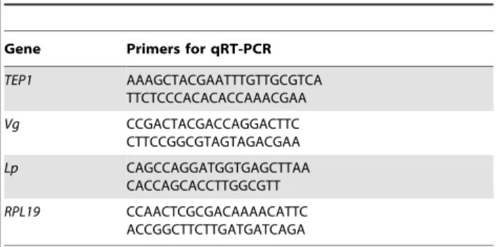
M-MLV enzyme and random primers (Invitrogen). Specific primers (Table 1) were used at 300 nM for qRT-PCR reactions. Ribosomal protein L19 (RPL19) served as an internal control to normalize gene expression. The reactions were run on an Applied Biosystems 7500 Fast Real-Time PCR System using Power SYBR Green Mastermix (http://www.appliedbiosystems.com).
Fluorescence Microscopy
In order to count the surviving GFP-expressing parasites, mosquito midguts were dissected between 7 and 10 dpi and prepared as previously described [36,37] and observed under a fluorescence microscope. To assess TEP1 binding to ookinetes, mosquito midguts were dissected at 18, 24, and 48 hpi, fixed in 4% formaldehyde at room temperature for 45 min, then washed with phosphate buffered saline, and stained with anti-TEP1 antibodies as previously described [31,36]. Parasite numbers and TEP1 labeling were scored using a Zeiss fluorescence microscope (Axiovert 200M) equipped with a Zeiss Apotome module (http:// www.zeiss.com). GFP-expressing parasites were considered live while dead parasites were GFP negative. Differential TEP1 staining on ookinete were gauged at 18, 24, and 48 hpi. At least three independent experiments were conducted per treatment group with a minimum of five mosquito midguts per treatment. For each midgut, all ookinetes visible in 4 fields covering most of the midgut were scored. Table S1 summarizes the ookinete counts from three independent experiments.
MALDI Mass Spectrometry
Coomassie-stained protein bands excised from SDS-PAGE gels were digested with trypsin. Tryptic peptides eluted from the gel slices were subjected to MALDI mass measurement on an
Autoflex III Smartbeam (Bruker-Daltonik GmbH, Bremen, Germany) matrix-assisted laser desorption/ionization time-of-flight mass spectrometer (MALDI-TOF TOF) used in reflector positive mode. The resulting peptide mass fingerprinting data and peptide fragment fingerprinting data were combined by Biotools 3 software (Bruker Daltonik) and transferred to the search engine MASCOT (Matrix Science, London, UK). Peptide mass error was limited to 50 ppm. Proteins were identified by searching data against NCBI non-redundant protein sequence database.
Supporting Information
Figure S1 Prophenoloxidase but not TEP1 or LRIM1 associates with Lp particles. (A, top panel) Coomassie-stained polyacrylamide gel resolving mosquito proteins fraction-ated on a potassium bromide gradient. Molecular weight standards are indicated on the left. Lp subunits (ApoI and ApoII, circled red in lane 1) are the main proteins detectable in top gradient fractions. Fractions 1, 2, 3 are 10-fold concentrated compared to fractions 4, 5, 6. (A, middle and bottom panel) Western blotting with anti-TEP1 and LRIM1 antibodies reveal TEP1 and LRIM1 proteins only in higher density fractions. TEP1-F, full-length TEP1; TEP1-C, C-terminal TEP1 fragment. (B) Western blotting analysis of KBr fractions using anti-PPO2 antibody. A fraction of PPO fractionates with Lp particles. (C) Immunoblotting analysis of Lp particles purified by immunopre-cipitation 0, 4, or 14 d after aP. bergheiinfection (dpi) with mouse anti-Lp (ApoLpII) monoclonal antibody. Non-specific mouse antibody (NS) is used as an immunoprecipitation control. TEP1 does not associate with purified Lp and is found only in post-IP (unbound) supernatants.
Found at: doi:10.1371/journal.pbio.1000434.s001 (0.92 MB TIF)
Figure S2 Lp and Vg proteins are readily visualized by Coomassie staining. Mosquitoes were injected with dsLacZ, dsLp, or dsVgas indicated and offered a blood meal 4 d later to induce Vg expression. Hemolymph was collected 24 h after a blood meal from clipped mosquito proboscises. Hemolymph from the equivalent of 4 mosquitoes as well as 5- and 10-fold dilutions of the controldsLacZhemolymph (2 lanes at the right of the gel) was resolved by electrophoresis on a 7% SDS-PAGE gel and transferred to a PVDF membrane. The membrane was subjected to staining with Coomassie brilliant blue (top panel). Proteins were subsequently revealed with the indicated antibodies (lower panels). Molecular weight markers are indicated on the right. Protein bands revealed by the antibodies superpose perfectly with the protein bands revealed by Coomassie staining. The protein identities were confirmed by a mass spectrometric analysis. The intensities of antibody signals in the 5- and 10-fold diluted sample indicate that residual Vg and Lp protein levels are less than 10% of the control level in the corresponding RNAi samples.
Found at: doi:10.1371/journal.pbio.1000434.s002 (2.34 MB TIF)
Figure S3 (A) Three additional repeats of the experiment shown in Figure 1A. (See Figure 1A for legend.) (B) Three additional repeats of the experiment shown in Figure 1B. (See Figure 1B for legend.) (C) Two additional repeats of the experiment shown in Figure 2A. (See Figure 2A for legend.)
Found at: doi:10.1371/journal.pbio.1000434.s003 (0.35 MB TIF)
Table S1 The table summarizes the parasite scores for three independent repeats of the experiment shown in Figure 2C.Shown are parasite percentages in each of the three possible classes (live, GFP positive; dying, GFP+TEP1 positive; dead, TEP1 positive). The total number of ookinetes scored for each treatment group is given in parentheses next to the injected Table 1.Primers used for qRT-PCR.
Gene Primers for qRT-PCR
TEP1 AAAGCTACGAATTTGTTGCGTCA TTCTCCCACACACCAAACGAA
Vg CCGACTACGACCAGGACTTC
CTTCCGGCGTAGTAGACGAA
Lp CAGCCAGGATGGTGAGCTTAA
CACCAGCACCTTGGCGTT
RPL19 CCAACTCGCGACAAAACATTC ACCGGCTTCTTGATGATCAGA
dsRNA.pvalues were obtained by chi-square analysis comparing parasite scores in dsLp and dsLacZ-injected mosquitoes or comparing parasite scores in dsLp-Vg and dsLacZ-injected mosquitoes. For this analysis, we summed all TEP1-positive
ookinetes (dead + dying). Figure 2C was generated with
Experiment 3.
Found at: doi:10.1371/journal.pbio.1000434.s004 (0.04 MB XLS)
Text S1 The supplemental text describes lipophorin particle purification from adult mosquitoes by potassi-um bromide gradient fractionation or immuno-precip-itation and a search for immune factors that co-purify with lipophorin.
Found at: doi:10.1371/journal.pbio.1000434.s005 (0.10 MB DOC)
Acknowledgments
The authors thank J. Soichot, M.E. Moritz, and C. Kappler for help with the mosquito colony and parasite cultures; C. Guillet and P. Hammann (IBMC mass spectrometry platform) for protein identification; V. Andres and N. Jung for hybridoma cultures and subcloning; and F. Catteruccia and members of her and our group for fruitful discussions and critical reading of the manuscript.
Author Contributions
The author(s) have made the following declarations about their contributions: Conceived and designed the experiments: MKR MMW EAL EM. Performed the experiments: MKR MMW EM. Analyzed the data: MKR MMW EAL EM. Contributed reagents/materials/analysis tools: MOA. Wrote the paper: MKR EAL EM.
References
1. Sinden RE (1999)Plasmodiumdifferentiation in the mosquito. Parassitologia 41: 139–148.
2. Blandin S, Shiao SH, Moita LF, Janse CJ, Waters AP, et al. (2004) Complement-like protein TEP1 is a determinant of vectorial capacity in the malaria vector Anopheles gambiae. Cell 116: 661–670.
3. Habtewold T, Povelones M, Blagborough AM, Christophides GK (2008) Transmission blocking immunity in the malaria non-vector mosquito Anopheles quadriannulatus species A. PLoS Pathog 4: doi:10.1371/journal.ppat.1000070. 4. Blandin SA, Marois E, Levashina EA (2008) Antimalarial responses in Anopheles gambiae: from a complement-like protein to a complement-like pathway. Cell Host Microbe 3: 364–374.
5. Levashina EA, Moita LF, Blandin S, Vriend G, Lagueux M, et al. (2001) Conserved role of a complement-like protein in phagocytosis revealed by dsRNA knockout in cultured cells of the mosquito,Anopheles gambiae. Cell 104: 709–718. 6. Baxter RH, Chang CI, Chelliah Y, Blandin S, Levashina EA, et al. (2007) Structural basis for conserved complement factor-like function in the antimalarial protein TEP1. Proc Natl Acad Sci U S A 104: 11615–11620. 7. Fraiture M, Baxter RH, Steinert S, Chelliah Y, Frolet C, et al. (2009) Two
mosquito LRR proteins function as complement control factors in the TEP1-mediated killing of Plasmodium. Cell Host Microbe 5: 273–284.
8. Povelones M, Waterhouse RM, Kafatos FC, Christophides GK (2009) Leucine-rich repeat protein complex activates mosquito complement in defense against Plasmodium parasites. Science 324: 258–261.
9. Osta MA, Christophides GK, Kafatos FC (2004) Effects of mosquito genes on Plasmodiumdevelopment. Science 303: 2030–2032.
10. Cohuet A, Osta MA, Morlais I, Awono-Ambene PH, Michel K, et al. (2006) AnophelesandPlasmodium: from laboratory models to natural systems in the field. EMBO Rep 7: 1285–1289.
11. Riehle MM, Markianos K, Niare O, Xu J, Li J, et al. (2006) Natural malaria infection inAnopheles gambiaeis regulated by a single genomic control region. Science 312: 577–579.
12. Riehle MM, Xu J, Lazzaro BP, Rottschaefer SM, Coulibaly B, et al. (2008) Anopheles gambiae APL1 is a family of variable LRR proteins required for Rel1-mediated protection from the malaria parasite, Plasmodium berghei. PLoS ONE 3: doi:10.1371/journal.pone.0003672.
13. Attardo GM, Hansen IA, Raikhel AS (2005) Nutritional regulation of vitellogenesis in mosquitoes: implications for anautogeny. Insect Biochem Mol Biol 35: 661–675.
14. Hansen IA, Attardo GM, Park JH, Peng Q, Raikhel AS (2004) Target of rapamycin-mediated amino acid signaling in mosquito anautogeny. Proc Natl Acad Sci U S A 101: 10626–10631.
15. Raikhel AS, Dhadialla TS (1992) Accumulation of yolk proteins in insect oocytes. Annu Rev Entomol 37: 217–251.
16. Tufail M, Takeda M (2008) Molecular characteristics of insect vitellogenins. J Insect Physiol 54: 1447–1458.
17. Canavoso LE, Jouni ZE, Karnas KJ, Pennington JE, Wells MA (2001) Fat metabolism in insects. Annu Rev Nutr 21: 23–46.
18. Arrese EL, Canavoso LE, Jouni ZE, Pennington JE, Tsuchida K, et al. (2001) Lipid storage and mobilization in insects: current status and future directions. Insect Biochem Mol Biol 1: 7–17.
19. Atella GC, Silva-Neto MA, Golodne DM, Arefin S, Shahabuddin M (2006) Anopheles gambiae lipophorin: characterization and role in lipid transport to developing oocyte. Insect Biochem Mol Biol 36: 375–386.
20. Pana´kova´ D, Sprong H, Marois E, Thiele C, Eaton S (2005) Lipoprotein particles are required for Hedgehog and Wingless signalling. Nature 435: 58–65. 21. Vaisar T, Pennathur S, Green PS, Gharib SA, Hoofnagle AN, et al. (2007) Shotgun proteomics implicates protease inhibition and complement activation in the antiinflammatory properties of HDL. J Clin Invest 117: 746–756. 22. Pays E, Vanhollebeke B (2009) Human innate immunity against African
trypanosomes. Curr Opin Immunol 21: 493–498.
23. Vlachou D, Schlegelmilch T, Christophides GK, Kafatos FC (2005) Functional genomic analysis of midgut epithelial responses inAnophelesduringPlasmodium invasion. Curr Biol 15: 1185–1195.
24. Cheon HM, Shin SW, Bian G, Park JH, Raikhel AS (2006) Regulation of lipid metabolism genes, lipid carrier protein lipophorin, and its receptor during immune challenge in the mosquito Aedes aegypti. J Biol Chem 281: 8426–8435. 25. Mendes AM, Schlegelmilch T, Cohuet A, Awono-Ambene P, De Iorio M, et al. (2008) Conserved mosquito/parasite interactions affect development of Plasmo-dium falciparum in Africa. PLoS Pathog 4: doi:10.1371/journal.ppat.1000069. 26. Atella GC, Bittencourt-Cunha PR, Nunes RD, Shahabuddin M, Silva-Neto MA (2009) The major insect lipoprotein is a lipid source to mosquito stages of malaria parasite. Acta Trop.
27. Zhu J, Chen L, Raikhel AS (2003) Posttranscriptional control of the competence factor betaFTZ-F1 by juvenile hormone in the mosquito Aedes aegypti. Proc Natl Acad Sci U S A 100: 13338–13343.
28. Zhu J, Chen L, Sun G, Raikhel AS (2006) The competence factor beta Ftz-F1 potentiates ecdysone receptor activity via recruiting a p160/SRC coactivator. Mol Cell Biol 26: 9402–9412.
29. Bian G, Shin SW, Cheon HM, Kokoza V, Raikhel AS (2005) Transgenic alteration of Toll immune pathway in the female mosquito Aedes aegypti. Proc Natl Acad Sci U S A 102: 13568–13573.
30. Meister S, Kanzok SM, Zheng XL, Luna C, Li TR, et al. (2005) Immune signaling pathways regulating bacterial and malaria parasite infection of the mosquitoAnopheles gambiae. Proc Natl Acad Sci U S A 102: 11420–11425. 31. Frolet C, Thoma M, Blandin S, Hoffmann JA, Levashina EA (2006) Boosting
NF-kappaB-dependent basal immunity ofAnopheles gambiaeaborts development ofPlasmodium berghei. Immunity 25: 677–685.
32. Franke-Fayard B, Trueman H, Ramesar J, Mendoza J, van der Keur M, et al. (2004) APlasmodium bergheireference line that constitutively expresses GFP at a high level throughout the complete life cycle. Mol Biochem Parasitol 137: 23–33. 33. Bose SG, Raikhel AS (1988) Mosquito vitellogenin subunits originate from a
common precursor. Biochem Biophys Res Commun 155: 436–442. 34. Sappington TW, Raikhel AS (1998) Molecular characteristics of insect
vitellogenins and vitellogenin receptors. Insect Biochem Mol Biol 28: 277–300. 35. Dhadialla TS, Raikhel AS (1990) Biosynthesis of mosquito vitellogenin. J Biol
Chem 265: 9924–9933.
36. Blandin S, Levashina EA (2004) Mosquito immune responses against malaria parasites. Curr Opin Immunol 16: 16–20.
37. Shiao SH, Hansen IA, Zhu J, Sieglaff DH, Raikhel AS (2008) Juvenile hormone connects larval nutrition with target of rapamycin signaling in the mosquito Aedes aegypti. J Insect Physiol 54: 231–239.
38. Jaramillo-Gutierrez G, Rodrigues J, Ndikuyeze G, Povelones M, Molina-Cruz A, et al. (2009) Mosquito immune responses and compatibility between Plasmo-dium parasites and anopheline mosquitoes. BMC Microbiol 9: 154. 39. Dong Y, Aguilar R, Xi Z, Warr E, Mongin E, et al. (2006)Anopheles gambiae
immune responses to human and rodent Plasmodium parasite species. PLoS Pathog 2: e52. doi:10.1371/journal.ppat.0020052.
40. Li Z, Zhang S, Liu Q (2008) Vitellogenin functions as a multivalent pattern recognition receptor with an opsonic activity. PLoS ONE 3: doi:10.1371/ journal.pone.0001940.
41. Garver LS, Dong Y, Dimopoulos G (2009) Caspar controls resistance to Plasmodium falciparum in diverse anopheline species. PLoS Pathog 5: doi:10.1371/journal.ppat.1000335.
42. Ahmed AM, Maingon R, Romans P, Hurd H (2001) Effects of malaria infection on vitellogenesis inAnopheles gambiaeduring two gonotrophic cycles. Insect Mol Biol 10: 347–356.
43. Hopwood JA, Ahmed AM, Polwart A, Williams GT, Hurd H (2001) Malaria-induced apoptosis in mosquito ovaries: a mechanism to control vector egg production. J Exp Biol 204: 2773–2780.
45. Brandt SM, Schneider DS (2007) Bacterial infection of fly ovaries reduces egg production and induces local hemocyte activation. Dev Comp Immunol 31: 1121–1130.
46. Zerofsky M, Harel E, Silverman N, Tatar M (2005) Aging of the innate immune response in Drosophila melanogaster. Aging Cell 4: 103–108.
47. St Groth FS, Scheidegger D (1980) Production of monoclonal antibodies: strategy and tactics. J Immunol Methods 35: 1–21.