Hepatoprotective Effect of Wheat-Based
Solid-State Fermented
Antrodia cinnamomea
in Carbon Tetrachloride-Induced Liver Injury
in Rat
Huan-Wen Chiu1, Kuo-Feng Hua1,2*
1Department of Biotechnology and Animal Science, National Ilan University, Ilan, Taiwan,2Department of Pathology, Tri-Service General Hospital, National Defense Medical Center, Taipei, Taiwan
*kuofenghua@gmail.com
Abstract
Antrodia cinnamomea(A.cinnamomea) is an indigenous medical fungus in Taiwan and has multiple biological functions, including hepatoprotective and immune-modulatory
effects. Currently, the commercially availableA.cinnamomeaare mainly liquid- and
solid-state fermentedA.cinnamomea. However, the hepatoprotective effect of solid-state
fer-mentedA.cinnamomeahas never been reported. Here we evaluate the ability of air-dried,
ground and non-extracted wheat-based solid-state fermentedA.cinnamomea(WFAC) to
protect against carbon tetrachloride (CCl4)-induced hepatic injury in vivo. The results
showed that oral administration of WFAC dose dependently (180, 540 and 1080 mg/kg) ameliorated the increase in plasma aspartate aminotransferase and alanine
aminotransfer-ase levels caused by chronic repeated CCl4intoxication in rats. WFAC significantly reduced
the CCl4-induced increase in hepatic lipid peroxidation levels and hydroxyproline contents, as well as reducing the spleen weight and water content of the liver. WFAC also restored the hepatic soluble protein synthesis and plasma albumin concentration in CCl4-intoxicated rats, but it did not affect the activities of superoxide dismutase, catalase, or glutathione per-oxidase. In addition, a hepatic morphological analysis showed that the hepatic fibrosis and
necrosis induced by CCl4were significantly ameliorated by WFAC. Furthermore, the body
weights of control rats and WFAC-administered rats were not significantly different, and no adverse effects were observed in WFAC-administered rats. These results indicate that
WFAC is a nontoxic hepatoprotective agent against chronic CCl4-induced hepatic injury.
Introduction
Antrodia cinnamomea(A.cinnamomea; synonym:Antrodia camphorata) is an indigenous medicinal fungus in Taiwan.A.cinnamomeahas been demonstrated to possess diverse medici-nal and pharmacological activities, particularly its anti-tumor [1–5], anti-inflammatory [6–10], anti-diabetic [11], anti-hypertension [12], anti-bacterial [13], renoprotective [14–16],
OPEN ACCESS
Citation:Chiu H-W, Hua K-F (2016)
Hepatoprotective Effect of Wheat-Based Solid-State FermentedAntrodia cinnamomeain Carbon Tetrachloride-Induced Liver Injury in Rat. PLoS ONE 11(4): e0153087. doi:10.1371/journal.pone.0153087
Editor:Jordi Gracia-Sancho, IDIBAPS - Hospital Clinic de Barcelona, SPAIN
Received:August 22, 2015
Accepted:March 23, 2016
Published:April 5, 2016
Copyright:© 2016 Chiu, Hua. This is an open access article distributed under the terms of the
Creative Commons Attribution License, which permits unrestricted use, distribution, and reproduction in any medium, provided the original author and source are credited.
Data Availability Statement:All the raw data and statistical analysis of this paper are available from the Dryad database (accession number(s) doi:10.5061/ dryad.p066m).
neuroprotective [17–19], and hepatoprotective effects [20,21]. Although the greatest number of publications are in regards to the anti-tumor activity ofA.cinnamomea, the Food and Drug Administration has not approved anyA.cinnamomeaextracts or purified compounds for clini-cal anti-tumor applications. Although the therapeutic effects need further investigation,A. cin-namomeais commonly used as a food supplement and is believed to preserve human vitality and promote longevity [22].
Among the diverse biological functions ofA.cinnamomea, the most recognized function is its hepatoprotective activity [23] becauseA.cinnamomeawas traditionally used in Taiwan by the aborigines as a traditional prescription for the discomforts caused by drinking alcohol or exhaustion [24]. The fruiting body ofA.cinnamomeaprotected livers against alcohol-induced liver damage in rats [25,26] and ameliorated carbon tetrachloride (CCl4)-induced hepatic
injury in mice [27]. TheA.cinnamomeafruiting body grows very slowly on only the inner cav-ity of the endemic speciesCinnamomum kanehirae(Bull camphor tree) Hayata (Lauraceae) in Taiwan. The Taiwanese government has protectedCinnamomum kanehiraefrom forest-denu-dation because this tree species is relatively rare in nature. Field gathering of theA. cinnamo-meafruiting body is also prohibited [24]. Therefore, theA.cinnamomeafruiting body is expensive because of its limited availability. Currently, most of the commercially availableA.
cinnamomeaproducts come from the submerged liquid or solid-state mycelia cultures. Extracts of mycelium fromA.cinnamomeain submerged liquid culture protect against alco-hol-induced liver injury in vitro [28] and in vivo [29]. In addition, fermented filtrates fromA.
cinnamomeain submerged liquid culture protect against CCl4-induced hepatic toxicity in rats
[30]. However, there are as yet no published reports demonstrating the hepatoprotective activ-ity of solid-state culturedA.cinnamomea.
In the present study, we examined the hepatoprotective activity of wheat-based solid-state fermentedA.cinnamomea(WFAC) in chronic CCl4-induced liver injury in rats. In particular,
to mimic our regular consumption ofA.cinnamomeaproducts, we fed the rats with air-dried and ground WFAC but not with the extracted WFAC.
Materials and Methods
Chemicals
Silymarin, carboxymethyl cellulose, trichloroacetic acid,σ-phthalaldehyde, thiobarbituric acid,
p-dimethylaminobenzoaldehyde, n-butanol, pyridine, catalase, hematoxylin and eosin, Sirius red and other chemicals were purchased from Sigma (St. Louis, MO, USA). The protein assay kit was purchased from Bio-Rad Laboratories Inc. (CA, USA)
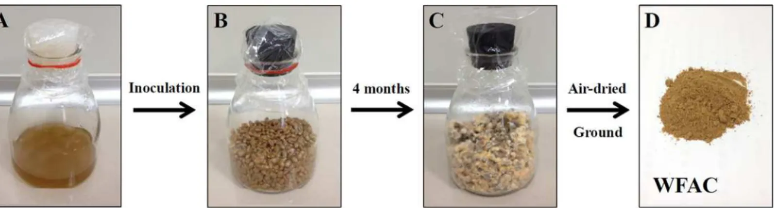
Preparation of WFAC
The WFAC was obtained from Ruei Sen Biotech Co., Ltd (Taipei, Taiwan). Briefly, WFAC was prepared by inoculating 10 ml of liquid fermentedA.cinnamomeawith 100 g of sterilized wheat pre-mixed with 100 ml of medium (2% sugar, 0.5% malt extract, 0.5% yeast extract in ddH2O) for 4 months at 25 ± 2°C. Air-dried WFAC was ground and stored at 4°C before
feed-ing it to the rats (Fig 1).
Ethics statement
All animal manipulations were performed in the Laboratory Animal Center of National Ilan University (Ilan, Taiwan) in accordance with the National Ilan University guide for the care and use of laboratory animals. The procedures used were approved by the Animal Care and
Use Committee of the National Ilan University (Approval number: 102–7). All manipulations were performed under isoflurane anesthesia, and all efforts were made to minimize suffering.
CCl
4-induced liver injury in rats
Male Wistar rats were obtained from BioLASCO Taiwan Co., Ltd and allowed free access to a standard laboratory chow and tap water ad libitum. The animals were maintained under a 12-h light/dark cycle in an air-conditioned room at 22 ± 2°C. When the rats reached 250–300 g, they were used for the experiments. Rats were divided randomly into six groups: (1) normal control (PBS); (2) vehicle (0.5% carboxymethyl cellulose) plus CCl4treatment; (3) 180 mg/kg
WFAC plus CCl4treatment; (4) 540 mg/kg WFAC plus CCl4treatment; (5) 1080 mg/kg
WFAC plus CCl4treatment; and (6) 200 mg/kg silymarin plus CCl4treatment. Liver injury
was induced in rats by an intraperitoneally (i.p.) administration of 2 ml/kg of 20% CCl4
(diluted in olive oil) twice a week for 8 weeks [31]. WFAC-treated rats were given oral adminis-trations of 180, 540, or 1080 mg/kg WFAC, respectively, every day for 8 weeks (started at one day before the administration of CCl4). Normal control and CCl4-treated rats were treated
with PBS or 0.5% carboxymethyl cellulose every day for 8 weeks. The time interval between CCl4and WFAC administrations was 5 h to avoid disturbing the absorption of each other.
Blood samples were collected from the tail vein at the end of the first, third, sixth and eighth weeks. Spleens and livers were obtained immediately after the rats were sacrificed at the end of the eighth week, and the weights of the livers and spleens were measured.
Analyses of plasma aspartate aminotransferase (AST), alanine
aminotransferase (ALT) and albumin
AST and ALT activities in the plasma were measured using AST and ALT kits according to the manufacturer’s instructions (Roche Applied Science, Mannheim, DE). The plasma albumin concentration was determined using an ELISA kit (Abcam, Cambridge, MA).
Analyses of hepatic glutathione (GSH) and lipid peroxidation levels
For the hepatic GSH analysis, 0.5 g of liver tissue was homogenized in 5 ml 1.15% KCl buffer and mixed with 5 ml 10% trichloroacetic acid. After centrifugation at 3000 g for 15 min, 0.01 ml supernatant was mixed with 0.18 ml phosphate-EDTA and 0.01 mlσ-phthalaldehyde (1
mg/ml methanol). After 15 min, the fluorescence at 350 nm excitation and 420 nm emission was read against a blank that contained deionized water to replace the liver tissue. The Fig 1. Preparation of WFAC.(A) Liquid fermentedA.cinnamomea. (B) Sterilized wheat beforeA.cinnamomeainoculation. (C) 4 months fermentedA. cinnamomeain wheat. (D) Ground WFAC.
concentration of GSH was determined using a GSH standard to replace the liver tissue. The results were expressed asμmol/g tissue. For the hepatic lipid peroxidation level analysis, 0.5 g
of liver tissue was homogenized in 5 ml 1.15% KCl buffer. After centrifugation at 4,000 rpm for 5 min at 4°C, the supernatant was mixed with the same volume of thiobarbituric acid (0.5% [w/v] thiobarbituric acid in 50% [v/v] acetic acid). Samples were boiled and extracted with n-butanol/pyridine (15:1) buffer and centrifuged for 10 min at 10,000 rpm. The butanol layer containing the thiobarbituric acid-reactive substances (malonaldehyde, MDA) was read at 532 nm. The results are expressed as nmol MDA/mg protein.
Analysis of hepatic superoxide dismutase (SOD) activity
The method of Xia et al was followed with slight modifications [32]. The SOD activity was measured using the SOD activity assay kit according to the manufacturer’s instructions (RAN-DOX Lab. Ltd. UK). The method of the SOD activity assay employs xanthine and xanthine oxi-dase to generate superoxide radicals which react with 2-(4-iodophenyl)-3-(4-nitrophenol)-5-phenyltetrazolium chloride to form a red formazan dye. The superoxide dismutase activity is then measured by the degree of inhibition of this reaction. One unit of SOD is that which causes a 50% inhibition of the rate of reduction of 2-(4-iodophenyl)-3-(4-nitrophenol)-5-phe-nyltetrazolium chloride under the conditions of the assay. The specific activity of SOD is expressed as U/mg protein.
Analysis of hepatic catalase activity
The method of Aebi et al was followed with slight modifications [33]. 0.5 g of liver tissue was homogenized in 5 ml 1.15% KCl buffer and centrifuged at 10,000 rpm for 10 min at 4°C. Then the supernatant (5μl) was mixed with 995μl of a 30 mM H2O2solution prepared in potassium
phosphate buffer. Commercially available catalase was used as a standard. A change in the absorbance was monitored at 240 nm for 25 seconds. The specific activity of catalase is expressed asK/mg protein. The following equation was generated to calculate the rate constant (K):K= (2.3/Δt) x log (A1/A2).Δt: the reaction time interval; A1: the absorbance at 0 second; A2: the absorbance at 25 second.
Analysis of hepatic glutathione peroxidase (GSH-Px) activity
The method of Xia et al was followed with slight modifications [32]. The GSH-Px activity was measured using the Ransel GSH-Px activity assay kit according to the manufacturer’s instruc-tions (RANDOX Lab. Ltd. UK). GSH-Px catalyzes the oxidation of GSH by cumene hydroper-oxide. In the presence of glutathione reductase and NADPH the oxidized glutathione is immediately converted to the reduced form with a concomitant oxidation of NADPH to NADP+. The decrease in absorbance at 340 nm is measured. The specific activity of GSH-Px is expressed as mU/mg protein.
Analysis of hepatic soluble protein levels and hydroxyproline contents
Protein contents of liver homogenates were determined by the Bio-Rad method using bovine serum albumin as a standard. The results are expressed as mg/g tissue. For hydroxyproline con-tents, dried liver tissue after hydrolysis was oxidized by H2O2and colored by
p-dimethylami-nobenzoaldehyde, and the absorbance was determined at 540 nm. The amount of
hydroxyproline is expressed asμg/g tissue. The percentage of water content of liver was
Liver histopathology
Rats were sacrificed at the end of the eighth week. The livers were immediately fixed in a 10% buffered formalin phosphate solution, embedded in paraffin, cut into 4–5μm thick sections,
and stained with hematoxylin and eosin and Sirius red. Liver fibrosis and necrosis were graded according to the guidelines of hepatoprotective assessment of the Food and Drug Administra-tion, Ministry of Health and Welfare, Taiwan.
Index compounds of
A
.
cinnamomea
in WFAC
Eight signature triterpenoids compounds ofA.cinnamomeain WFAC were determined and quantitated by quantitative LC-MS/MS. The data was provided by ABM International Lab Inc. (Pingtung, Taiwan).
Statistical analysis
Data are presented as the mean ± SD. All other experimental data, except the pathological find-ings, were analyzed using a one-way ANOVA with Dunnett’s test. Liver histopathological examination data were analyzed by the Kruskall-Wallis non-parametric test followed by a Mann-Whitney U-test.
Results
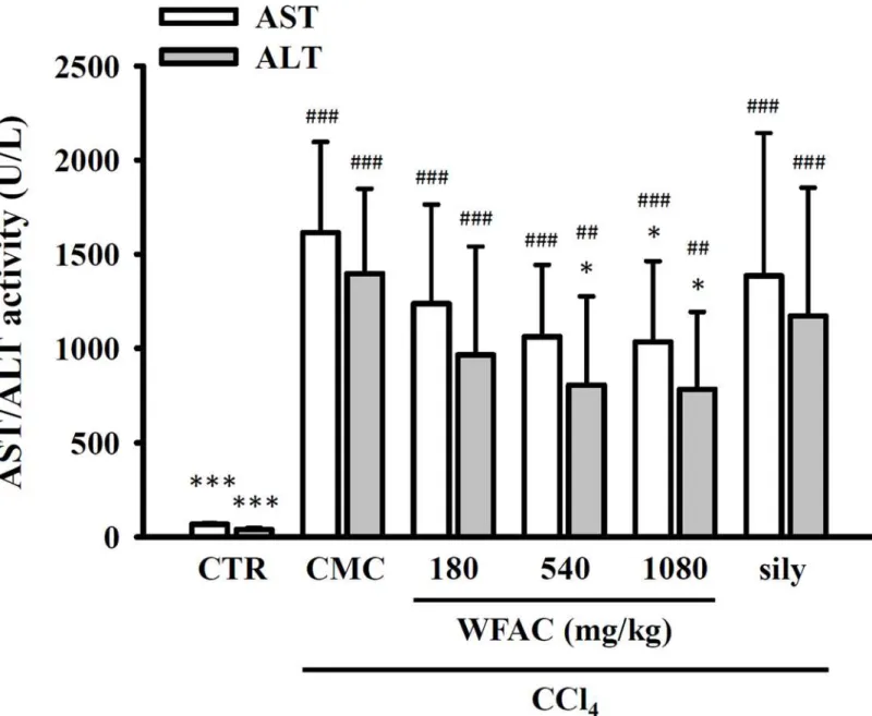
WFAC reduces plasma AST and ALT formation and increases the
plasma albumin concentration in CCl
4-intoxicated rats
As shown inFig 2, the plasma levels of AST and ALT were elevated by the oral administration of CCl4at the end of the first week and then increased to 1615.0 ± 482.0 and 1396.7 ± 450.8 U/
L by the eighth week, respectively. In the control group (PBS treatment), plasma AST and ALT levels were 70.5 ± 8.3 and 39.4 ± 7.9 U/L, at the end of the eighth weeks, respectively. Oral administration of WFAC (180, 540 and 1080 mg/kg) reduced plasma AST and ALT levels dose dependently at the end of the first to eighth weeks. The WFAC (1080 mg/kg) markedly reduced plasma AST and ALT levels to 1034.3 ± 429.0 and 784.0 ± 409.6 at the end of the eighth week in CCl4-intoxicated rats. However, oral administration of silymarin (200 mg/kg) also reduced
plasma ALT and AST levels in CCl4-intoxicated rats, but it did not reach statistical significance.
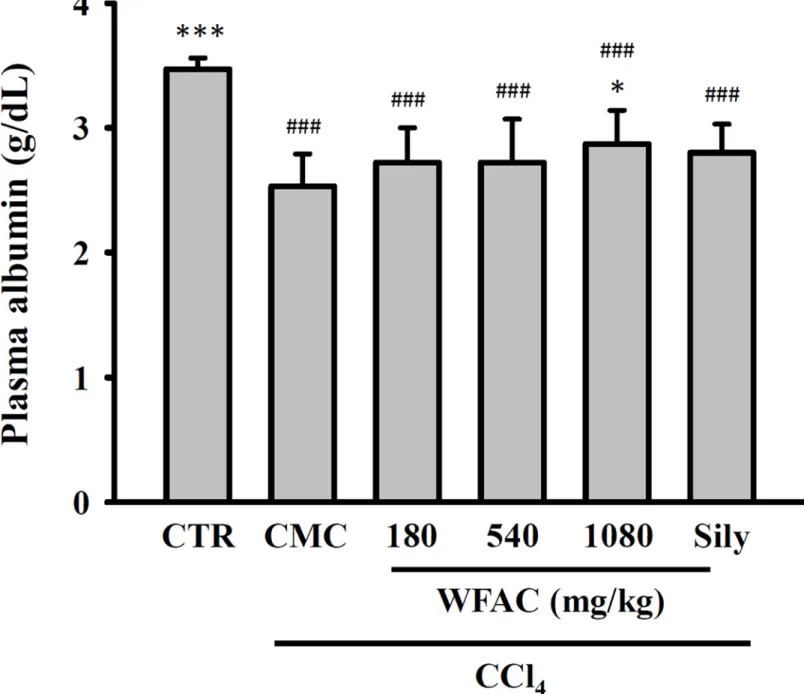
Albumin is a protein made by the liver, and lower-than-normal levels of plasma albumin may be a sign of liver injury. The plasma albumin concentration was significantly reduced by CCl4
intoxication at the end of the eighth week, and WFAC (1080 mg/kg) significantly increased the plasma albumin concentration (Fig 3). Again, silymarin (200 mg/kg) increased the plasma albumin concentration in CCl4-intoxicated rats, but it did not reach statistical significance (Fig
3). Additionally, the changes in body weights of the CCl4-intoxicated or WFAC-treated rats
were similar to those of the control rats, although the body weights of the CCl4-intoxicated rats
at the end of the first and sixth weeks was temporarily reduced (Table 1). These results sug-gested that the administration of WFAC did not have any adverse effects.
WFAC reduces hepatic lipid peroxidation levels in CCl
4-intoxicated rats
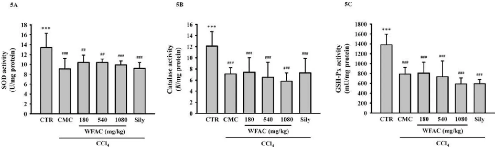
Lipid peroxidation is one of the consequences of uncontrolled oxidative stress in organ injuries caused by oxidative damage. As shown inFig 4A, CCl4intoxication increased the level ofglutathione levels have also been used as a determinate of oxidative status. However, CCl4
intoxication and WFAC administration did not affect the hepatic glutathione levels (Fig 4B). Furthermore, although CCl4intoxication reduced the activities of hepatic anti-oxidant
enzymes, such as SOD, catalase, and GSH-Px, WFAC and silymarin did not restore the levels of these enzymes (Fig 5A–5C).
WFAC increases hepatic soluble protein levels and reduces hepatic
hydroxyproline contents in CCl
4-intoxicated rats
The hepatic soluble protein levels will be reduced in liver injury. We found that the hepatic sol-uble protein levels (mg/g tissue) significantly decreased from 194.8 ± 13.5 in control rats to Fig 2. Effect of WFAC on plasma AST and ALT activities in CCl4-intoxicated rat.Blood samples collected at the end of the eighth weeks after CCl4
intoxication. All values are the means±S.D. (n = 10). ##p<0.01 and ###p<0.001 compared with the control group.*p<0.05 and***p<0.001 compared
with the CCl4+ CMC group. CTR: control; Sily: silymarin (200 mg/kg).
149.7 ± 17.7 CCl4in CCl4-intoxicated rats at the end of the eighth week. WFAC (1080 mg/kg)
significantly increased the hepatic soluble protein levels to 183.1 ± 37.3 (mg/g tissue) in CCl4
-intoxicated rats. Silymarin (200 mg/kg) also increased the hepatic soluble protein, but it did not reach statistical significance (Fig 6A). In addition, we asked whether WFAC ameliorated the liver fibrosis induced by CCl4intoxication. To investigate liver fibrosis, the total collagen
present in the liver was determined by estimating the hydroxyproline content, a product of col-lagen metabolism. We found that the hydroxyproline content (μg/g protein) increased from
309.9 ± 49.0 in control rats to 807.3 ± 192.5 in CCl4-intoxicated rats. WFAC (1080 mg/kg)
Fig 3. Effect of WFAC on plasma albumin concentration in CCl4-intoxicated rat.Blood samples collected at the end of the eighth weeks after CCl4
intoxication. All values are the means±S.D. (n = 10). ###p<0.001 compared with the control group.*p<0.05 and***p<0.001 compared with the CCl4
+ CMC group. CTR: control; Sily: silymarin (200 mg/kg).
significantly decreased the hydroxyproline content to 572.8 ± 72.0 (μg/g protein) in CCl4
-intoxicated rats. Silymarin (200 mg/kg) also decreased the hydroxyproline content, but it did not reach statistical significance (Fig 6B).
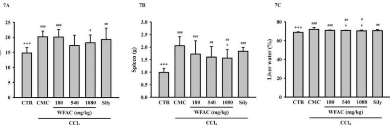
WFAC reduces spleen weight and liver water content in CCl
4-intoxicated
rats
As shown inFig 7, In control rats, the weight of the liver (Fig 7A) and spleen (Fig 7B) were 14.8 ± 1.8 and 0.99 ± 0.16 g, respectively. The weight of the liver and spleen of CCl4-intoxicated
rats increased to 20.2 ± 1.9 and 2.05 ± 0.36 g, respectively, at the end of the eighth week. Table 1. Effect of WFAC on body weights in CCl4-intoxicated rat.
Body weight (g)
Drugs Doses
(mg/kg)
Before CCl4
Week 1 Week 2 Week 3 Week 4 Week 5 Week 6 Week 7 Week 8
Control - 265.1±10.9 293.9±23.5 314.6±28.1 327.5±25.2 350.5±17.1 371.5±19.2 388.4±16.6 408.8±18.4 416.5±25.2
CCl4+ CMC - 265.0±9.0 266.4±17.1# 305.8±20.0 316.7±18.5 330.5±26.9 351.9±23.6 357.5±10.7# 380.9±22.0 389.7±20.9
CCl4+ WFAC 180 267.5±11.7 274.0±16.2 317.7±23.2 326.8±27.6 344.5±30.6 358.9±13.0 363.9±36.6 388.3±38.4 399.2±35.0
CCl4+ WFAC 540 272.0±13.8 282.1±13.4 324.0±19.4 333.5±19.1 343.4±17.1 362.6±14.9 359.0±20.8 391.8±14.0 404.5±20.3
CCl4+ WFAC 1080 268.6±8.4 271.1±12.2 310.4±21.1 322.0±27.4 330.0±36.0 343.9±36.3 347.3±35.1 365.8±35.0 384.5±28.3
CCl4+ silymarin 200 265.9±7.8 265.6±16.3 292.0±33.8 303.1±33.8 338.3±26.3 353.1±26.0 359.4±24.8 378.6±25.4 394.6±24.5
All values are the means±S.D. (n = 10).
#p<0.05 compared with the control group.
doi:10.1371/journal.pone.0153087.t001
Fig 4. Effect of WFAC on the hepatic levels of lipid peroxidation and glutathione in CCl4-intoxicated rat.Liver tissue samples collected at the end of the eighth weeks after CCl4intoxication. (A) Levels of lipid peroxidation. (B) Levels of glutathione. All values are the means±S.D. (n = 10). #p<0.05, ##
p<0.01 and ###p<0.001 compared with the control group.*p<0.05 and***p<0.001 compared with the CCl4+ CMC group. CTR: control; Sily: silymarin
(200 mg/kg).
WFAC (1080 mg/kg) significantly decreased the spleen weight to 1.56 ± 0.34 g in CCl4
-intoxi-cated rats. However, WFAC at 180 and 540 mg/kg did not decrease the spleen weight. WFAC and silymarin did not decrease the liver weight in CCl4-intoxicated rats. In addition, as shown
inFig 7C, the water content of the liver increased from 68.8 ± 0.7% in control rats to
72.1 ± 1.9% in CCl4-intoxicated rats. WFAC at 540 and 1080 mg/kg significantly reduced the
water content of the liver to 70.8 ± 0.3 and 70.4 ± 0.9%, respectively, in CCl4-intoxicated rats.
Silymarin reduced the water content of the liver to 70.6 ± 1.1, but it did not reach statistical significance.
Fig 5. Effect of WFAC on hepatic SOD, catalase and GSH-Px activities in CCl4-intoxicated rat.Liver tissue samples collected at the end of the eighth weeks after CCl4intoxication. (A) SOD activity. (B) Catalase activity. (C) GSH-Px activity. All values are the means±S.D. (n = 10). ##p<0.01 and ###
p<0.001 compared with the control group.***p<0.001 compared with the CCl4+ CMC group. CTR: control; Sily: silymarin (200 mg/kg).
doi:10.1371/journal.pone.0153087.g005
Fig 6. Effect of WFAC on hepatic soluble protein levels and hydroxyproline contents in CCl4-intoxicated rat.Liver tissue samples collected at the end of the eighth weeks after CCl4intoxication. (A) Soluble protein levels. (B) Hydroxyproline contents. All values are the means±S.D. (n = 10). ###p<0.001
WFAC reduces liver fibrosis and necrosis in CCl
4-intoxicated rats
Histopathological changes of necrotic and lipid-laden hepatocytes of liver sections were assessed at the end of the eighth week after CCl4intoxication. The typical intense centrilobularnecrosis of hepatotoxicity and inflammatory cell infiltration were observed in CCl4-intoxicated
rats (Fig 8A). Marked macro- and microvesicular fatty changes of hepatocytes around the cen-tral vein and parenchymal disarrangement were also found in CCl4-intoxicated rats (Fig 8A).
WFAC administration (1080 mg/kg) improved these pathological changes in the liver tissue (Fig 8A). In addition, Sirius-red staining was performed to detect hepatic fibrosis. In the Fig 7. Effect of WFAC on the weights of the liver and spleen and the water content of livers in CCl4-intoxicated rat.Liver tissue samples collected at the end of the eighth weeks after CCl4intoxication. (A) Liver. (B) Spleen. (C) Liver water content. All values are the means±S.D. (n = 10). #p<0.05, ##
p<0.01 and ###p<0.001 compared with the control group.*p<0.05 and***p<0.001 compared with the CCl4+ CMC group. CTR: control; Sily: silymarin
(200 mg/kg).
doi:10.1371/journal.pone.0153087.g007
Fig 8. Hematoxylin/eosin and Sirius red staining showing the effect of the WFAC on hepatic histopathology in CCl4-intoxicated rat.Liver tissue samples collected at the end of the eighth weeks after CCl4intoxication. (A) Hematoxylin and eosin staining of a representative liver tissue. (B) Sirius red
CCl4-intoxicated rat, the liver showed fibrous connective tissue proliferation and fiber interval
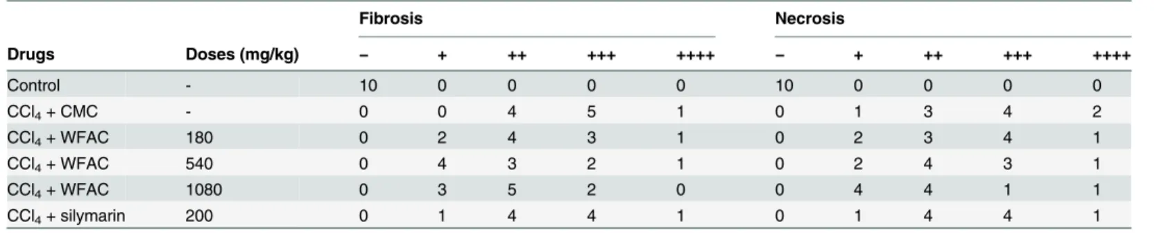
formation, which was associated with a disorder of the lobular structure in the portal area. Most rat livers appeared to have pseudo lobules (Fig 8B). In the WFAC administered rats (1080 mg/kg), livers appeared to have fibrous connective tissue proliferation, the formation of a few fiber intervals in the portal area, and the occasional pseudo lobule (Fig 8B). Scoring of these histopathological changes showed that WFAC administration (1080 mg/kg) significantly reduced the liver necrosis and fibrosis, but silymarin did not (Table 2).
Index compounds of A. cinnamomea in WFAC
Eight signature triterpenoids compounds ofA.cinnamomeain WFAC were determined and quantitated by LC-MS/MS as shown inTable 3. The compounds could be divided into two chemical groups: (1) triterpenoids and steroids [dehydroeburicoic acid (1.43 ppm), dehydro-sulfurenic acid (75.64 ppm), antcin A (1.68 ppm), antcin B (1.00 ppm), antcin C (0.84 ppm), antcin H (non-detectable), antcin K (2.70 ppm)]; (2) benzenoids [1,4-dimethoxy-2,3-methyle-nedioxy-5-methylbenzene (2747 ppm)].
Discussion
A.cinnamomeais a unique and endemic mushroom of Taiwan and is one of the most popular heath foods used in Asia, particularly in Taiwan.A.cinnamomeahas been well-known for its Table 2. Effect of WFAC on hepatic histopathological changes in CCl4-intoxicated rat.
Fibrosis Necrosis
Drugs Doses (mg/kg) − + ++ +++ ++++ − + ++ +++ ++++
Control - 10 0 0 0 0 10 0 0 0 0
CCl4+ CMC - 0 0 4 5 1 0 1 3 4 2
CCl4+ WFAC 180 0 2 4 3 1 0 2 3 4 1
CCl4+ WFAC 540 0 4 3 2 1 0 2 4 3 1
CCl4+ WFAC 1080 0 3 5 2 0 0 4 4 1 1
CCl4+ silymarin 200 0 1 4 4 1 0 1 4 4 1
Grade designation of the histologicalfindings; (−) normal, (+) very slight, (++) slight, (+++) moderate, (++++) severe. Each value is the number of animals with grading changes.
*p<0.05 compared with the CCl4+ CMC group forfibrosis.
#p<0.05 compared with the CCl4+ CMC group for necrosis.
doi:10.1371/journal.pone.0153087.t002
Table 3. Index compounds ofA.cinnamomeain WFAC.
Compound name Content1(ppm) Bioactivities
1,4-dimethoxy-2,3-methylenedioxy-5-methylbenzene 2747 anti-tumor [54]
Dehydroeburicoic acid 1.43 anti-diabetic [53]; hepatoprotective [47]; anti-inflammation [8]; anti-tumor [52]
Dehydrosulphurenic acid 75.64 anti-tumor [55]
Antcin A 1.68 anti-inflammation [57]
Antcin B 1.00 anti-tumor [55]
Antcin C 0.84 anti-oxidant and Hepatoprotective [43]
Antcin H N. D.2
Antcin K 2.70 anti-tumor [56]
medicinal properties, particularly with regard to liver complaints [20]. However, theA. cinna-momeafruiting body is expensive, therefore, its application is limited. Currently, most of the commercially availableA.cinnamomeacomes from artificial cultures, such as submerged liq-uid and solid-state mycelia cultures. In this study, we investigated the hepatoprotective effect of
A.cinnamomeaobtained by wheat-based solid-state fermentation. For an appropriate conver-sion of drug doses from humans to rats, we converted the human dose to an animal dose based on the body surface area, and not by a simple conversion based on body weight, because body surface area correlates well with several physiological parameters in mammals, including blood volume, circulating plasma proteins, basal metabolism, oxygen utilization, caloric expenditure, and renal function [33]. In this study, the animal dose for the rat was calculated by considering the suggested daily usage doses of solid-state fermentedA.cinnamomea(2 g/ day in human). The dose calculation was performed on the basis of body surface area using a conversion factor of 0.018 (200 g rat/70 kg human) [34]. Therefore, rats were administered 180 mg/ kg WFAC, which is equal to 2 g/70 kg in humans. However, 180 mg/kg WFAC was ineffective in prevent-ing liver cirrhosis induced by CCl4in rats. To get significant hepatoprotective effect, WFAC
requires a dose at 1080 mg/kg. It should be noted that WFAC is an un-extracted raw material and is mainly composed of wheat. Although the concentration of signature triterpenoids com-pounds ofA.cinnamomeain WFAC is not so high, WFAC is a cost-efficiency way of produc-ing these rare bioactive compounds. These results provide the scientific evidence that human daily usage dose of WFAC should be increased to get the significant hepatoprotective effect. Otherwise, partial purification of these rare bioactive compounds from WFAC is needed to reduce the daily usage dose of WFAC.
AST and ALT are enzymes found mainly in the liver, and they are released into the blood-stream when the liver is diseased or damaged, causing the levels of the enzymes in the plasma to rise. Therefore, the amount of AST and ALT in the plasma is directly related to the extent of the liver damage. We found that administering WFAC significantly reduced the plasma AST and ALT levels in CCl4-intoxicated rats, indicating that WFAC is able to ameliorate CCl4
-induced liver injury. Although plasma AST and ALT levels are a valuable aid primarily in the diagnosis of liver disease, they are not specific for liver disease because AST and ALT are also found in red blood cells, heart cells, muscle tissue and other organs, such as the pancreas and the kidneys [35]. To further confirm the hepatoprotective effect of WFAC, the plasma albumin concentration in CCl4-intoxicated rats was measured. Albumin is the major plasma protein
produced in the liver that circulates in the bloodstream. Albumin is essential for maintaining the oncotic pressure in the vascular system. Decreased plasma albumin levels indicate chronic liver failure caused mainly by cirrhosis, a late stage of hepatic fibrosis that results in widespread distortion of the normal hepatic architecture [36]. Our data showed that decreased plasma albumin levels were observed at the end of the sixth and eight weeks after CCl4intoxication,
and these effects were partially recovered in WFAC-treated rats. In addition, WFAC also increased the CCl4-induced decrease in hepatic soluble protein levels. These results indicated
that WFAC improved the liver function in CCl4-intoxicated rats, including increasing protein
synthesis.
Cirrhosis or liver fibrosis lead to an abnormally high blood pressure in the portal vein called portal hypertension. The spleen becomes enlarged as the increased pressure interferes with blood flow from the spleen into the portal blood vessels [37]. CCl4intoxication caused spleen
enlargement in rats, and this effect was inhibited by WFAC treatment. This result indicates that WFAC administration ameliorated portal hypertension in CCl4-intoxicated rats. CCl4
intoxication causes liver damage and inflammation, and liver regeneration can occur in rats, which increases liver weight [38]. In this study, we found that CCl4intoxication increased the
WFAC treatment, the water content of the liver was significantly reduced. These results suggest that WFAC reduced the inflammation in the liver. Liver fibrosis is a wound response to severe liver damage and occurs in many forms of chronic liver damage, including CCl4intoxication
[39]. Hydroxyproline is detected specifically in collagen, which plays a major role in liver fibro-sis and appears to reflect the degree of liver fibrofibro-sis [40]. WFAC treatment decreased the CCl4
-induced increase in liver hydroxyproline levels, indicating that WFAC ameliorated liver fibro-sis. The anti-fibrosis effect of WFAC was also confirmed by histopathological staining with Sir-ius-red. It has been demonstrated that CCl4-induced liver fibrosis occurs via free
radical-mediated lipid peroxidation [41]. CCl4intoxication increased hepatic lipid peroxidation, and
this effect was reduced by WFAC, indicating that oxidative stress in the liver was reduced. Although CCl4intoxication reduced the activities of hepatic antioxidant enzymes, including
SOD, catalase, and GSH-Px, WFAC did not restore the activities of these enzymes. The level of glutathione, an important antioxidant in the liver, was also not changed by WFAC. It has been suggested that liver injury caused by CCl4through lipid peroxidation and protein oxidation,
which involves cytochrome P450-dependent formation of trichloromethyl free radicals and ROS [42]. It has been showed that antcin C fromA.cinnamomea protect liver cells from oxida-tive stress and cell death via Nrf2/ARE activation [43]. We suggested that WFAC reduced the lipid peroxidation level in the CCl4-intoxicated rats might through inducing Nfr2/ARE
activa-tion or other anti-oxidative pathways; however, the detailed mechanism needs further investigation.
Although both the fruiting body and fermented mycelia ofA.cinnamomeahave been dem-onstrated to exhibit hepatoprotective activity in various hepatic injury models [27,44,45], the bioactive components are not fully understand. Antrodin B, a maleimide derivative isolated from the fruiting body ofA.cinnamomea, inhibited transforming growth factor-β1-induced fibrosis in hepatic stellate cells [46]. Antcin C (from the fruiting body) and antroquinonol (from mycelium) protect hepatic cells from free radical- and ethanol-induced oxidative stress, respectively, through Nrf-2 activation [28,43]. In addition, eburicoic acid and dehydroeburi-coic acid isolated from the fruiting body ofA.cinnamomeawere identified as hepatoprotective compounds in CCl4-intoxicated mice [47]. Furthermore, a neutral polysaccharide isolated from
the mycelium ofA.cinnamomeaamelioratedPropionibacterium acnesand lipopolysaccharide induced hepatic injury in mice [48].A.cinnamomeapolysaccharides also have been demon-strated to exhibit anti-hepatitis B virus effects and immune modulation properties [49–51]. WFAC contains Antcin C and dehydroeburicoic acid, which might be response for its hepato-protective effect [43,47]. Dehydroeburicoic acid not only exhibits hepatoprotective effect but also shows anti-inflammation [8], anti-tumor [52] and antidiabetic activities [53]. WFAC also contains 1,4-dimethoxy-2,3-methylenedioxy-5-methylbenzene, dehydrosulphurenic acid, antcin B and antcin K, which exhibit anti-tumor activities [54–57]. In addition, anti-inflammatory com-pound antcin A in WFAC might contribute to its hepatoprotective effect [58].
In this study, we used silymarin as a positive control for ameliorating liver injury. However, silymarin only reduced the CCl4-induced increase in lipid peroxidation and had no effect on
other liver injury markers. Some clinical review papers show that silymarin was not able to improve the mortality, histopathological changes, or liver function indexes in patients with chronic liver disease [59,60]. Although there are many papers showing that silymarin amelio-rates chronic liver injury in animal models, some papers indicate that silymarin is useless for chronic liver injury [61–63]. These results may come from the low bioavailability of silymarin [64]. Therefore, Letteron et al used high dose silymarin (800 mg/kg i.p.) to prevent CCl4
In conclusion, we demonstrated that WFAC has several benefits in CCl4-intoxicated rats,
including: 1) reducing plasma AST and ALT levels; 2) increasing plasma albumin and hepatic sol-uble protein levels; 3) reducing spleen weight and water content of the liver; 4) and reducing lipid peroxidation and fibrosis of liver. This is the first report to show that the raw material of heat-based solid-state fermentedA.cinnamomeaprotects against CCl4-induced hepatic injury in vivo.
Author Contributions
Conceived and designed the experiments: KFH. Performed the experiments: HWC. Analyzed the data: HWC KFH. Contributed reagents/materials/analysis tools: KFH. Wrote the paper: HWC KFH.
References
1. Lee WT, Lee TH, Cheng CH, Chen KC, Chen YC, Lin CW. Antroquinonol fromAntrodia Camphorata suppresses breast tumor migration/invasion through inhibiting ERK-AP-1- and AKT-NF-κB-dependent MMP-9 and epithelial-mesenchymal transition expressions. Food Chem Toxicol. 2015; 78:33–41. doi:
10.1016/j.fct.2015.01.012PMID:25656647
2. Wang SC, Lee TH, Hsu CH, Chang YJ, Chang MS, Wang YC, et al. Antroquinonol D, isolated from Antrodia camphorata, with DNA demethylation and anticancer potential. J Agric Food Chem. 2014; 62 (24):5625–35. doi:10.1021/jf4056924PMID:24784321
3. Yeh CT, Huang WC, Rao YK, Ye M, Lee WH, Wang LS, et al. A sesquiterpene lactone antrocin from Antrodia camphoratanegatively modulates JAK2/STAT3 signaling via microRNA let-7c and induces apoptosis in lung cancer cells. Carcinogenesis. 2013; 34(12):2918–28. doi:10.1093/carcin/bgt255
PMID:23880305
4. Yang PY, Hu DN, Liu FS. Cytotoxic effect and induction of apoptosis in human cervical cancer cells by Antrodia camphorata. Am J Chin Med. 2013; 41(5):1169–80. doi:10.1142/S0192415X13500791
PMID:24117076
5. Park DK, Lim YH, Park HJ. Antrodia camphorata grown on germinated brown rice inhibits HT-29 human colon carcinoma proliferation through inducing G0/G1 phase arrest and apoptosis by targeting theβ-catenin signaling. J Med Food. 2013; 16(8):681–91. doi:10.1089/jmf.2012.2605PMID:
23957353
6. Tsai TC, Tung YT, Kuo YH, Liao JW, Tsai HC, Chong KY, et al. Anti-inflammatory effects ofAntrodia camphorata, a herbal medicine, in a mouse skin ischemia model. J Ethnopharmacol. 2015; 159:113– 21. doi:10.1016/j.jep.2014.11.015PMID:25449461
7. Huang GJ, Deng JS, Chen CC, Huang CJ, Sung PJ, Huang SS, et al. Methanol extract ofAntrodia camphorataprotects against lipopolysaccharide-induced acute lung injury by suppressing NF-κB and MAPK pathways in mice. J Agric Food Chem. 2014; 62(23):5321–9. doi:10.1021/jf405113gPMID:
24849405
8. Deng JS, Huang SS, Lin TH, Lee MM, Kuo CC, Sung PJ, et al. Analgesic and anti-inflammatory bioac-tivities of eburicoic acid and dehydroeburicoic acid isolated fromAntrodia camphorataon the inflamma-tory mediator expression in mice. J Agric Food Chem. 2013; 61(21):5064–71. doi:10.1021/jf303820k
PMID:23495748
9. Liaw CC, Chen YC, Huang GJ, Tsai YC, Chien SC, Wu JH, et al. Anti-inflammatory lanostanoids and lactone derivatives fromAntrodia camphorata. J Nat Prod. 2013; 76(4):489–94. doi:10.1021/ np300443pPMID:23517145
10. Park DK, Park HJ. Ethanol extract ofAntrodia camphoratagrown on germinated brown rice suppresses inflammatory responses in mice with acute DSS-induced colitis. Evid Based Complement Alternat Med. 2013b; 2013:914524.
11. Kuo YH, Lin CH, Shih CC. Ergostatrien-3β-ol fromAntrodia camphoratainhibits diabetes and hyperlip-idemia in high-fat-diet treated mice via regulation of hepatic related genes, glucose transporter 4, and AMP-activated protein kinase phosphorylation. J Agric Food Chem. 2015; 63(9):2479–89. doi:10. 1021/acs.jafc.5b00073PMID:25693659
12. Liu DZ, Liang YC, Lin SY, Lin YS, Wu WC, Hou WC, et al. Antihypertensive activities of a solid-state cul-ture ofTaiwanofungus camphoratus(Chang-chih) in spontaneously hypertensive rats. Biosci Biotech-nol Biochem. 2007; 71(1):23–30. PMID:17213674
13. Lien HM, Tseng CJ, Huang CL, Lin YT, Chen CC, Lai YY. Antimicrobial activity ofAntrodia camphorata extracts against oral bacteria. PLoS One. 2014; 9(8):e105286. doi:10.1371/journal.pone.0105286
14. Yang SM, Ka SM, Hua KF, Wu TH, Chuang YP, Lin YW, et al. Antroquinonol mitigates an accelerated and progressive IgA nephropathy model in mice by activating the Nrf2 pathway and inhibiting T cells and NLRP3 inflammasome. Free Radic Biol Med. 2013; 61:285–97. doi:10.1016/j.freeradbiomed. 2013.03.024PMID:23567192
15. Tsai PY, Ka SM, Chang JM, Lai JH, Dai MS, Jheng HL, et al. Antroquinonol differentially modulates T cell activity and reduces interleukin-18 production, but enhances Nrf2 activation, in murine accelerated severe lupus nephritis. Arthritis Rheum. 2012; 64(1):232–42. doi:10.1002/art.33328PMID:21905011
16. Tsai PY, Ka SM, Chao TK, Chang JM, Lin SH, Li CY, et al. Antroquinonol reduces oxidative stress by enhancing the Nrf2 signaling pathway and inhibits inflammation and sclerosis in focal segmental glo-merulosclerosis mice. Free Radic Biol Med. 2011; 50(11):1503–16. doi:10.1016/j.freeradbiomed.2011. 02.029PMID:21376112
17. Lu MK, Cheng JJ, Lai WL, Lin YJ, Huang NK. FermentedAntrodia cinnamomeaextract protects rat PC12 cells from serum deprivation-induced apoptosis: the role of the MAPK family. J Agric Food Chem. 2008; 56(3):865–74. doi:10.1021/jf072828bPMID:18186605
18. Lu MK, Cheng JJ, Lai WL, Lin YR, Huang NK. Adenosine as an active component ofAntrodia cinnamo-meathat prevents rat PC12 cells from serum deprivation-induced apoptosis through the activation of adenosine A(2A) receptors. Life Sci. 2006; 79(3):252–8. PMID:16443241
19. Chen CC, Shiao YJ, Lin RD, Shao YY, Lai MN, Lin CC, et al. Neuroprotective diterpenes from the fruit-ing body ofAntrodia camphorata. J Nat Prod. 2006; 69(4):689–91. PMID:16643055
20. Yue PY, Wong YY, Wong KY, Tsoi YK, Leung KS. Current evidence for the hepatoprotective activities of the medicinal mushroomAntrodia cinnamomea. Chin Med. 2013; 8(1):21. doi: 10.1186/1749-8546-8-21PMID:24180549
21. Ao ZH, Xu ZH, Lu ZM, Xu HY, Zhang XM, Dou WF. Niuchangchih (Antrodia camphorata) and its poten-tial in treating liver diseases. J Ethnopharmacol. 2009; 121(2):194–212. doi:10.1016/j.jep.2008.10.039
PMID:19061947
22. Geethangili M, Tzeng YM. Review of pharmacological effects ofAntrodia camphorataand its bioactive compounds. Evid Based Complement Alternat Med. 2011; 2011:212641. doi:10.1093/ecam/nep108
PMID:19687189
23. Liu YW, Lu KH, Ho CT, Sheen LY. Protective effects ofAntrodia cinnamomeaagainst liver injury. J Tra-dit Complement Med. 2012; 2(4):284–94. PMID:24716143
24. Wu SH, Ryvarden L, Chang TT.Antrodia camphorate(‘niu-chang-chih’), new combination of a medici-nal fungus in Taiwan. Botanical Bulletin of Academia Sinica. 1997; 38(4):273–5.
25. Wu MT, Tzang BS, Chang YY, Chiu CH, Kang WY, Huang CH, et al. Effects ofAntrodia camphorataon alcohol clearance and antifibrosis in livers of rats continuously fed alcohol. J Agric Food Chem. 2011; 59(8):4248–54. doi:10.1021/jf104561hPMID:21401100
26. Huang CH, Chang YY, Liu CW, Kang WY, Lin YL, Chang HC, et al. Fruiting body of Niuchangchih (Antrodia camphorata) protects livers against chronic alcohol consumption damage. J Agric Food Chem. 2010; 58(6):3859–66. doi:10.1021/jf100530cPMID:20192205
27. Hsiao G, Shen MY, Lin KH, Lan MH, Wu LY, Chou DS, et al. Antioxidative and hepatoprotective effects ofAntrodia camphorataextract. J Agric Food Chem. 2003; 51(11):3302–8. PMID:12744658
28. Kumar KJ, Chu FH, Hsieh HW, Liao JW, Li WH, Lin JC, et al. Antroquinonol from ethanolic extract of mycelium ofAntrodia cinnamomeaprotects hepatic cells from ethanol-induced oxidative stress through Nrf-2 activation. J Ethnopharmacol. 2011; 136(1):168–77. doi:10.1016/j.jep.2011.04.030PMID:
21540101
29. Lu ZM, Tao WY, Xu HY, Ao ZH, Zhang XM, Xu ZH. Further studies on the hepatoprotective effect of Antrodia camphoratain submerged culture on ethanol-induced acute liver injury in rats. Nat Prod Res. 2011; 25(7):684–95. doi:10.1080/14786410802525487PMID:20623423
30. Song TY, Yen GC. Protective effects of fermented filtrate fromAntrodia camphoratain submerged cul-ture against CCl4-induced hepatic toxicity in rats. J Agric Food Chem. 2003; 51(6):1571–7. PMID:
12617586
31. Chen X, Ying X, Zhang W, Chen Y, Shi C, Hou Y, Zhang Y. The hepatoprotective effect of fraxetin on carbon tetrachloride induced hepatic fibrosis by antioxidative activities in rats. Int Immunopharmacol. 2013; 17(3):543–7. doi:10.1016/j.intimp.2013.08.006PMID:23994349
32. Xia E, Rao G, Van Remmen H, Heydari AR, Richardson A. Activities of antioxidant enzymes in various tissues of male Fischer 344 rats are altered by food restriction. J Nutr. 1995; 125(2):195–201. PMID:
7861246
33. Aebi H. Catalase in vitro. Methods Enzymol. 1984; 105:121–6. PMID:6727660
35. Sturgill MG, Lambert GH. Xenobiotic-induced hepatotoxicity: mechanisms of liver injury and methods of monitoring hepatic function. Clin Chem. 1997; 43(8 Pt 2):1512–26. PMID:9265903
36. Muir AJ. Understanding the complexities of cirrhosis. Clin Ther. 2015;pii: S0149-2918(15)00846-2. 37. Garbuzenko DV. Contemporary concepts of the medical therapy of portal hypertension under liver
cir-rhosis. World J Gastroenterol. 2015; 21(20):6117–26. doi:10.3748/wjg.v21.i20.6117PMID:26034348
38. Palmes D, Spiegel HU. Animal models of liver regeneration. Biomaterials. 2004; 25(9):1601–11. PMID:
14697862
39. Bataller R, Brenner DA. Liver fibrosis. J Clin Invest 2005; 115:209–18. PMID:15690074
40. Toyoki Y, Sasaki M, Narumi S, Yoshihara S, Morita T, Konn M. Semiquantitative evaluation of hepatic fibrosis by measuring tissue hydroxyproline. Hepatogastroenterology. 1998; 45(24):2261–4. PMID:
9951907
41. Camps J, Bargallo T, Gimenez A, Alie S, Caballeria J, Pares A, et al. Relationship between hepatic lipid peroxidation and fibrogenesis in carbon tetrachloride-treated rats: effect of zinc administration. Clin Sci (Lond). 1992; 83(6):695–700.
42. McCay PB, Lai EK, Poyer JL, DuBose CM, Janzen EG. Oxygen- and carbon-centered free radical for-mation during carbon tetrachloride metabolism. Observation of lipid radicals in vivo and in vitro. J Biol Chem. 1984; 259(4):2135–43. PMID:6321461
43. Gokila Vani M, Kumar KJ, Liao JW, Chien SC, Mau JL, et al. Antcin C fromAntrodia cinnamomea pro-tects liver cells against free radical-induced oxidative stress and apoptosis in vitro and in vivo through Nrf2-dependent mechanism. Evid Based Complement Alternat Med. 2013; 2013:296082. doi:10.1155/ 2013/296082PMID:24391672
44. Lu ZM, Tao WY, Zou XL, Fu HZ, Ao ZH. Protective effects of mycelia ofAntrodia camphorataand Armillariella tabescensin submerged culture against ethanol-induced hepatic toxicity in rats. J Ethno-pharmacol. 2007; 110(1):160–4. PMID:17092673
45. Wang LC, Kuo IU, Tsai TY, Lee CL.Antrodia camphorata-fermented product cultured in deep ocean water has more liver protection against thioacetamide-induced fibrosis. Appl Microbiol Biotechnol. 2013; 97(23):9955–67. doi:10.1007/s00253-013-5214-1PMID:24061418
46. Geng Y, Wang J, Sun Q, Xie M, Lu ZM, Xu HY, et al. Identification of antrodin B fromAntrodia camphor-ataas a new anti-hepatofibrotic compound using a rapid cell screening method and biological evalua-tion. Hepatol Res. 2015 Mar 9. doi:10.1111/hepr.12516[Epub ahead of print]
47. Huang GJ, Deng JS, Huang SS, Lee CY, Hou WC, Wang SY, et al. Hepatoprotective effects of eburi-coic acid and dehydroeburieburi-coic acid fromAntrodia camphoratain a mouse model of acute hepatic injury. Food Chem. 2013; 141(3):3020–7. doi:10.1016/j.foodchem.2013.03.061PMID:23871054
48. Han HF, Nakamura N, Zuo F, Hirakawa A, Yokozawa T, Hattori M. Protective effects of a neutral poly-saccharide isolated from the mycelium ofAntrodia cinnamomeaonPropionibacterium acnesand lipo-polysaccharide induced hepatic injury in mice. Chem Pharm Bull (Tokyo). 2006; 54(4):496–500. 49. Lee IH, Huang RL, Chen CT, Chen HC, Hsu WC, Lu MK. Antrodia camphorata polysaccharides exhibit
anti-hepatitis B virus effects. FEMS Microbiol Lett. 2002; 209(1):63–7. PMID:12007655
50. Liu KJ, Leu SJ, Su CH, Chiang BL, Chen YL, Lee YL. Administration of polysaccharides fromAntrodia camphoratamodulates dendritic cell function and alleviates allergen-induced T helper type 2
responses in a mouse model of asthma. Immunology. 2010; 129(3):351–62. doi:10.1111/j.1365-2567. 2009.03175.xPMID:19909376
51. Meng LM, Pai MH, Liu JJ, Yeh SL. Polysaccharides from extracts ofAntrodia camphoratamycelia and fruiting bodies modulate inflammatory mediator expression in mice with polymicrobial sepsis. Nutrition. 2012; 28(9):942–9. doi:10.1016/j.nut.2012.01.006PMID:22541057
52. Du YC, Chang FR, Wu TY, Hsu YM, El-Shazly M, Chen CF, et al. Antileukemia component, dehydroe-buricoic acid fromAntrodia camphoratainduces DNA damage and apoptosis in vitro and in vivo mod-els. Phytomedicine. 2012; 19(8–9):788–96. doi:10.1016/j.phymed.2012.03.014PMID:22516893
53. Kuo YH, Lin CH, Shih CC. Antidiabetic and antihyperlipidemic properties of a triterpenoid compound, dehydroeburicoic acid, fromAntrodia camphoratain vitro and in streptozotocin-induced mice. J Agric Food Chem. 2015; 63(46):10140–51. doi:10.1021/acs.jafc.5b04400PMID:26503742
54. Lien HM, Lin HW, Wang YJ, Chen LC, Yang DY, Lai YY, et al. Inhibition of anchorage-independent pro-liferation and G0/G1 cell-cycle regulation in human colorectal carcinoma cells by 4,7-dimethoxy-5-methyl-l,3-benzodioxole isolated from the fruiting body ofAntrodia camphorate. Evid Based Comple-ment Alternat Med. 2011; 2011:984027. doi:10.1093/ecam/nep020PMID:19293251
56. Hsieh YC, Rao YK, Whang-Peng J, Huang CY, Shyue SK, Hsu SL, et al. Antcin B and its ester deriva-tive fromAntrodia camphoratainduce apoptosis in hepatocellular carcinoma cells involves enhancing oxidative stress coincident with activation of intrinsic and extrinsic apoptotic pathway. J Agric Food Chem. 2011; 59(20):10943–54. doi:10.1021/jf202771dPMID:21916504
57. Huang YL, Chu YL, Ho CT, Chung JG, Lai CI, Su YC, et al. Antcin K, an active triterpenoid from the fruit-ing bodies of basswood-cultivatedAntrodia cinnamomea, inhibits metastasis via suppression of integ-rin-mediated adhesion, migration, and invasion in human hepatoma cells. J Agric Food Chem. 2015; 63 (18):4561–9. doi:10.1021/jf5059304PMID:25911944
58. Chen YC, Liu YL, Li FY, Chang CI, Wang SY, Lee KY, et al. Antcin A, a steroid-like compound from Antrodia camphorata, exerts anti-inflammatory effect via mimicking glucocorticoids. Acta Pharmacol Sin. 2011; 32(7):904–11. doi:10.1038/aps.2011.36PMID:21602840
59. Dhiman RK, Chawla YK. Herbal medicines for liver diseases. Dig Dis Sci. 2005; 50(10):1807–12. PMID:16187178
60. Jacobs BP, Dennehy C, Ramirez G, Sapp J, Lawrence VA. Milk thistle for the treatment of liver disease: a systematic review and meta-analysis. Am J Med. 2002; 113(6):506–15. PMID:12427501
61. Muriel P, Moreno MG, Hernández Mdel C, Chávez E, Alcantar LK. Resolution of liver fibrosis in chronic CCl4administration in the rat after discontinuation of treatment: effect of silymarin, silibinin, colchicine
and trimethylcolchicinic acid. Basic Clin Pharmacol Toxicol. 2005; 96(5):375–80. PMID:15853930
62. Hall PM, Plummer JL, Ilsley AH, Ahern MJ, Cmielewski PL, Williams RA. The pathology of liver injury induced by the chronic administration of alcohol and 'low-dose' carbon tetrachloride in Porton rats. J Gastroenterol Hepatol. 1994; 9(3):250–6. PMID:8054523
63. Paulová J, Dvorák M, Kolouch F, Vánová L, Janecková L. Verification of the hepatoprotective and ther-apeutic effect of silymarin in experimental liver injury with tetrachloromethane in dogs. Vet Med (Praha). 1990; 35(10):629–35.
64. Luper S. A review of plants used in the treatment of liver disease: part 1. Altern Med Rev. 1998; 3 (6):410–21. PMID:9855566