Full paper published online: August 31, 2007 ISSN 1678-9199.
HISTOLOGICAL AND BIOCHEMICAL EFFECTS INDUCED BY SUBLETHAL
DOSES OF Bothrops jararacussu VENOM IN MICE
ZENI A. L. B. (1), BECKER A. (1), KRUG M. (1), ALBUQUERQUE C. A. C. (1)
(1) Department of Natural Sciences, Center of Exact and Natural Sciences, Regional University of Blumenau, Blumenau, Santa Catarina State, Brazil.
ABSTRACT: Snake venom is characterized by hemorrhagic, coagulant, proteolytic and myotoxic activities which in Bothrops jaracussu venom are related to intraspecific variations. In the present study, female Swiss mice were divided into two groups: treated with 25μg or 50μg venom. These were subdivided into three groups of six animals each, according to blood collection: 2, 4 or 24h after venom injection. Animals were anesthetized using diethyl-ether inhalation and 1ml of blood was collected by heart puncture. Then, the following organs were removed: spleen, skeletal muscle, kidneys, liver and lungs; histological sections were obtained and stained with hematoxylin-eosin (HE). The following biochemical parameters were analyzed: aspartate aminotransferase (AST/GOT), alanine aminotransferase (ALT/GPT), total lactate dehydrogenase (LDH), glucose, creatinine and urea levels, and total protein content. Results showed significant alterations in AST, LDH, glucose and urea levels, and total protein content, as well as important tissue alterations in the liver, kidneys and lungs. It could be concluded that, even using sublethal doses of venom, there were significant changes in almost all the tested biochemical parameters as well as tissue alterations in the kidneys and lungs.
KEY WORDS: Bothrops jararacussu, snake venoms, sublethal doses, in vivo studies, biochemical parameters, histopathological alterations.
CONFLICTS OF INTEREST: There is no conflict.
CORRESPONDENCE TO:
ANA LÚCIA BERTARELLO ZENI, Departamento de Ciências Naturais, Centro de
Ciências Naturais e Exatas, Universidade Regional de Blumenau, Rua Antônio da
Veiga, 140, 89012-900, Blumenau, SC, Brasil. Phone: 55 21 47 3321 0272.
INTRODUCTION
In the Brazilian territory, snakes of the Bothrops genus account for a total of twenty
species and are responsible for 90% of the snakebite accidents. According to the
Ministry of Health, more than 17000 bothropic accidents occur every year in the
country, with a fatality rate of approximately 0.6% treated cases.
Bothropic venom is very complex. It contains twenty or more different compounds
and over 90% of its dry weight is constituted of proteins encompassing a large variety
of components such as lecithin, metalloproteinases, serine proteinases, disintegrins,
phospholipases and peptides such as bradykinin and angiotensin (7). Non-protein
fractions are represented by carbohydrates, lipids, metals, and biogenic enzymes
(10).
The venom from Bothrops snakes presents a complex mixture of toxins with different
toxic or enzymatic properties. Proteases apparently act by degrading tissue proteins
in a non-specific manner (26) and by cleaving plasmatic proteins through hydrolysis,
interfering in the homeostasis of the organism (16, 21). Such substances can have
hemorrhagic, coagulant, proteolytic and myotoxic activities that result in inflammatory
process and tissue destruction during damage to blood vessel walls or in pain,
edema, ecchymosis, abscess formation, and necrosis (12, 18, 20, 23, 27).
The aim of the present work was to study in mice the local and systemic effects of
sublethal doses of venom from B. jararacussu specimens commonly found in tropical
forests of Santa Catarina, Brazil. Histological alterations as well as changes in serum
levels of several enzymes and metabolites were analyzed.
MATERIALS AND METHODS
Venom
Venom was obtained from an adult B. jararacussu in the animal facility of the
Regional University of Blumenau, Santa Catarina, Brazil. It was preserved at –20°C
until use, when the lyophilized venom was dissolved in phosphate buffered saline
(PBS), pH 7.2, to obtain the doses 25μg and 50μg.
Animals
Female Swiss mice (20–25g) were divided into two groups: treated with 25μg or 50μg
subdivided into three subgroups of six animals each. The fasted animals were
intraperitoneally (i.p.) injected with venom and anesthetized through diethyl-ether
inhalation after 2, 4 or 24h for blood collection (about 1ml) by cardiac puncture.
Control animals received 100μl PBS, i.p. The present experiment was approved by
the Committee of Ethics in Research with Animals 009/04.
Histological Analysis
Animals were sacrificed by ether inhalation and the following organs were removed:
spleen, skeletal muscle, kidneys, liver and lungs, which were then fixed in 10%
formalin, dehydrated and included in paraffin wax for 5μm sections that were stained
with HE and observed under light microscope.
Biochemical Analysis
After blood collection, serum was separated by centrifugation and the following tests
were carried out: AST and ALT levels determination, which was based on the
formation of 2,4-dinitrophenyl phosphate; LDH levels determination, which was based
on the formation of NADH (one unit results in the formation of 1μmol NADH per
minute); total protein content determination using the Biuret method; glucose levels
determination, which was based on the formation of quinoneimine dye; creatinine
levels determination using the creatinine picrate method; and urea levels
determination, which was based on the formation of indophenol blue, using a
BioSystems BTS 310 photocolorimeter and Standard BioSystems reagents.
Statistical Analysis
Data of normal distribution were expressed as means ± S.E.M. The values obtained
were evaluated by analysis of variance (ANOVA), followed by sufficient post hoc
tests. Each treatment was considered an independent variable. In all cases, the
considered statistical significance level was p<0.05. The Graph Pad Prism®
program, version 3.0, was used for obtaining graphs and statistical analysis of the
RESULTS
HistologicalFindings
Figure 1 shows some tissues of organs from treated animals, compared with those
from control animals. Cellular alteration was verified on the lungs, kidneys and liver,
but not in the spleen or skeletal muscle. Figure 1A shows a histological section of
liver 4h after injection of 25μg venom. There are leukocyte aggregations near blood
vessels and evident vascular congestion. Figure 1B displays a histological section of
liver from a control mouse. The center-lobe vein has normal morphological
characteristics. Figures 1C and 1D present lung sections 4h after injection of 25μg
venom. Cellular alterations were noticed due to the presence of inflammatory cells in
the inter-alveolar spaces and inside pulmonary alveoli as well as abnormal
accumulation of erythrocytes and leukocytes. Control group presented normal lung
characteristics (Figure 1E).
Figures 1F and 1G show the cortical region of kidneys 4h after injection of 50 and
25μg venom, respectively. Evident glomerular alterations were noticed as renal
glomeruli necrosis and vascular congestion (Figure 1F). A renal corpuscle with
leukocyte infiltration was observed in the glomerular capillaries and, more discretely,
in the periarterial areas and between the renal tubules (Figure 1G). The renal
corpuscle and the renal tubule in the control group (Figure 1H) were normal.
Venom-Induced Biochemical Alterations
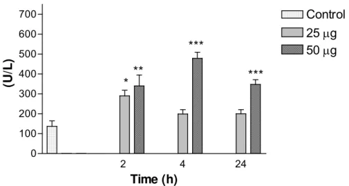
An increase in AST activity was observed 4h after injection of 25μg venom,
compared with the control group; 2h after injection of 50μg venom, it was also
significant (Figure 2). On the other hand, ALT levels did not change significantly with
venom doses at each time interval (results not shown). Serum levels of LDH were
high at all studied periods and 50μg venom caused significant increase, whereas
25μg caused significant changes only at 2h (Figure 3).
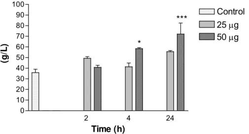
Regarding the enzymatic determination of total proteins, the main increases were
observed with 50μg at 4h and 24h and were considered extremely significant (Figure
4). Serum levels of glucose with 50μg at 2h were considered very significant
Creatinine levels were not significant in relation to the control group (results not
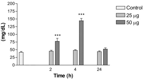
shown). Urea levels at 2h and 4h with 50μg venom were extremely significant
compared with control (Figure 6).
Figure 1. A: Center-lobe vein in the liver of an animal treated with 25μg venom (4h);
B: Liver of a control animal (4h); C and D: Alveolar spaces and pulmonary alveoli in
the lungs of animals treated with 25μg venom (4h); E: Lung of a control animal; F
and G: Cortical region in the kidneys with glomeruli of animals treated with 50μg and
25μg venom, respectively (4h); H: Kidney of a control animal (4h). 400X
magnification.
→: Vascular congestion.
2 4 24 0
250 500 750
Control 25μg 50μg *
**
Time (h)
(U
/L
)
Figure 2. Variations in aspartate aminotransferase (AST/GOT) levels in mice
intraperitoneally injected with 25 or 50μg of Bothrops jararacussu venom diluted with
0.1ml PBS. Each bar represents the mean and vertical lines, the standard error of
mean (S.E.M.) of results obtained from 6 animals.
Asterisks denote the significance levels when compared with control group: * p<0.05,
** p<0.01.
2 4 24
0 100 200 300 400 500 600
700 Control
25μg 50μg
**
***
***
Time (h)
(U
/L
)
*
Figure 3: Variations in lactate dehydrogenase (LDH) levels in mice intraperitoneally
injected with 25 or 50μg of Bothrops jararacussu venom diluted with 0.1ml PBS.
Each bar represents the mean and vertical lines, the standard error of mean (S.E.M.)
of results obtained from 6 animals.
Asterisks denote the significance levels when compared with control group: * p<0.05,
2 4 24 0
10 20 30 40 50 60 70 80 90 100
Control 25μg 50μg *
***
Time (h)
(g
/L
)
Figure 4. Variations in total protein levels in mice intraperitoneally injected with 25 or
50μg of Bothrops jararacussu venom diluted with 0.1ml PBS. Each bar represents
the mean and vertical lines, the standard error of mean (S.E.M.) of results obtained
from 6 animals.
Asterisks denote the significance levels when compared with control group: * p<0.05,
*** p<0.001.
2 4 24
0 50 100 150 200
Control 25μg 50μg **
Time (h)
(
m
g/
dL)
Figure 5. Variations in glucose levels in mice intraperitoneally injected with 25 or
50μg of Bothrops jararacussu venom diluted with 0.1ml PBS. Each bar represents
the mean and vertical lines, the standard error of mean (S.E.M.) of results obtained
from 6 animals.
Asterisks denote the significance levels when compared with control group: **
2 4 24 0
25 50 75 100 125 150 175 200
Control 25μg 50μg
***
***
Time (h)
(m
g/
dL)
Figure 6: Variations in urea levels in mice intraperitoneally injected with 25 or 50μg of
Bothrops jararacussu venom diluted with 0.1ml PBS. Each bar represents the mean
and vertical lines, the standard error of mean (S.E.M.) of results obtained from 6
animals.
Asterisks denote the significance levels when compared with control group: ***
p<0.001.
DISCUSSION
Bothrops jararacussu has one of the most lethal venoms among Bothrops species. In
a review of 29 snakebite accidents caused by B. jararacussu, several patients
developed shock and oliguria few hours after the bite and three victims died (18).
Pathological alterations (local effects provoked by snakebites) have been extensively
reported due to the large variety of enzymes and other compounds present in snake
venoms. However, pathophysiological alterations, the role of enzymes, and the
systemic complications induced by sublethal doses are not very clear for B.
jararacussu venom.
Figures 1A–H present important information about tissue alterations in some organs
of animals treated or not with venom, corroborating the obtained serum data;
however, such alterations were not dose and time-dependent. In Figure 1A, tissue
alterations in the liver are visible as there is high concentration of perivascular
leukocytes, indicating an inflammatory process, besides intense vascular congestion
in the center-lobular vein.
Alterations in striated muscle were not visible, probably because the chosen
system, not restricting it to only one site. A snakebite accident is more similar to
subcutaneous injection of venom (15, 17), allowing the observation of systemic signs
and modified tissue characteristics in the liver and pulmonary parenchyma, together
with renal hemorrhage. Zamunér et al. (28) also reported effective myonecrosis by B.
jararacussu and B. moojeni venoms.
Hemorrhagic toxins and other components impair or increase the vascular
permeability of endothelial cells and the basal membrane, allowing the blood to
escape towards the neighboring tissues (2, 25). Liver cells presented altered
metabolic activity, which was verified by blood biochemical analysis, suggesting
hepatocyte death. Inflammatory focus was observed in all the liver parenchyma,
principally in theperivascularregion.
The metalloproteinases found in the venom presented hemorrhagic effect and were
capable of inducing the release of inflammation mediators such as cytokines,
intensifying the inflammatory response (25), which may justify the occurrence of a
large quantity of defense cells in the pulmonary parenchyma. This effect collaborates
to coagulation in microcirculation, promoting disseminated intravascular coagulation,
which leads to hemorrhage and pulmonary edema.
In lung histological sections, morphological alterations were evident with 25μg
venom, which caused increased inflammatory cells and intra-alveolar erythrocytes as
well as edema, indicating compromised pulmonary functions (Figure 1D). The
increase in pulmonary damage depends on the local lesion but its evolution is not
frequent in human envenomation by B. jararacussu (4).
In animals treated with 25 or 50μg of venom, histological analysis of the kidneys
confirmed the diagnosis of increased renal workload resultant from the high protein
levels observed in serum analysis, which can lead to cellular and/or tissue alterations
that compromise the organ function and cause renal glomeruli destruction, vascular
congestion, microvascular hemorrhagic lesion, and leukocyte aggregation.
Renal failure is the major complication in envenomation by B. jararacussu and other
Bothrops species (18). Cortical renal necrosis can be related to intravascular
coagulation or directly to renal endothelium or even vasospasm toxic effect (1).
In the liver, alterations were not clearly visible under the microscope and were only
verified by leukocyte aggregation and intravascular coagulation. Figure 2 shows that
observed in hepatitis, other hepatic diseases associated with necrosis after
administration of some classes of medication or after myocardial infarction, diseases
of the muscular-skeletal system, acute pancreatitis, hemolytic diseases, and others
(5, 11).
The levels of ALT were also altered, although not significantly. The fact that AST
levels increased more with dose than ALT levels indicates not only cell destruction,
but also mitochondrial disruption (3). The results obtained for AST and ALT levels
were similar to those obtained by Chavez et al. (6) with B. asper, which in turn were
different from those reported by Teibler et al. (27) with B. alternatus: all two
parameters significantly changed.
Regarding LDH, the increase in its activity was significant at 2, 4 and 24h with 50μg
and only at 2h with 25μg venom; in the study of Chavez et al. (6), it differed only at
6h. LDH activity is increased in hepatic diseases, renal alterations, myocardial
infarction, and progressive muscular dystrophy and in any case of hemolysis (5, 11).
These alterations and ALT levels indicated skeletal-muscle (6) and, possibly,
myocardial damage (3), which was also reported by Benvenuti et al. (4) about a
woman bitten by B. jararacussu. The glucose levels observed in mice were within the
normal range of 62–175mg/dl (13).
Total protein content presented dose and time-dependent alterations and urea levels
significantly increased, showing intense protein degradation, increased renal
workload and liver necrosis. Renal damages with different types of lesions
(glomerular, tubular, interstitial or vascular) increased urea serum levels (19).
Results suggested that the increase in total protein content and urea levels was
related to the myotoxic (14, 18, 22, 23) and proteolytic activities (4, 9) of B.
jararacussu venom and to the decrease in the liver activity. Milani et al. (18)
described 29 snakebite accidents caused by this species, in which several patients
developed shock and oliguria few hours after the bite. This also explains the data
found in the present study.
ACKNOWLEDGEMENTS
This work was supported by Support Foundation of Scientific and Technological
Janene de Oliveira Lima (in memorian) for the pathological analysis and Professor
Alessandra Beirith for the general review of the paper.
REFERENCES
1 AMARAL CFS., SILVA OA., GODOY P., MIRANDA D. Renal cortical necrosis
following Bothrops jararaca and B. jararacussu snakebite. Toxicon, 1985, 23, 877-85.
2 ARTASHES VA., SILVA VA. Histopathological changes induced by the venom of
the snake Vipera raddei (Armenian adder). Toxicon, 2005, 47, 141-3.
3 BABCOCK JL., SUBER RL., FRITH CH., GEREN CR. Systemic effect in mice of
venom apparatus extract and toxin from the brown recluse spider (Loxosceles
reclusa). Toxicon, 1981, 19, 463-71.
4 BENVENUTI LA., FRANÇA OSF., BARBARO KC., NUNES JR., CARDOSO JLC.
Pulmonary haemorrhge causing rapid death after Bothrops jararacussu snakebite: a
case report. Toxicon, 2003, 42, 331-4.
5 BURTIS CA., ASHWOOD ER. Tietz Textbook of Clinical Chemistry. 5.ed.
Philadelphia: WB Saunders, 2001.
6 CHAVES F., GUITIÉRREZ JM., LOMONTE B., CERDAS L. Histopathological and
biochemical alterations induced by intramuscular injection of Bothrops asper
(terciopelo) venom in mice. Toxicon, 1989, 27, 1085- 93.
7 CLISSA PB. Caracterização do efeito da jararagina sobre a produção e liberação
de citocinas pró-inflamatórias em modelo murino. São Paulo: Universidade de São
Paulo, Instituto de Ciências Biomédicas, 2002. 109p. [PhD Thesis].
8 FEITOSA NETO AC. Verificação da resposta imunológica do veneno de serpente
Bothrops jararacussu frente ao inoculo de ovoalbumina em camundongos Swiss.
Blumenau: Universidade Regional de Blumenau, 2003. 89p. [End of Course Paper –
Biological Sciences].
9 FRANCISCHETTI IMB., CASTRO HC., ZINGALLI RB., CARLINI CR.,
GUIMARÃES JA. Bothrops sp. Snake venoms: comparison of some biochemical and
physiochemical properties and interference in platelet functions. Comp. Biochem.
Physiol., 1998, 119C, 21-9.
10 FRANÇA FOS., CARDOSO JLC. Estudo retrospectivo da evolução de acidentes
botrópicos. Rev. Soc. Bras. Med. Trop., 1987, 20, 56.
11 FRIEDMAN RB., YOUNG DS. Effects of disease on clinical laboratory tests. 3.ed.
12 GUIMARÃES AQ., CRUZ-HÖFLING MA., FERREIRA DE ARAÚJO PM., BON C.,
LÔBO DE ARAÚJO A. Pharmacological and histopathological characterization of
Bothrops lancelatus (Fer de lance) venom-induced edema. Inflamm. Res, 2004, 53,
284-91.
13 HARKNESS JE., WAGNER JE. Biologia e clínica de coelhos e roedores. 3.ed.
São Paulo: Roca, 1993. 238p.
14 JORGE MT., RIBEIRO LA., O’CONNELL JL. Prognostic factors for amputation in
the case of envenoming by snakes of the genus Bothrops (Viperidae). Ann. Trop.
Med. Parasitol., 1999, 93, 401-8.
15 KAMIGUTI AS., THEAKSTON RDG., DESMOND H., HUTTON A. Systemic
haemorrhage in rats induced by a haemorrhagic fraction from Bothrops jararaca
venom. Toxicon, 1991, 29, 1097-105.
16 MATSUI T., FUGIMURA Y., TITANI K Snake venom proteases affecting
haemostasis and thrombosis. Biochim. Biophys. Acta, 2000, 1477, 146-56.
17 MELO PA., HOMSI-BRANDEBURGO MI., GIGLIO JR., SUAREZ-KURTZ G.
Antagonism of the myotoxic effects of Bothrops jararacussu venom and
bothropstoxin by polyanions. Toxicon, 1993, 31, 285-91.
18 MILANI R., JORGE MT., CAMPOS FPF., MARTINS FP., BOUSSO A.,
CARDOSO JLC., RIBEIRO LA., FAN HW., FRANÇA FOS., SANO MARTINS IS.,
CARDOSO D., FERNANDES IDOF., FERNANDES JC., ALDRED VL., SANDOVAL
MP., PUORTO G., THEAKSTON RDG., WARRELL DA. Snake bites by jararacuçu
(Bothrops jararacussu): clinicopathological studies of 29 proven cases in São Paulo,
Brazil. Q. J. Med, 1997, 90, 323-34.
19 MOTTA VT. Bioquímica clínica para laboratório: princípios e interpretações. Porto
Alegre: Editora Médica Missau, 2003. 419 p.
20 OSHIMA-FRANCO Y., ALVES CMV., ANDRÉO FILHO N., GERENUTTI M.,
CINTRA ACO., LEITE GB., RODRIGUES–SIMIONI L., SILVA MG. Neutralization of
the neuromuscular activity of Bothopstoxin-I, a myotoxin from Bothrops jararacussu
snake venom, by a hydroalcoholic extract of Caseria sylvestris Sw. (Guaçatonga), J.
Venom. Anim. Toxins incl. Trop. Dis., 2005, 11, 465-78.
21 OSHIMA-FRANCO Y., HYSLOP S., CINTRA ACO., GIGLIO JR.,
CRUZ-HÖFLING MA., RODRIGUES-SIMIONI L. Neutralizing capacity of commercial
bothropic antivenom against Bothrops jararacussu venom and bothropstoxin-I.
22 OSHIMA-FRANCO Y., LEITE GB., VALERIO AA., HYSLOP S., ESCARSO SHA.,
GIGLIO JR., PRADO-FRANCESCHI J., CRUZ-HÖFLING MA.,
RODRIGUES-SIMIONI L. Rabbit antivenom efficacy against myotoxic and neurotoxic activities of
Bothrops jararacussu venom and Bothropstoxin-I. J. Venom. Anim. Toxins, 2002, 8,
226-43.
23 QUEIRÓZ LS., SANTO NETO H., RODRIGUES-SIMIONI L.,
PRADO-FRANCESCHI J. Muscle necrosis and regeneration after envenomation by Bothrops
jararacussu snake venom. Toxicon, 1984, 22, 339-46.
24 SHERLOCK S. Drugs and Liver. In: SHERLOCK S. Diseases of the liver and
biliary system. 7.ed. London: Blackwell, 1985: 304-33.
25 SILVEIRA KSO., BOECHEM NT., NASCIMENTO SMD., MURAKAMI YLB.,
BARBOSA AP., MELO PA., MORAES VLG., ROCCO PRM., ZIN WA. Pulmonary
mechanics and lung histology in acute lung injury induced by Bothrops jararaca
venom. Resp. Physiol. Neurobiol., 2004, 139, 167-77.
26 SOARES AM., RODRIGUES VM., BORGES MH., ESCARSO SHA., CUNHA
OAB., HOMSI-BRANDEBURGO MI., GIGLIO JR. Inhibition of proteases, myotoxins
and phospholipases A2 from Bothrops venoms by the heteromeric protein complex of
Didelphis albiventris opossum serum. Biochem. Mol. Biol. Int., 1997, 43, 1091-9.
27 TEIBLER P., ACOSTA DE PÉREZ O., MARUÑAK S., RUIZ R., KOSCINCZUK P.,
SÁNCHEZ NEGRETTE M., MUSSART DE COPPO N. Local and systemic lesions
induced by Bothrops alternatus venom (víbora de la cruz) of Argentine. Acta Toxicol.
Argentina, 1999, 7, 7-10.
28 ZAMUNÉR SR., PRADO-FRANCESCHI J., CRUZ-HÖFLING MA.,
RODRIGUES-SIMIONI L. Comparative myotoxic activities of bothropic venoms as assessed by