Interstitial lung disease in patients with progressive
systemic sclerosis. A study of 58 cases*
SERGIO FERNANDES DE OLIVEIRA JEZLER1, MITTERMAYER BARRETO SANTIAGO2,
THAMINE LESSA ANDRADE3, CÉSAR ARAUJO NETO4, HELIO BRAGA4, ÁLVARO AUGUSTO CRUZ5
ABSTRACT
Objective: To estimate the frequency of interstitial lung disease in a group of patients with progressive systemic sclerosis, and to describe the clinical, functional and radiological characteristics of the patients studied. Methods: Fifty-eight patients diagnosed with progressive systemic sclerosis were submitted to high-resolution computed tomography of the chest, pulmonary function tests and a blood test for anti-Scl 70 antibodies. Comparisons were drawn between patients with interstitial lung disease and those without. Logistic regression with multivariate analysis was used to identify factors predictive of interstitial lung disease. Results: Of the 58 patients evaluated, 51.7% presented interstitial lung disease on high-resolution computerized tomography scans. Dyspnea and cough were the most common symptoms (seen in 65.5% and 39.7%, respectively). Bronchiolectasis and honeycombing were the most common tomographic abnormalities (observed in 83.3% and 80%, respectively). When compared to individuals without interstitial lung disease, patients with the condition had a comparable frequency of pulmonary and extrapulmonary symptoms but presented progressive systemic sclerosis of longer duration, a higher frequency of crackling rales, higher rates of anti-Scl 70 positivity, lower vital capacity and reduced total lung capacity. Only forced vital capacity < 80% was found to be a predictor of interstitial lung disease. Conclusion: Interstitial lung disease was common in this group of patients with progressive systemic sclerosis. No correlation with symptoms was found, although interstitial lung disease was found to correlate with crackling rales and with anti-Scl 70 positivity. Nevertheless, only reduced forced vital capacity was found to be predictive of interstitial lung disease.
Keywords: Scleroderma, systemic/complications; Scleroderma, systemic/radiography; Lung diseases, interstitial/etiology; Lung diseases, interstitial/radiography; Pulmonary fibrosis; Tomography, X-ray computed; Thorax/radiography; Hypertension, pulmonary
* Study carried out at the Universidade Federal da Bahia (UFBA, Federal University of Bahia) School of Medicine, Salvador, Brazil.
1. Coordinator of the Interstitial Diseases Outpatient Clinic of the Universidade Federal da Bahia (UFBA, Federal University of Bahia) School of Medicine, Salvador, Brazil
2. Coordinator of the Rheumatology Sector of the Hospital Santa Izabel, Salvador, Brazil
3. Physician from the Interstitial Diseases Outpatient Clinic of the Universidade Federal da Bahia (UFBA, Federal University of Bahia) School of Medicine, Salvador, Brazil
4. Professor of Radiology at the Universidade Federal da Bahia (UFBA, Federal University of Bahia) School of Medicine, Salvador, Brazil
5. Assistant professor at the Universidade Federal da Bahia (UFBA, Federal University of Bahia) School of Medicine, Salvador, Brazil
Correspondence to: Sérgio F. O. Jezler. Rua Augusto Viana, s/n 3º andar. CEP: 40110-160, Salvador (BA), Brasil. Fax: 55 71 237-6679. E-mail: [email protected]
INTRODUCTION
Interstitial pulmonary involvement is the leading cause of death in cases of progressive systemic sclerosis (PSS).(1-3) Reports on the frequency of interstitial lung
disease (ILD) among PSS patients present varying results. This can be attributed to the different methods of detection used. However, this variation also results from differences in demographic variables, such as race, and in clinical and laboratory parameters, such as the presence of anti-Scl 70 antibodies, among the populations studied.(4-6) In studies using high-resolution
computed tomography (HRCT), ILD has been reported in up to 91% of PSS cases.(7-8)
In the only Brazilian study using HRCT in PSS patients, signs of ILD were reported in 59% of the 22 individuals studied.(9) However, that study was limited by the sample size, as well as by the fact that 95% of the subjects were Caucasian, which certainly gave the sample an ethnic profile different from that typically found in our milieu.
The main objective of the present study was to determine, through the use of HRCT of the chest, the frequency of ILD among individuals with PSS in a sample of outpatients. Secondarily, we sought to characterize this group of patients from a clinical, laboratory, functional and radiological viewpoint in the attempt to identify variables correlated with the presence of ILD.
METHODS
Between April of 2002 and December of 2003, patients diagnosed with PSS(10) were consecutively
referred to the interstitial diseases outpatient clinics of the Hospital Santa Izabel (Santa Izabel Hospital) and the Hospital Universitário Professor Edgard Santos (Professor Edgard Santos University Hospital) of the Universidade Federal da Bahia (Federal University of Bahia) for evaluation. These university hospitals are integrated into the network of the national Sistema Único de Saúde (Unified Health System). Patients were evaluated regardless of the presence of symptoms or signs of pulmonary involvement. The interval between the stages of the evaluation of each patient never exceeded 90 days. Pregnant women and individuals with a history of environmental or occupational exposure recognized as causes of ILD, as well as patients presenting worsening of the symptoms related to
PSS in the last 30 days or presenting another concomitant connective tissue disease, were excluded from the study. The study was evaluated and approved by the Ethics in Human Research Committee of the Santa Izabel Hospital, and all participants gave written informed consent.
Clinical evaluation consisted of completing a standardized questionnaire designed to collect demographic and clinical data on the patient. Duration of PSS was defined as time elapsed since the onset of the first symptom related to PSS, and the criteria established by LeRoy et al. was used to characterize the type of skin involvement.(11) Patients
were categorized as active smokers if they had smoked cigarettes within the last six months, as former smokers if they had quit smoking more than six months prior and as nonsmokers if they had never smoked. Intensity of smoking was defined in pack-years. Dyspnea was scored using the baseline dyspnea index score described by Mahler et al., which ranges from 0 (worst score) to 12 points (best score).(12)
Blood tests for anti-Scl 70 antibodies were performed using the enzyme immunoassay technique (VAR ELISA Kit - Spain).All patients were submitted to HRCT of the chest in a volumetric (helical) CT device, model CT HISPEED LX/i (General Electric Medical Systems, Milwaukee, WI, USA). The images were obtained with thin (1-mm) cuts using a high-spatial resolution filter (bone filter) to reconstruct the image, in 20-mm increments, in dorsal decubitus and (when indicated for excluding the presence of opacities in the basal segments, resulting from the effects of gravity) ventral decubitus, examining the lungs from the apex to the base, in the inspiratory apnea phase, and complementing this with three sections in the maximum expiratory apnea phase at the following levels: aortic arch, tracheal bifurcation and immediately above the right diaphragmatic dome. We used 120 kV and 200 mA, with one second of cutting time in 360°. The images were obtained and reconstructed in a 512 X 512 matrix and photographed with a window opening that ranged around 1500 H, opening level of approximately -750 H, for the evaluation of the lung fields, and a window opening of 600 H and -40 H for the analysis of the mediastinum.
absence of ILD based on the evaluation of abnormalities in any of the compartments of the pulmonary interstitium. Abnormalities that were not correlated to PSS, such as scar processes of infectious granulomatous diseases, were not considered in the final analysis. In addition, the frequencies of the following findings were reported: consolidation, honeycombing cysts, ground-glass opacity, bronchiolectasis and septal thickening, the findings being defined as previously reported.(13) When there
was discordance between the final impressions of the two radiologists, a third radiologist was summoned in order to decide the points in question. Pulmonary function tests were performed using a Vmax 22 device (Sensor Medics, Yorba Linda, CA, USA) according to the guidelines established by the American Thoracic Society for the acceptance of spirometric curves.(14) Measurement of the lung
volumes was performed using the technique of nitrogen lavage with multiple respirations in open circuit, and measurement of the diffusing capacity for carbon monoxide (DLCO) was performed using the single-breath technique, in which the breath is held for ten seconds. Values were expressed as percentage of predicted for age, gender and height, using equations previously described.(15-16) We
recorded the proportions of patients presenting reduced forced vital capacity (FVC), reduced total lung capacity (lower than 80% of predicted) and a reduced ratio between forced expiratory volume in one second and FVC, as well as DLCO lower than 75% of predicted, were reported.
Scalar or continuous variables were expressed as means and standard deviations or as medians, and categorical variables were expressed as proportions. For comparison purposes, individuals were divided into two groups based on the presence or the absence of ILD (Groups 1 and 2, respectively). The Mann-Whitney test was used for the analysis of scalar or continuous variables, and the chi-square test (or Fisher's exact test, when necessary) was used to test differences in proportions. In the evaluation of the HRCT scans, the Kappa coefficient value was estimated in order to determine the concordance between the findings of the observers.
We constructed a model of multivariate analysis, using multiple logistic regression in order to determine the independent impact of each variable on the capacity to determine the presence of ILD. We determined odds ratios and their respective 95%
confidence intervals.(17) In these tests, values of p <
0.05 were considered statistically significant (for the rejection of the null hypothesis). Data were entered into a database and processed using the Statistical Package for Social Science (SPSS) program, version 9.0 (SPSS Inc., Chicago, IL, USA, 1998).
RESULTS
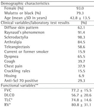
A total of 63 individuals were initially evaluated. Of those, 5 were excluded from the study because they presented PSS related to exposure to silica. The final sample analyzed consisted of 58 patients, whose general characteristics are shown in Table 1. Ages ranged from 13 to 69 years (mean ± standard deviation: 42.8 ± 13.5 years), disease duration ranged from 0.3 to 30 years (median, 4 years), and 63.8% of the patients presented diffuse cutaneous disease. Dyspnea was the most often reported respiratory symptom (38 cases, 65.5%), followed by cough and chest pain. Cracking rales were detected in only 9 individuals (15.5%). Anti-Scl 70 antibodies were detected in only 17 patients (29.3%).
presented FVC > 80% of predicted, and only 26.7% presented DLCO < 40% of predicted.
Table 4 shows the results of the multivariate analysis. Of the variables studied, only FVC < 80% of predicted demonstrated a tendency to be predictive of ILD in the group evaluated (odds ratio = 2.99), with borderline statistical significance in the multivariate analysis. However, this variable presented low sensitivity and specificity for the identification of ILD in cases of PSS.
DISCUSSION
Early detection of ILD seems to be relevant for the treatment of PSS patients, and the clinical approach may be insufficient.(18) Similar to what
has been seen in other studies,(8,19-20) dyspnea was
the most often reported respiratory symptom in our group, with similar frequency and intensity in patients with and without ILD, which reduced its usefulness in detecting this complication. This finding might be explained by the fact that a portion of the individuals with ILD probably present an incipient form of the disease (not yet radiologically detectable), which would in turn explain the high proportion of patients with normal FVC in this group. However, cracking rales were found exclusively in patients with significant ILD, a correlation previously reported in other studies. (8-9,19) This indication, which is highly specific for
the diagnosis of ILD in individuals with PSS, lacks sensitivity since it was found in only 30% of the ILD patients in our study.
As has been reported in other studies, ILD patients presented significantly reduced FVC and total lung capacity.(8,13) Although lower, DLCO values
and the proportion of patients with a significant reduction in DLCO (less than 40%) were not significantly different in ILD patients, representing a divergence from the findings of other studies.(8,13)
The presence of pulmonary vascular disease in patients without pulmonary fibrosis may, in addition to making tomographic detection possible in cases of incipient ILD, reduce DLCO, which explains why there was no difference in DLCO between the groups of PSS patients with and without ILD.(21)
In the present study, the frequency of ILD was high despite being lower than that found in most reports involving HRCT scans of PSS patients. In a prospective study evaluating 23 PSS patients, it was demonstrated that 91% of the individuals presented signs of ILD.(7) The population evaluated
in that study presented longer disease duration than did our group. Similar high frequencies were also reported in three other studies.(22-24) In the
study with the greatest number of patients, ILD was reported in 63.7% of the 91 individuals evaluated, this value being closer to the results of most of the studies currently available.(8-9,26-28) In
that study, the authors only included patients who presented respiratory symptoms and were,
TABLE 1
General characteristics of the 58 patients with progressive systemic sclerosis
TABLE 2
Frequency of abnormalities detected in the 30 patients in whom alterations were seen on the
high-resolution computed tomography scan of the chest
Demographic characteristics
Female (%) 93.0
Mulatto or black (%) 79.3 Age (mean ±SD in years) 42.8 ± 13.5 Clinical variables/laboratory test results (%)
Diffuse skin pattern 62.1 Raynaud’s phenomenon 91.4 Sclerodactylia 98.3
Arthralgia 84.5
Teleangiectasis 58.6 Current or former smoker 15.9
Dyspnea 65.5
Cough 39.7
Chest pain 37.9
Crackling rales 15.5
Hissing 6.9
Anti-Scl 70 positive 29.3 Functional variables**
FVC 77.2 ± 15.5
DLCO 56.7 ± 20.6
TLC* 74.8 ± 14.6
RV* 80.8 ± 31.1
*n = 43; **% of predicted (mean ± SD)
Abnormality Frequency (%) Bronchiolectasis 83.3
Honeycombing 80.0
therefore, more likely to present pulmonary involvement. In the only study in which a lower frequency of ILD than that of the present study was found, interstitial involvement was detected in only 39.5% of the 43 cases evaluated.(29)
The difference between the frequency found in this series and those found in other studies may be due to the heterogeneity of demographic characteristics (such as race), as well as of the
clinical and immunogenetic characteristics, of the individuals evaluated. All of the studies previously mentioned evaluated almost exclusively Caucasian individuals. Despite having evaluated patients with different characteristics from those evaluated in the present study, the Brazilian study reported a higher frequency of ILD.(9) In that study, 95% of
the patients were white and presented more prolonged ILD, which can result in different clinical
TABLE 3
Demographic, clinical and functional characteristics - Comparison between patients with interstitial lung disease and those without
Parameters ILD no ILD p
(n = 30) (n = 28) Demographic, clinical or laboratory test variables
Age (years) 45.1 40.3 NS
Duration of PSS (years) 8.5 6.1 0.05
Diffuse skin pattern (%) 73.3 53.6 NS
Dyspnea (%) 70.0 60.7 NS
Cough (%) 43.3 35.7 NS
Dysphagia (%) 46.7 50 NS
Regurgitation (%) 48.0 43.3 NS
Baseline dyspnea index (score) 7.8 8.1 NS
Crackling rales (%) 30 0 0.001
Anti-Scl 70 positive (%) 53.3 3.6 0.0003 Functional characteristics
FVC* 72.7 80.1 0.02
FEV1* 70.1 77.5 NS
DLCO* 53.8 59.8 NS
TLC* 69.2 80.1 0.01
FVC < 80%** 66.7 35.7 0.02
TLC < 80%** 71.4 54.5 NS
DLCO < 75%** 86.7 81.5 NS
*% of predicted (mean ± standard deviation); **frequency (%)
ILD: interstitial lung disease; PSS: progressive systemic sclerosis; NS: not significant; FVC: forced vital capacity; FEV1: forced expiratory volume in one second; DLCO: lung diffusing capacity for carbon monoxide; TLC: total lung capacity
TABLE 4
Potential predictors of interstitial lung disease in the 58 progressive systemic sclerosis patients studied - Logistic regression analysis
Variable Univariate Multivariate OR Sensitivity Specificity (95% CI) (%) (%)
DLCO < 50% 0.17 NS ND ND ND FVC < 80% 0.06 0.05** 2.99 64 60
(1.12-6.95)
TLC < 80%* 0.36 NS ND ND ND BDI score < 6 0.42 NS ND ND ND PSS duration > 4 years 0.05 NS ND ND ND
*n = 43; **p values
expressions of the disease.(4-5) A close correlation
between human leukocyte antigen haplotypes and expression of autoantibodies such as anti-Scl 70 has been described.(4,30) The anti-Scl 70 antibody
was detected in a third of our group and seems to be correlated with the presence of ILD.(4,31-32)
However, this marker also presented low sensitivity since it was positive in only half of the ILD patients. It is unclear whether anti-Scl 70 is only a marker of involvement or an etiopathogenic determinant. The identification of factors predictive of ILD in PSS patients may be important for clinical management. Multivariate analysis revealed that the clinical and functional variables studied were of little use in the prediction of ILD, which underscores the importance of radiological evaluation using HRCT. Among the variables studied, only abnormal FVC (< 80% of predicted) tended to be predictive of ILD in this group of PSS patients, with borderline significance. The confidence interval correlated with this variable was wide and sensitivity and specificity were not high. The correlation between reduced FVC and interstitial involvement is frequent but not constant. Evaluation with HRCT was routinely performed in a group of patients that were not selected and therefore may have presented an incipient form of ILD, with few repercussions for the functional evaluation. In addition, restriction patterns may also occur in patients without ILD,(19)
resulting from the dysfunction of the respiratory muscles or alterations in chest cavity compliance. Despite the more prolonged PSS duration seen among the ILD patients, multivariate analysis failed to demonstrate a significant correlation between PSS duration and ILD. This relationship has been described in previous studies,(8,26) in which the
existence of more prolonged PSS in patients presenting interstitial pulmonary involvement was also mentioned. In a prospective cohort study, severe pulmonary fibrosis was reported to occur more often in the three first years of disease. Therefore, we should evaluate our findings with caution since they result from an evaluation made at a specific point in time.
In summary, the frequency of ILD was high in our group of PSS patients. From a practical viewpoint, the data presented in our study may indicate that PSS patients who present reduced FVC, presence of anti-Scl 70 and cracking rales
should be monitored more rigorously, using, for example, HRCT. However, it is important to emphasize that the present study was carried out in tertiary-care facilities and that, therefore, our sample may not be representative of the total population of PSS patients. In future studies, the inclusion of patients from primary-care facilities and private clinics should be encouraged in order to improve the sample representativeness and thereby establish a frequency of ILD that is more reliable in view of the broad clinical spectrum of PSS patients. The presence of ILD on HRCT scans does not necessarily indicate clinically significant disease, and this study did not evaluate the magnitude of the tomographic findings. Longitudinal, long-term follow-up studies are necessary in order to determine the impact of these findings on the prognosis and clinical evolution of PSS patients.
REFERENCES
1. Steen VD, Medsger TA Jr. Severe organ involvement in systemic sclerosis with diffuse scleroderma. Arthritis Rheum. 2000;43(11):2437-44.
2. Steen VD, Conte C, Owens GR, Medsger TA Jr. Severe restrictive lung disease in systemic sclerosis. Arthritis Rheum. 1994;37(9):1283-89.
3. McCarthy DS, Baragar FD, Dhingra S, Sigurdson M, Sutherland JB, Rigby M et al. The lungs in systemic sclerosis (scleroderma): A review and new information. Semin Arthritis Rheum. 1988;17(4):271-83.
4. Briggs DC, Vaughan RW, Welsh KI, Myers A, Du Bois RM, Black CM. Immunogenetic prediction of pulmonary fibrosis in systemic sclerosis. Lancet. 1991;338-(8768):661-2.
5. Reveille JD, Fischbach M, McNearney T, Friedman AW, Aguilar MB, Lisse J et al. Systemic sclerosis in three US ethnic groups: a comparison of clinical, sociodemographic, serologic and immunogenetic determinants. Semin Arthritis Rheum. 2001;30(5):332-46.
6. White B. Interstitial lung disease in scleroderma. Rheum Dis Clin North Am. 2003;29(2):371-90.
7. Schurawitzki H, Stiglbauer R, Graninger W, Herold C, Polzleitner D, Burghuber OC, et al. Interstitial lung disease in progressive systemic sclerosis: high resolution CT versus radiography. Radiology. 1990;176(3):755-9. 8. Harrison NK, Granville AR, Strickland B, Haslam PL, Corrin B, Addis BJ, et al. Pulmonary involvement in systemic sclerosis: the detection of early changes by thin section CT scan, bronchoalveolar lavage and 99m-Tc -DTPA clearance. Respir Med. 1989;83(5):403-14.
10. Preliminary criteria for the classification of systemic sclerosis (scleroderma). Subcommittee for scleroderma criteria of the American Rheumatism Association Diagnosis and Therapeutic Criteria committee. Arthritis Rheum. 1980;23(5):581-90.
11. LeRoy EC, Black CM, Fleischmajer R, Jablonska S, Krieg T, Medsger TA Jr, et al. Scleroderma (systemic sclerosis): classification, subsets and pathogenesis. J Rheumatol. 1988;15(2):202-5.
12. Mahler DA, Weinberg DH, Wells CK, Feinstein AR. The measurement of dyspnea. Contents, interobserver agreement, and physiologic correlates of two new clinical indexes. Chest. 1984;85(6):751-8.
13. Remy-Jardin M, Remy J, Wallaert B, Bataille D, Hatron PY. Pulmonary involvement in progressive systemic sclerosis: sequential evaluation with CT, pulmonary function tests and bronchoalveolar lavage. Radiology. 1993;188(2):499-506. 14. American Thoracic Society ( ATS ). Standardization of
spirometry. 1994 update. Am J Respir Crit Care Med. 1995;152(3):1107-36.
15. Quanjer PH, Tammeling GJ, Cotes JE, Pedersen R, Peslin R, Yearnault JC. Lung volumes and forced ventilatory flows. Report of working party, standardization of lung function tests. European Community for steel and coal. Official Statement of the European Respiratory Society. Eur Respir J Suppl. 1993;16:5-40.
16. Crapo RO, Morris AH. Standardized single breath normal values for carbon monoxide diffusing capacity. Am Rev Respir Dis. 1981;123(2):185-9.
17. Hosmer DW, Lemeshow S. Applied logistic regression. New York: Wiley; 1989.
18. Steen VD, Lanz JK Jr, Conte C, Owens GR, Medsger TA Jr. Therapy for severe interstitial lung disease in systemic sclerosis: A retrospective study. Arthritis Rheum. 1994;37(9):1290-6.
19. Steen VD, Owens GR, Fino GJ, Rodnan GP, Medsger TA Jr. Pulmonary involvement in systemic sclerosis (scleroderma). Arthritis Rheum. 1985;28(7):759-67.
20. Morelli S, Barbieri C, Sgreccia A, Ferrante L, Pittoni V, Conti et al. Relationship between cutaneous and pulmonary involvement in systemic sclerosis. J Rheumatol. 1997;24(1):81-5.
21. Steen V, Medsger TA Jr. Predictors of isolated pulmonary hypertension in patients with systemic sclerosis and limited cutaneous involvement. Arthritis Rheum. 2003;48(2):516-22.
22. Warrick JH, Bhalla M, Schabel SI, Silver R. High resolution computed tomography in early scleroderma lung disease. J Rheumatol. 1991;18(10):1520-8.
23. Garber SJ, Wells AU, duBois RM, Hansell DM. Enlarged mediastinal lymph nodes in the fibrosing alveolitis of systemic sclerosis. Br J Radiol. 1992;66(77):983-6. 24. Ooi GC, Mok MY, Tsang KW, Wong Y, Khong PL, Fung
PC, et al. Interstitial lung disease in systemic sclerosis. An HRCT - clinical correlative study. Acta Radiol. 2003; 44(3):258-64.
25. Devenyi K, Czirjak L. High resolution computed tomography for the evaluation of lung involvement in 101 patients with scleroderma. Clin Rheumatol. 1995;14(6):633-40.
26. Shahin AA, Sabri YY, Mostafa HA, Sabry EY, Hamid MA, Gamal H, et al. Pulmonary function tests, high re s o l u t i o n c o m p u t e r i z e d t o m o g r a p h y, a l p h a - 1 antitrypsin measurement, and early detection of pulmonary involvement in patients with systemic sclerosis. Rheumatol Int. 2001;20(3):95-100. 27. Wechsler RJ, Steiner RM, Spirn PW, Rubin S, Salazar
AM, Shah R, et al. The relationship of thoracic lymphadenopathy to pulmonary interstitial disease in diffuse and limited systemic sclerosis: CT findings. AJR Am J Roentgenol. 1996;167(1):101-4. 28. Pignone A, Matucci-Cerinic M, Lombardi A, Fedi R,
Fargnoli R, De Dominicis R, et al. High resolution computed tomography in systemic sclerosis. Real diagnostic utilities in the assessment of pulmonary involvement and comparison with other modalities of lung investigation. Clin Rheumatol. 1992;11(4):465-72. 29. Marie I, Dominique S, Levesque H, Ducrotté P, Denis P, Hellot MF, et al. Esophageal involvement and pulmonary manifestations in systemic sclerosis. Arthritis Rheum. 2001;45(4):346-54.
30. Tan FK. Systemic sclerosis: the susceptible host (genetics and environment). Rheum Dis Clin North Am. 2003; 29(2):211-37.
31. Manoussakis MN, Constantopoulos SH, Gharavi AE, Moutsopoulos HM. Pulmonary involvement in systemic sclerosis. Association with anti-Scl 70 antibody and digital pitting. Chest. 1987;92(3):509-13.