Cop
yright
© ABE&M t
odos os dir
eit
os r
eser
vados
.
Thyroid autoimmunity in patients
with hyperprolactinemia:
an observational study
Autoimunidade tireoidiana em pacientes com hiperprolactinemia: um estudo observacional
Eda Demir Onal1, Fatma Saglam1, Muhammed Sacikara1, Reyhan Ersoy1, Bekir Cakir1
ABSTRACT
Objective: To establish whether there is a relationship between hyperprolactinemia and primary thyroid disorders, focusing on patients with autoimmune features. Materials and methods: The medical records of 100 patients with hyperprolactinemia (HPRL) were retrospectively examined. Records of thyroid ultrasonography (USG), basal serum levels of thyroid stimulating hormone, cir-culating free thyroxine, free triiodothyronine, antithyroglobulin (anti-Tg), and antithyroperoxidase (anti-TPO) antibodies were analyzed. In 100 control subjects, matched by age and gender with HPRL patients, thyroid USG, thyroid function tests (TFTs), and autoantibody panel were obtained. Results: The median PRL in patients was 93 ng/mL (range: 37-470). Twenty-ive patients (25%) and 22 controls (22%) had positive anti-Tg and/or anti-TPO titers (P = 0.739). The median serum PRL was 98 (37-470) ng/mL in patients with positive thyroid autoantibodies, and 92 (40-470) ng/mL in patients who were negative (P = 0.975). Among the individuals with autoantibody positivity TFTs abnormalities were more frequent in HPRL patients (60%, out of 25 patients, 14 with subclinical hypothyroidism and one with hyperthyroidism) than in controls (9.1%, out of 22 patients, 2 with subclinical hyperthyroidism) (P < 0.001). Twenty-seven patients with HPRL and 31 controls had goiter (27 vs. 31%, P = 0.437). Forty-six patients (46%) and 50 (50%) controls had one or more of the features of thyroid disorder, which were goiter, positive thyroid autoantibody, and thyroid function abnormality (P = 0.888). Conclusion: HPRL may be associated with more severe thyroid dysfunction in patients with thyroid autoimmunity.
Arq Bras Endocrinol Metab. 2014;58(1):48-52
Keywords
Hyperprolactinemia; thyroiditis; autoimmunity; goiter
RESUMO
Objetivo: Veriicar se existe uma relação entre a hiperprolactinemia e distúrbios primários da tireoi-de, focando em pacientes com características autoimunes. Materiais e métodos: Os prontuários de 100 pacientes com hiperprolactinemia (HPRL) foram examinados retrospectivamente. Foram analisa-dos registros de ultrassonograia da tireoide (USG), níveis séricos basais de hormônio tireoestimu-lante, tiroxina livre, triiodotironina livre e anticorpos antitireoglobulina (anti-Tg) e antitireoperoxida-se (anti-TPO). Foram obtidos de 100 controles, pareados por idade e antitireoperoxida-sexo com pacientes com HPRL, USG, testes de função da tireoide (TFTs) e painel de autoanticorpos. Resultados: A média de PRL em pacientes foi de 93 ng/mL (variação: 37-470). Vinte e cinco pacientes (25%) e 22 controles (22%) foram positivos para títulos de anti-Tg e/ou anti-TPO (P = 0,739). A mediana de PRL sérica foi de 98 (37-470) ng/mL em pacientes positivos para autoanticorpos tiroidianos e 92 (40-470) ng/mL em pa-cientes negativos (P = 0,975). Entre os indivíduos positivos para autoanticorpos, as anormalidades da TFTs foram mais frequentes em pacientes HPRL (60%; de 25 pacientes, 14 com hipotireoidismo subc-línico e um com hipertireoidismo) do que nos controles (9,1%; de 22 pacientes, 2 com hipertireoidis-mo subclínico) (P < 0,001). Vinte e sete pacientes com HPRL e 31 controles apresentavam bócio (27 contra 31%; P = 0,437). Quarenta e seis pacientes (46%) e 50 (50%) controles tiveram uma ou mais das características de problemas de tireoide, como bócio, autoanticorpos antitireoide e anormalidades da função tiroidiana (P = 0,888). Conclusão: A HPRL pode estar associada à disfunção da tireoide mais grave em pacientes com autoimunidade contra a tireoide. Arq Bras Endocrinol Metab. 2014;58(1):48-52
Descritores
Hiperprolactinemia; tireoidite; autoimunidade; bócio
1 Yildirim Beyazit University Medical
School Ataturk Teaching and Research Hospital, Department of Endocrinology and Metabolism, Bilkent, Ankara, Turkey
Correspondence to:
Eda Demir Onal
Ceyhun Atuf Kansu Cad Ehl-i Beyt Mah 1268,
Sok 10/6 TR-06520,
Balgat, Cankaya, Ankara, Turkey [email protected]
Cop
yright
© ABE&M t
odos os dir
eit
os r
eser
vados
.
INTRODUCTION
Prolactin (PRL) is a polypeptide hormone discovered more than 70 years ago to be a pituitary factor that stimulates mammary gland development and lactation (1,2). It is not only produced by lactotrophic cells of the anterior pituitary gland, but also by various extra-pituitary sites as neurons, prostate, decidua, mammary epithelium, skin, and immune cells (3). Over the past decade, PRL has been linked to an extended family of proteins, referred to as hematopoietic cytokines (3). The classiication of PRL as a cytokine has been based on several molecular and functional evidences. First, its receptor is a member of the cytokine receptor superfamily. Second, PRL is predicted to adopt the up-up-down-down four α-helix bundle fold characteristic of hematopoietic cytokines (4,5). Third, similar to well-recognized cytokines, PRL was shown to act on cells of the immune system (6,7).
For the time being, the only characterized disorder related to PRL is hyperprolactinemia (HPRL), which is responsible for almost 25% of menstrual cycle disorders in women. It can also be argued that PRL should be included in the list of factors favoring the proliferation of certain tumors, e.g. breast and prostate cancer (8,9). Besides these diseases linked to abnormal levels of circulating PRL, a role in the pathogenesis of autoimmune diseases has been suggested. Hyper-prolactinemia has been often observed in some non-organ-speciic autoimmune diseases, such as systemic lupus erithematosus, rheumatoid artritis, systemic sclerosis, and Sjogren syndrome; moreover, it has been also described in some organ-speciic autoimmune diseases, such as type 1 diabetes mellitus, Graves’ diseases (GD), Hashimoto’s thyroiditis (HT), Addison’s disease (AD), lymphocytic hypophysitis (LYH), celiac disease (CD), and multiple sclerosis (MS) (10-19). But there is still considerable controversy concerning the true immunomodulatory role of PRL, and the subject deserves further investigation.
In this study, we attempted to establish whether there is a relationship between HPRL and primary thyroid disorders, focusing on patients with autoimmune features.
MATERIALS AND METHODS
This is a retrospective study in endocrinology outpa-tient clinics of a tertiary reference center in the period from January 2005 to April 2013. All HPRL cases
with a records of thyroid ultrasonography (USG) and at least one measurement of basal serum thyroid sti-mulating hormone (TSH), circulating free thyroxine (fT4), free triiodothyronine (fT3), antithyroglobulin (anti-Tg), and/or antithyroperoxidase (anti-TPO) an-tibodies, were selected for inclusion. Women who were breastfeeding, using oral contraceptives, or pregnant were not enrolled. All patients participated in the stu-dy after signing an informed consent form. The local Ethics Committee of our hospital approved the study.
Venous blood samples were drawn between 8 am and 9 am from all the patients while fasting. Serum PRL, fT4, fT3, TSH, anti-Tg, and anti-TPO levels were measured by immunoassay using commercial kits. The normal ranges were: fT3: 1.8-4.6 pg/mL, fT4: 0.9-1.7 ng/ dL, TSH: 0.27-4.2 uIU/mL; anti-Tg: 0-115 IU/mL and anti-TPO: 0-34 IU/mL. Anti-Tg and anti-TPO titers that showed three standard deviations above the mean were considered positive. HPRL was deined as serum prolactin level > 29.9 ng/mL, according to the reference assay provided by the manufacturer. The thyroid USG was performed with a Logic 200 scanner using a 7.5 MHz linear array transducer and direct contact technique by an experienced endocrinologist. Goiter was diagnosed when the anteroposterior diameter of both lobes was ≥ 20 mm, or when any nodule was detected. The presence of a diffuse hypoechoic pattern with or without hyperechoic lines was considered suggestive of thyroiditis. A patient was deined as having primary thyroid disorders by one or more of the following diagnostic criteria: (i) goiter, (ii) positive anti-thyroid antibodies, and/or (iii) primary thyroid function abnormalities.
A control group, matched by age and gender with HPRL patients, was used for this study. All subjects denied having known thyroid diseases or relatives with any kind of thyroid disorders. In the control group, TFTs and autoantibody panel were obtained and thyroid USG was performed.
Cop
yright
© ABE&M t
odos os dir
eit
os r
eser
vados
.
RESULTS
Group 1 included 100 patients (mean age: 37.1 ± 10.9 years, range 17-63) and group 2 included 100 control subjects (mean age: 39.9 ± 10.5 years, range 21-68). There were 17 males (17%) and 83 females (83%) in each group. There were no signiicant differences between the two groups with respect to sex and age (P
> 0.05). The median PRL in patients was 93 ng/mL (range: 37-470). The median duration of HPRL was 3 years (range: 1-20). HPRL was caused by prolactinoma in 93 (93%), antipsychotic drugs in 4 (4%), and stalk compression by nonfunctional pituitary adenoma in 3 (3%) patients. HPRL was successfully controlled by cabergoline in 85 (85%), bromocriptine in 4, and by the combination of the two agents in 11 patients.
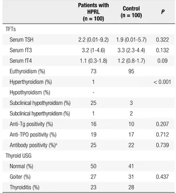
The results of the evaluation of TFTs and thyroid au-toantibody panel are shown in table 1. The fT4 was sig-niicantly lower in group 1 (P = 0.009), but the median fT4 levels was still within normal range in both groups (Table 1). The proportion of patients with subclini-cal hypothyroidism was signiicantly higher in group 1 compared with group 2 (25% vs. 3%, P < 0.001). There
Table 1. Thyroid function tests and autoantibody panel of the patients with
hyperprolactinemia (HPRL) and control subjects
Patients with HPRL (n = 100)
Control
(n = 100) P
TFTs
Serum TSH 2.2 (0.01-9.2) 1.9 (0.01-5.7) 0.322
Serum fT3 3.2 (1-4.6) 3.3 (2.3-4.4) 0.132
Serum fT4 1.1 (0.3-1.8) 1.2 (0.8-1.7) 0.09
Euthyroidism (%) 73 95
Hyperthyroidism (%) 1 < 0.001
Hypothyroidism (%)
-Subclinical hypothyroidism (%) 25 3
Subclinical hyperthyroidism (%) 1 2
Anti-Tg positivity (%) 16 10 0.207
Anti-TPO positivity (%) 19 17 0.712
Antibody positivity (%)a 25 22 0.739
Thyroid USG
Normal (%) 50 41
Goiter (%) 27 31 0.437
Thyroiditis (%) 23 28
TSH: thyroid stimulating hormone; fT3: free triiodothyronine; fT4: free thyroxine; anti-Tg: antithyroglobulin antibody; anti-TPO: antithyroperoxidase antibody; TFTs: thyroid function tests; USG: ultrasonography. Parametric values was expressed as means ± S.D. Statistical signiicance was set at a P value of 5%.
Normal values: fT3: 1.8-4.6 pg/mL, fT4: 0.9-1.7 ng/dL, TSH: 0.27-4.2 uIU/mL.
a Positivity of anti-Tg and/or anti-TPO.
were no signiicant differences between the two groups with respect to autoantibody positivity and the propor-tion of patients with ultrasonographic evidence of goiter or thyroiditis (P > 0.05). On the other hand, TFTs ab-normalities were more frequent in HPRL patients (60%, out of 25 patients, 14 subclinical hypothyroidism and one hyperthyroidism) than in controls (9.1%, out of 22 patients, 2 subclinical hyperthyroidism) when indivi-duals with autoantibody positivity were considered (P
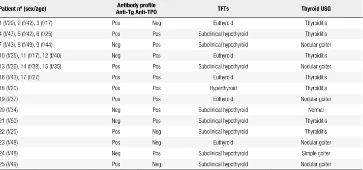
< 0.001). Characteristics of 25 HPRL patients who had evidence of thyroid autoimmunity during follow-up are shown in table 2. The median serum PRL was 98 (37-470) ng/mL in patients with positive thyroid autoanti-bodies and 92 (40-470) ng/mL in patients who tested negative for thyroid autoantibodies (P = 0.975). Forty-six patients (46%) and 50 (50%) control subjects had one or more of the features of thyroid disorder, which were goiter, positive thyroid autoantibody, and thyroid function abnormality (P = 0.888). The median serum PRL was 98.1 (37-470) ng/mL in patients with any fea-ture of primary thyroid disorder and 91 (40-470) ng/ mL in the remaining patients in group 1 (P = 0.691).
DISCUSSION
Cop
yright
© ABE&M t
odos os dir
eit
os r
eser
vados
.
Table 2. Characteristics of 25 patients with hyperprolactinemia who had evidence of thyroid autoimmunity during follow-up
Patient nº (sex/age) Antibody proile
Anti-Tg Anti-TPO TFTs Thyroid USG
1 (f/29), 2 (f/42), 3 (f/17) Pos Neg Euthyroid Thyroiditis
4 (f/47), 5 (f/42), 6 (f/25) Pos Pos Subclinical hypothyroid Thyroiditis
7 (f/43), 8 (f/49), 9 (f/44) Neg Pos Subclinical hypothyroid Nodular goiter
10 (f/35), 11 (f/17), 12 (f/40) Neg Pos Euthyroid Thyroiditis
13 (f/36), 14 (f/38), 15 (f/35) Pos Pos Subclinical hypothyroid Nodular goiter
16 (f/43), 17 (f/27) Pos Pos Euthyroid Thyroiditis
18 (f/20) Pos Pos Hyperthyroid Thyroiditis
19 (f/37) Pos Pos Euthyroid Nodular goiter
20 (f/34) Neg Pos Subclinical hypothyroid Normal
21 (f/50) Neg Pos Subclinical hypothyroid Thyroiditis
22 (f/25) Pos Neg Subclinical hypothyroid Thyroiditis
23 (f/48) Pos Neg Euthyroid Nodular goiter
24 (f/48) Neg Pos Subclinical hypothyroid Simple goiter
25 (f/49) Pos Neg Subclinical hypothyroid Nodular goiter
TFTs: thyroid function tests; f: female; m: male; anti-Tg: antithyroglobulin antibody; anti-TPO: antithyroperoxidase antibody; pos: positive; neg: negative.
There are several clinical studies that demonstrate a relationship between HPRL and autoimmune thyroid disease (15,22-24). Giusti and cols. showed that the prevalence of thyroid ultrasonographic alterations including goiter and chronic thyroiditis (30.8% vs. 15.5%) and the prevalence of autoantibodies (29.6%
vs. 14.3%) were signiicantly higher in HPRL than in controls (15). Another report by Ferrari and cols. reported comparable results: in a group of 82 women with HPRL, 20% had anti-Tg and 12% had anti-microsomal antibodies (22). Most patients were euthyroid in this study. In a series by Kramer and cols., positivity for any of the thyroid autoantibodies was 57.1% in untreated HPRL, 10% in HPRL on treatment, and 3.6% in the control group (23). Last, Poyraz and cols. analyzed a group of schizophrenic patients and observed that the prevalence of HPRL was signiicantly higher in patients positive for thyroid autoantibodies (81%), when compared with patients negative for them (49%). Serum levels of PRL were also signiicantly higher in patients with positivity for thyroid autoantibodies in that series (24). Despite the studies mentioned above, literature is still scarce with respect to primary thyroid disorders in patients with HPRL.
Anti-TG and anti-TPO are indicators of thyroid inlammation and detection of these autoantibodies is a
Cop
yright
© ABE&M t
odos os dir
eit
os r
eser
vados
.
relationship between HPRL and autoimmune diseases may not be observed on every occasion because of the complexity of the immune system. Although PRL abnormalities have been reported in immune system disorders, it has not been possible to inluence the course of these diseases by manupilating PRL release in any randomized controlled trial.
The prevalence of thyroid ultrasonographic altera-tions including goiter and thyroiditis was not more prev-alent in our patients with HPRL. Endocrinologists are more inclined to screen patients with HPRL under their care for thyroid dysfunction and thyroid nodules than general practitioners. For this reason we think that such a relationship can only be demonstrated by randomized, prospective, double-blind, placebo-controlled trials.
In conclusion, our study results did show a remarkably high prevalence of thyroid dysfunction in patients with HPRL and thyroid autoimmunity, but thyroid autoimmunity and goiter were not more frequent. Autoimmunity is a complex process and the results of similar series in the literature should be cautiously interpreted. Future prospective studies on larger populations are needed to provide irrefutable evidence to support a direct causative link between HPRL and autoimmune thyroid disease.
Disclosure: no potential conlict of interest relevant to this article was reported.
REFERENCES
1. Stricker P, Grueter R. Action du lobe ant’erieur de l’hypophyse sur la mont’ee laiteuse. C R Soc Biol. 1928;99:1978-80.
2. Riddle O, Bates RW, Dykshorn SW. The preparation, identiication and assay of prolactin – a hormone of the anterior pituitary. Am J Physiol. 1933;105:191-216.
3. Ben-Jonathan N, Mershon JL, Allen DL, Steinmetz RW. Extrapituitary prolactin: distribution, regulation, functions, and clinical aspects. Endocr Rev. 1996;17(6):639-69.
4. Horseman ND, Yu-Lee LY. Transcriptional regulation by the helix bundle peptide hormones: growth hormone, prolactin, and hematopoietic cytokines. Endocr Rev. 1994;15(5):627-49.
5. GofinV, Martial JA, Summers NL. Use of a model to understand prolactin and growth hormone speciicities. Protein Eng. 1995;8(12):1215-31.
6. Nagy E, Berczi I, Wren GE, Asa SL, Kovacs K. Immunomodulation by bromocriptine. Immunopharmacology. 1983;6(3):231-43. 7. Bernton EW, Meltzer MS, Holaday JW. Suppression of macrophage
activation and T-lymphocyte function in hypoprolactinemic mice. Science. 1989;239(4838):401-4.
8. Llovera M, Pichard C, Bernichtein S, Jeay S, Touraine P, Kelly PA, et al. Human prolactin (hPRL) antagonists inhibit hPRL-activated
signaling pathways involved in breast cancer cell proliferation. Oncogene. 2000;19(41):4695-705.
9. Leav I, Merk FB, Lee KF, Loda M, Mandoki M, McNeal JE, et al. Prolactin receptor expression in the developing human prostate and in hyperplastic, dysplastic, and neoplastic lesions. Am J Pathol. 1999;154(3):863-70.
10. Jara LJ, Vera-Lastra O, Miranda JM, Alcala M, Alvarez-Nemegyei J. Prolactin in human systemic lupus erythematosus. Lupus. 2001;10(10):748-56.
11. Chikanza IC, Petrou P, Chrousos G, Kingsley G, Panayi GS. Excessive and dysregulated secretion of prolactin in rheumatoid arthritis: immunopathogenetic and therapeutic implications. Br J Rheumatol. 1993;32(6):445-8.
12. El Miedany YM, Ahmed I, Moustafa H, El Baddini M. Hyperprolactinemia in Sjogren’s syndrome: a patient subset or a disease manifestation? Joint Bone Spine. 2004;71(3):203-8. 13. Neidhart M. Prolactin in autoimmune diseases. Proc Soc Exp Biol
Med. 1998;217(4):408-19.
14. Li J, Teng W, Shan Z. Effects of prolactin on HLA-DR and CD40 expressions by human thyrocytes. Chin Med J. 2001;114(11):1151-6. 15. Giusti M, Foppiani L, Fazzuoli L, Molinari E, Guido R, Valenti
S, et al. An increased prevalence of thyroid echographic and autoimmune changes in hyperprolactinemic women on therapy with dopaminergic drugs. Recenti Prog Med. 1999;90(3):147-51. 16. Lever EG, McKerron CG. Auto-immune Addison’s disease
associated with hyperprolactinaemia. Clin Endocrinol. 1984;21(4):451-7.
17. Thodou E, Asa SL, Kontogeorgos G, Kovacs K, Horvath E, Ezzat S. Clinical case seminar: lymphocytic hypophysitis: clinicopathological indings. J Clin Endocrinol Metab. 1995;80(8):2302-11.
18. Kapur G, Patwari AK, Narayan S, Anand VK. Serum prolactin in celiac disease. J Trop Pediatr. 2004;50(1):37-40.
19. Azar ST, Yamout B. Prolactin secretion is increased in patients with multiple sclerosis. Endocr Res. 1999;25(2):207-14.
20. Matera L, Mori M, Geuna M, Buttiglieri S, Palestro G. Prolactin in autoimmunity and antitumor defence. J Neuroimmunol. 2000;109(1):47-55.
21. Yu M, Johnson JM, Tuohy VK. Generation of autonomously pathogenic neoautoreactive TH1 cells during the development of the determinant spreading cascade in murine autoimmune encephalomyelitis. J Neurosci. 1996;45(4):463-70.
22. Ferrari C, Boghen M, Paracchi A, Rampini P, Raiteri F, Benco R, et al. Thyroid autoimmunity in hyperprolactinaemic disorders. Acta Endocrinol (Copenh). 1983;104(1):35-41.
23. Kramer CK, Tourinho TF, de Castro WP, da Costa Oliveira M. Association between systemic lupus erythematosus, rheumatoid arthritis, hyperprolactinemia and thyroid autoantibodies. Arch Med Res. 2005;36(1):54-8.
24. Poyraz BC, Aksoy C, Balcioğlu I. Increased incidence of autoim-mune thyroiditis in patients with antipsychotic-induced hyper-prolactinemia. Eur Neuropsychopharmacol. 2008;18(9):667-72. 25. Rho MH, Kim DW, Hong HP, Park YM, Kwon MJ, Jung SJ, et
al. Diagnostic value of antithyroid peroxidase antibody for incidental autoimmune thyroiditis based on histopathologic results. Endocrine. 2012;42(3):647-52.